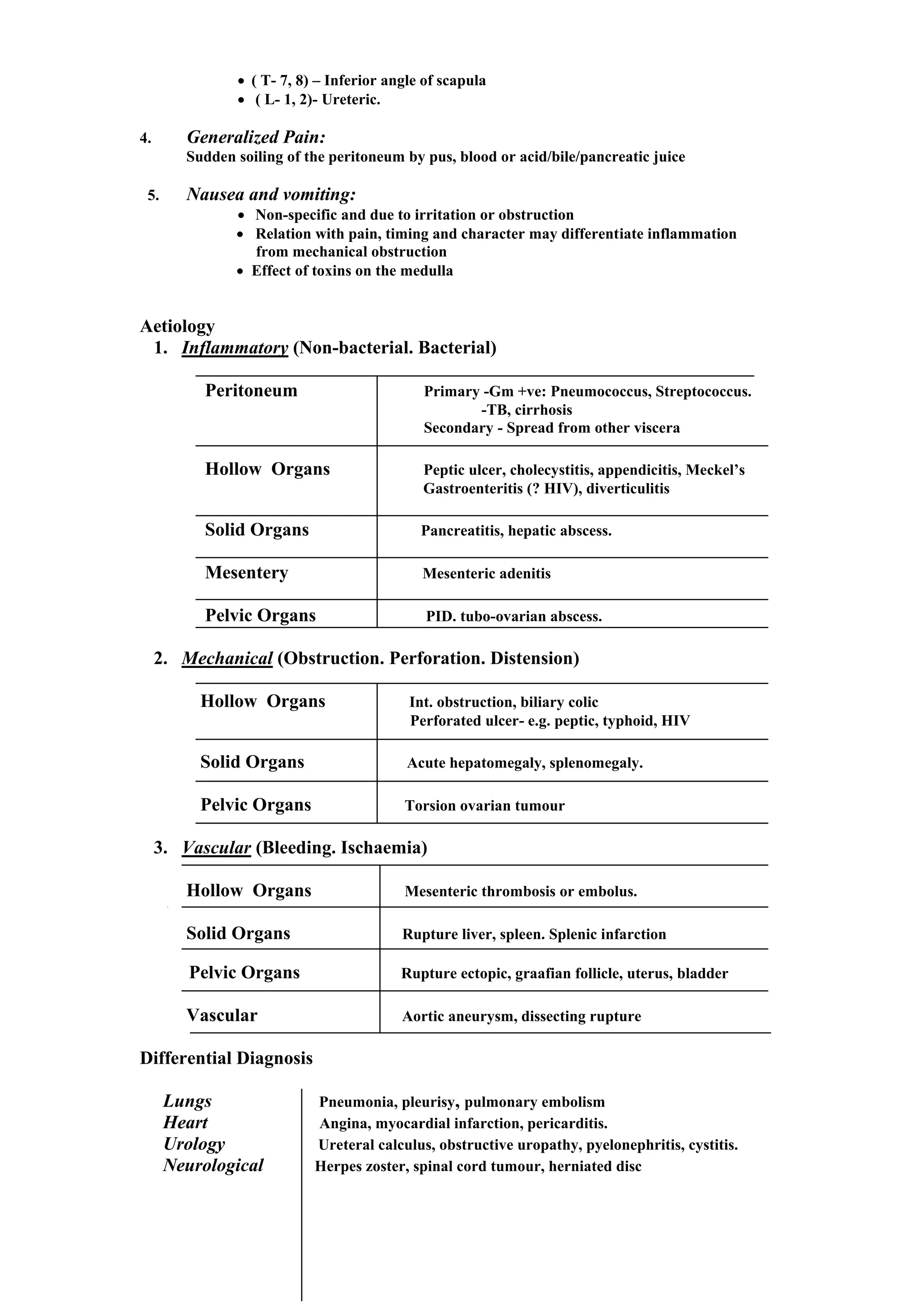
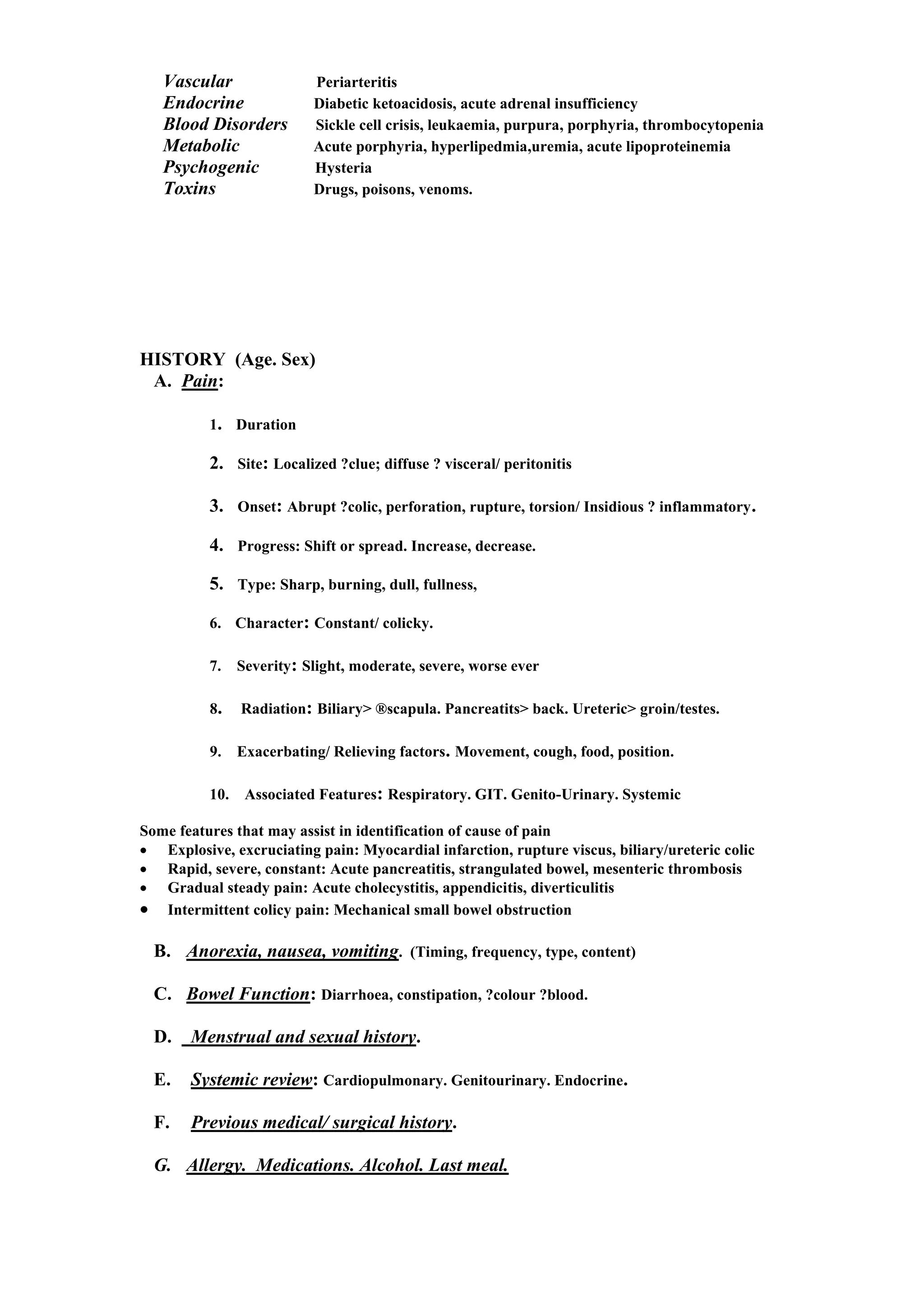
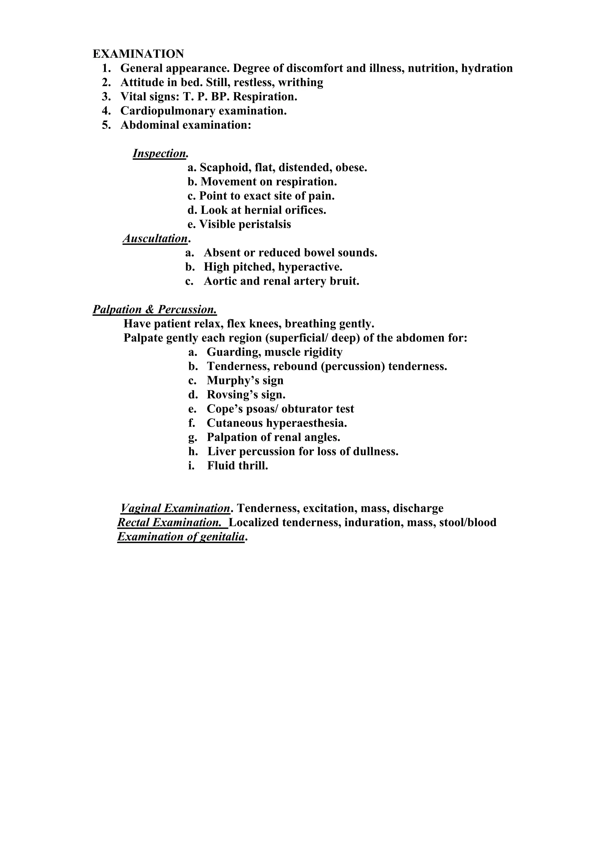
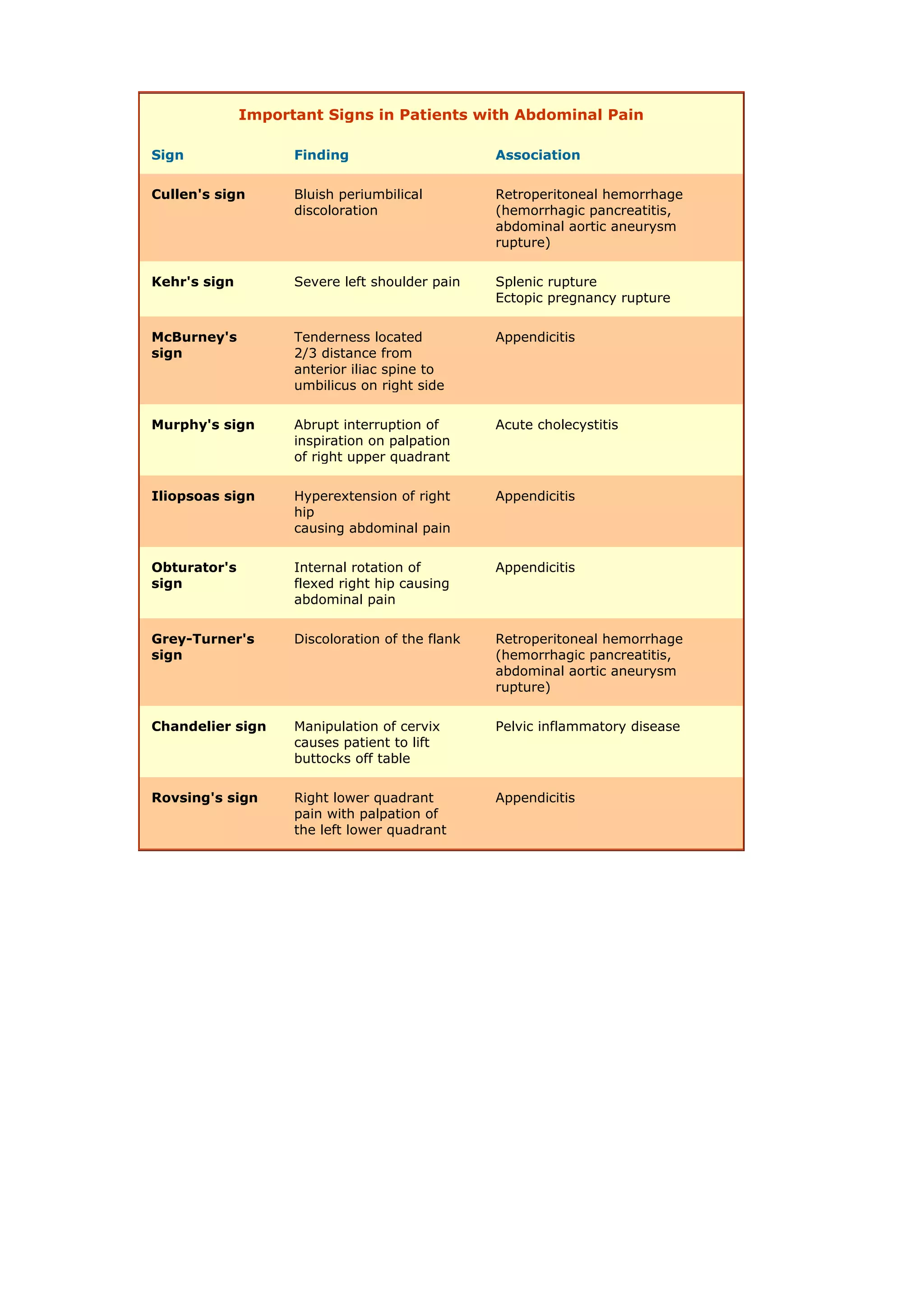
SURGERY
ACUTE ABDOMEN
DR. CHONGO SHAPI (BSc. HB, MBChB)
ACUTE ABDOMEN
Definition: Rapid onset of ABDOMINAL PAIN (in a patient previously well)
+/- NAUSEA and VOMITING.
“Acute abdomen” => Early diagnosis => Management
Types of Abdominal Pain ( thru sympathetic [splanchnic ] nerves)
1. Visceral Pain:
• Caused by stimulation of visceral afferents on distension, contraction,
ischaemia, chemical irritation
• Usually colicky and relieved by pressure.
• Diffuse, poorly localized, and difficult to describe.
• May be referred to a distant region.
• Midline:
Structure Nerves/ Sensory level Site
Foregut Coeliac ( T6-8) Epigastrium
Midgut Sup. Mesenteric ( T10-11) Umbilical
Hindgut Inf. Mesenteric (T11-L1) Hypogastrium
• May be associated with nausea, vomiting and reflex hypotension
2. Parietal Pain:
• Irritation of parietal peritoneum by blood, inflammatory exudates, content of
hollow viscus e.g. bile, acid, pancreatic juice
• Constant, sharp and localized to site of irritation.
• Aggravated by pressure and movement.
• Associated with reflex rigidity, reduced bowel activity.
3. Referred Pain:
Pain of visceral disease referred to a superficial area of body
derived from the same segment of cord as the viscus
• ( C- 3, 4, 5) - Diaphragm
• ( T- 7, 8) – Inferior angle of scapula
• ( L- 1, 2)- Ureteric.
4. Generalized Pain:
Sudden soiling of the peritoneum by pus, blood or acid/bile/pancreatic juice
5. Nausea and vomiting:
• Non-specific and due to irritation or obstruction
• Relation with pain, timing and character may differentiate inflammation
from mechanical obstruction
• Effect of toxins on the medulla
Aetiology
1. Inflammatory (Non-bacterial. Bacterial)
Peritoneum Primary -Gm +ve: Pneumococcus, Streptococcus.
-TB, cirrhosis
Secondary - Spread from other viscera
Hollow Organs Peptic ulcer, cholecystitis, appendicitis, Meckel’s
Gastroenteritis (? HIV), diverticulitis
Solid Organs Pancreatitis, hepatic abscess.
Mesentery Mesenteric adenitis
Pelvic Organs PID. tubo-ovarian abscess.
2. Mechanical (Obstruction. Perforation. Distension)
Hollow Organs Int. obstruction, biliary colic
Perforated ulcer- e.g. peptic, typhoid, HIV
Solid Organs Acute hepatomegaly, splenomegaly.
Pelvic Organs Torsion ovarian tumour
3. Vascular (Bleeding. Ischaemia)
Hollow Organs Mesenteric thrombosis or embolus.
Solid Organs Rupture liver, spleen. Splenic infarction
Pelvic Organs Rupture ectopic, graafian follicle, uterus, bladder
Vascular Aortic aneurysm, dissecting rupture
Differential Diagnosis
Lungs Pneumonia, pleurisy, pulmonary embolism
Heart Angina, myocardial infarction, pericarditis.
Urology Ureteral calculus, obstructive uropathy, pyelonephritis, cystitis.
Neurological Herpes zoster, spinal cord tumour, herniated disc
Vascular Periarteritis
Endocrine Diabetic ketoacidosis, acute adrenal insufficiency
Blood Disorders Sickle cell crisis, leukaemia, purpura, porphyria, thrombocytopenia
Metabolic Acute porphyria, hyperlipedmia,uremia, acute lipoproteinemia
Psychogenic Hysteria
Toxins Drugs, poisons, venoms.
HISTORY (Age. Sex)
A. Pain:
1. Duration
2. Site: Localized ?clue; diffuse ? visceral/ peritonitis
3. Onset: Abrupt ?colic, perforation, rupture, torsion/ Insidious ? inflammatory.
4. Progress: Shift or spread. Increase, decrease.
5. Type: Sharp, burning, dull, fullness,
6. Character: Constant/ colicky.
7. Severity: Slight, moderate, severe, worse ever
8. Radiation: Biliary> ®scapula. Pancreatits> back. Ureteric> groin/testes.
9. Exacerbating/ Relieving factors. Movement, cough, food, position.
10. Associated Features: Respiratory. GIT. Genito-Urinary. Systemic
Some features that may assist in identification of cause of pain
• Explosive, excruciating pain: Myocardial infarction, rupture viscus, biliary/ureteric colic
• Rapid, severe, constant: Acute pancreatitis, strangulated bowel, mesenteric thrombosis
• Gradual steady pain: Acute cholecystitis, appendicitis, diverticulitis
• Intermittent colicy pain: Mechanical small bowel obstruction
B. Anorexia, nausea, vomiting. (Timing, frequency, type, content)
C. Bowel Function: Diarrhoea, constipation, ?colour ?blood.
D. Menstrual and sexual history.
E. Systemic review: Cardiopulmonary. Genitourinary. Endocrine.
F. Previous medical/ surgical history.
G. Allergy. Medications. Alcohol. Last meal.
Stereotypical Localization of Abdominal Pain
Localization
of pain
Organs Embryonic
derivative
Nerves
Epigastrium • Stomach
• First two parts of the
duodenum
• Liver
• Gallbladder
• Pancreas
Foregut • Vagus nerve (parasympathetic)
• Greater thoracic splanchnic nerves
(sympathetic)
Periumbilical • Third and fourth parts of
duodenum
• Jejunum
• Ileum
• Cecum
• Appendix
• Ascending colon
• First two-thirds of
transverse colon
Midgut • Vagus nerve (parasympathetic)
• Greater thoracic splanchnic nerves
(sympathetic)
Hypogastrium • Distal one-third of
transverse colon
• Descending and sigmoid
colon
• Rectum and upper
portion of
anal canal
• Reproductive organs
(ovaries, fallopian
tubes, uterus, seminal
vesicles, prostate)
• Bladder
Hindgut,
genitourinary
• Pelvic splanchnic nerves
(parasympathetic)
• Lesser thoracic splanchnic nerves
(sympathetic
Stereotypes of Pain Onset and Associated Pathology
Sudden onset
(full pain in seconds)
• Perforated ulcer
• Mesenteric
infarction
• Ruptured
abdominal aortic
aneurysm
• Ruptured ectopic
pregnancy
• Ovarian torsion or
ruptured cyst
• Pulmonary
embolism
• Acute myocardial
infarction
Rapid onset
(initial sensation to full
pain over minutes or
hours)
• Strangulated hernia
• Volvulus
• Intussusception
• Acute pancreatitis
• Biliary colic
• Diverticulitis
• Ureteral and renal
colic
Gradual onset
(hours)
• Appendicitis
• Strangulated hernia
• Chronic pancreatitis
• Peptic ulcer disease
• Inflammatory bowel
disease
• Mesenteric
lymphadenitis
• Cystitis and urinary
retention
• Salpingitis and
prostatitis
Possible Causes of Pain by Location
Location of Pain Associated Diseases
Right upper
quadrant
(liver, kidney,
gallbladder)
Acute cholecystitis, biliary colic, acute hepatitis, duodenal ulcer, right
lower lobe pneumonia
Right lower
quadrant
(ascending colon,
appendix, ovary,
fallopian tube)
Appendicitis, cecal diverticulitis, ectopic pregnancy, tubo-ovarian
abcess, ruptured ovarian cyst, ovarian torsion
Left upper quadrant
(pancreas, spleen,
kidney)
Gastritis, acute pancreatitis, splenic pathology, left lower lobe
pneumonia
Left lower quadrant
(sigmoid and
descending colon,
ovary, fallopian
tube)
Diverticulitis, ectopic pregnancy, tubo-ovarian abcess, ruptured
ovarian cyst, ovarian torsion
Midline or
periumbilical
Appendicitis (early), gastroenteritis, mesenteric lymphadenitis,
myocardial ischemia or infarction, pancreatitis
Flank Abdominal aortic aneurysm, renal colic, pyelonephritis
Front to back Acute pancreatitis, ruptured abdominal aortic aneurysm, retrocecal
appendicitis, posterior duodenal ulcer
Suprapubic or lower
abdominal
Ectopic pregnancy, mittelschmerz, ruptured ovarian cyst, pelvic
inflammatory disease, endometriosis, urinary tract infection
EXAMINATION
1. General appearance. Degree of discomfort and illness, nutrition, hydration
2. Attitude in bed. Still, restless, writhing
3. Vital signs: T. P. BP. Respiration.
4. Cardiopulmonary examination.
5. Abdominal examination:
Inspection.
a. Scaphoid, flat, distended, obese.
b. Movement on respiration.
c. Point to exact site of pain.
d. Look at hernial orifices.
e. Visible peristalsis
Auscultation.
a. Absent or reduced bowel sounds.
b. High pitched, hyperactive.
c. Aortic and renal artery bruit.
Palpation & Percussion.
Have patient relax, flex knees, breathing gently.
Palpate gently each region (superficial/ deep) of the abdomen for:
a. Guarding, muscle rigidity
b. Tenderness, rebound (percussion) tenderness.
c. Murphy’s sign
d. Rovsing’s sign.
e. Cope’s psoas/ obturator test
f. Cutaneous hyperaesthesia.
g. Palpation of renal angles.
h. Liver percussion for loss of dullness.
i. Fluid thrill.
Vaginal Examination. Tenderness, excitation, mass, discharge
Rectal Examination. Localized tenderness, induration, mass, stool/blood
Examination of genitalia.
Important Signs in Patients with Abdominal Pain
Sign Finding Association
Cullen's sign Bluish periumbilical
discoloration
Retroperitoneal hemorrhage
(hemorrhagic pancreatitis,
abdominal aortic aneurysm
rupture)
Kehr's sign Severe left shoulder pain Splenic rupture
Ectopic pregnancy rupture
McBurney's
sign
Tenderness located
2/3 distance from
anterior iliac spine to
umbilicus on right side
Appendicitis
Murphy's sign Abrupt interruption of
inspiration on palpation
of right upper quadrant
Acute cholecystitis
Iliopsoas sign Hyperextension of right
hip
causing abdominal pain
Appendicitis
Obturator's
sign
Internal rotation of
flexed right hip causing
abdominal pain
Appendicitis
Grey-Turner's
sign
Discoloration of the flank Retroperitoneal hemorrhage
(hemorrhagic pancreatitis,
abdominal aortic aneurysm
rupture)
Chandelier sign Manipulation of cervix
causes patient to lift
buttocks off table
Pelvic inflammatory disease
Rovsing's sign Right lower quadrant
pain with palpation of
the left lower quadrant
Appendicitis
INVESTIGATIONS
Laboratory.
1. Hb, WBC, Platelet count, Blood group and match.
2. Urinalysis.
3. Creatinine & Electrolytes.
4. LFTs.
5. Serum amylase. Blood sugar
6. Pregnancy test for (F) in childbearing age.
Radiology.
1. Chest x-ray: upright
2. Abdominal x-rays: supine & erect (?decubitus)
3. USS, CT, MRI
1. Upper GI. Lower GI. X-rays ( water soluble contrast)
2. IVU.
3. Angiography.
Other Studies.
1. Endoscopy: Upper GI, lower GI.
2. Paracentesis or diagnostic peritoneal lavage (DPL)
3. Laparoscopy.
PLAN.
Initial Treatment/Resucitation.
1. Prompt and timely work up.
2. Nil by mouth (NBM).
3. Repeated evaluation and monitoring.
3 Ts - 1. Cannula and IV fluids.
2. Nasogastric tube. (NGT)
3. Foley’s catheter.
Management based on diagnosis:
1. Immediate surgery. Timing, incision, plan, ?antibiotics.
2. Admit. Serial evaluation and observe for possible surgery.
3. Conservative management.

Acute Abdomen.pdf

  • 1.
    SURGERY ACUTE ABDOMEN DR. CHONGOSHAPI (BSc. HB, MBChB)
  • 2.
    ACUTE ABDOMEN Definition: Rapidonset of ABDOMINAL PAIN (in a patient previously well) +/- NAUSEA and VOMITING. “Acute abdomen” => Early diagnosis => Management Types of Abdominal Pain ( thru sympathetic [splanchnic ] nerves) 1. Visceral Pain: • Caused by stimulation of visceral afferents on distension, contraction, ischaemia, chemical irritation • Usually colicky and relieved by pressure. • Diffuse, poorly localized, and difficult to describe. • May be referred to a distant region. • Midline: Structure Nerves/ Sensory level Site Foregut Coeliac ( T6-8) Epigastrium Midgut Sup. Mesenteric ( T10-11) Umbilical Hindgut Inf. Mesenteric (T11-L1) Hypogastrium • May be associated with nausea, vomiting and reflex hypotension 2. Parietal Pain: • Irritation of parietal peritoneum by blood, inflammatory exudates, content of hollow viscus e.g. bile, acid, pancreatic juice • Constant, sharp and localized to site of irritation. • Aggravated by pressure and movement. • Associated with reflex rigidity, reduced bowel activity. 3. Referred Pain: Pain of visceral disease referred to a superficial area of body derived from the same segment of cord as the viscus • ( C- 3, 4, 5) - Diaphragm
  • 3.
    • ( T-7, 8) – Inferior angle of scapula • ( L- 1, 2)- Ureteric. 4. Generalized Pain: Sudden soiling of the peritoneum by pus, blood or acid/bile/pancreatic juice 5. Nausea and vomiting: • Non-specific and due to irritation or obstruction • Relation with pain, timing and character may differentiate inflammation from mechanical obstruction • Effect of toxins on the medulla Aetiology 1. Inflammatory (Non-bacterial. Bacterial) Peritoneum Primary -Gm +ve: Pneumococcus, Streptococcus. -TB, cirrhosis Secondary - Spread from other viscera Hollow Organs Peptic ulcer, cholecystitis, appendicitis, Meckel’s Gastroenteritis (? HIV), diverticulitis Solid Organs Pancreatitis, hepatic abscess. Mesentery Mesenteric adenitis Pelvic Organs PID. tubo-ovarian abscess. 2. Mechanical (Obstruction. Perforation. Distension) Hollow Organs Int. obstruction, biliary colic Perforated ulcer- e.g. peptic, typhoid, HIV Solid Organs Acute hepatomegaly, splenomegaly. Pelvic Organs Torsion ovarian tumour 3. Vascular (Bleeding. Ischaemia) Hollow Organs Mesenteric thrombosis or embolus. Solid Organs Rupture liver, spleen. Splenic infarction Pelvic Organs Rupture ectopic, graafian follicle, uterus, bladder Vascular Aortic aneurysm, dissecting rupture Differential Diagnosis Lungs Pneumonia, pleurisy, pulmonary embolism Heart Angina, myocardial infarction, pericarditis. Urology Ureteral calculus, obstructive uropathy, pyelonephritis, cystitis. Neurological Herpes zoster, spinal cord tumour, herniated disc
  • 4.
    Vascular Periarteritis Endocrine Diabeticketoacidosis, acute adrenal insufficiency Blood Disorders Sickle cell crisis, leukaemia, purpura, porphyria, thrombocytopenia Metabolic Acute porphyria, hyperlipedmia,uremia, acute lipoproteinemia Psychogenic Hysteria Toxins Drugs, poisons, venoms. HISTORY (Age. Sex) A. Pain: 1. Duration 2. Site: Localized ?clue; diffuse ? visceral/ peritonitis 3. Onset: Abrupt ?colic, perforation, rupture, torsion/ Insidious ? inflammatory. 4. Progress: Shift or spread. Increase, decrease. 5. Type: Sharp, burning, dull, fullness, 6. Character: Constant/ colicky. 7. Severity: Slight, moderate, severe, worse ever 8. Radiation: Biliary> ®scapula. Pancreatits> back. Ureteric> groin/testes. 9. Exacerbating/ Relieving factors. Movement, cough, food, position. 10. Associated Features: Respiratory. GIT. Genito-Urinary. Systemic Some features that may assist in identification of cause of pain • Explosive, excruciating pain: Myocardial infarction, rupture viscus, biliary/ureteric colic • Rapid, severe, constant: Acute pancreatitis, strangulated bowel, mesenteric thrombosis • Gradual steady pain: Acute cholecystitis, appendicitis, diverticulitis • Intermittent colicy pain: Mechanical small bowel obstruction B. Anorexia, nausea, vomiting. (Timing, frequency, type, content) C. Bowel Function: Diarrhoea, constipation, ?colour ?blood. D. Menstrual and sexual history. E. Systemic review: Cardiopulmonary. Genitourinary. Endocrine. F. Previous medical/ surgical history. G. Allergy. Medications. Alcohol. Last meal.
  • 5.
    Stereotypical Localization ofAbdominal Pain Localization of pain Organs Embryonic derivative Nerves Epigastrium • Stomach • First two parts of the duodenum • Liver • Gallbladder • Pancreas Foregut • Vagus nerve (parasympathetic) • Greater thoracic splanchnic nerves (sympathetic) Periumbilical • Third and fourth parts of duodenum • Jejunum • Ileum • Cecum • Appendix • Ascending colon • First two-thirds of transverse colon Midgut • Vagus nerve (parasympathetic) • Greater thoracic splanchnic nerves (sympathetic) Hypogastrium • Distal one-third of transverse colon • Descending and sigmoid colon • Rectum and upper portion of anal canal • Reproductive organs (ovaries, fallopian tubes, uterus, seminal vesicles, prostate) • Bladder Hindgut, genitourinary • Pelvic splanchnic nerves (parasympathetic) • Lesser thoracic splanchnic nerves (sympathetic
  • 6.
    Stereotypes of PainOnset and Associated Pathology Sudden onset (full pain in seconds) • Perforated ulcer • Mesenteric infarction • Ruptured abdominal aortic aneurysm • Ruptured ectopic pregnancy • Ovarian torsion or ruptured cyst • Pulmonary embolism • Acute myocardial infarction Rapid onset (initial sensation to full pain over minutes or hours) • Strangulated hernia • Volvulus • Intussusception • Acute pancreatitis • Biliary colic • Diverticulitis • Ureteral and renal colic Gradual onset (hours) • Appendicitis • Strangulated hernia • Chronic pancreatitis • Peptic ulcer disease • Inflammatory bowel disease • Mesenteric lymphadenitis • Cystitis and urinary retention • Salpingitis and prostatitis
  • 7.
    Possible Causes ofPain by Location Location of Pain Associated Diseases Right upper quadrant (liver, kidney, gallbladder) Acute cholecystitis, biliary colic, acute hepatitis, duodenal ulcer, right lower lobe pneumonia Right lower quadrant (ascending colon, appendix, ovary, fallopian tube) Appendicitis, cecal diverticulitis, ectopic pregnancy, tubo-ovarian abcess, ruptured ovarian cyst, ovarian torsion Left upper quadrant (pancreas, spleen, kidney) Gastritis, acute pancreatitis, splenic pathology, left lower lobe pneumonia Left lower quadrant (sigmoid and descending colon, ovary, fallopian tube) Diverticulitis, ectopic pregnancy, tubo-ovarian abcess, ruptured ovarian cyst, ovarian torsion Midline or periumbilical Appendicitis (early), gastroenteritis, mesenteric lymphadenitis, myocardial ischemia or infarction, pancreatitis Flank Abdominal aortic aneurysm, renal colic, pyelonephritis Front to back Acute pancreatitis, ruptured abdominal aortic aneurysm, retrocecal appendicitis, posterior duodenal ulcer Suprapubic or lower abdominal Ectopic pregnancy, mittelschmerz, ruptured ovarian cyst, pelvic inflammatory disease, endometriosis, urinary tract infection
  • 8.
    EXAMINATION 1. General appearance.Degree of discomfort and illness, nutrition, hydration 2. Attitude in bed. Still, restless, writhing 3. Vital signs: T. P. BP. Respiration. 4. Cardiopulmonary examination. 5. Abdominal examination: Inspection. a. Scaphoid, flat, distended, obese. b. Movement on respiration. c. Point to exact site of pain. d. Look at hernial orifices. e. Visible peristalsis Auscultation. a. Absent or reduced bowel sounds. b. High pitched, hyperactive. c. Aortic and renal artery bruit. Palpation & Percussion. Have patient relax, flex knees, breathing gently. Palpate gently each region (superficial/ deep) of the abdomen for: a. Guarding, muscle rigidity b. Tenderness, rebound (percussion) tenderness. c. Murphy’s sign d. Rovsing’s sign. e. Cope’s psoas/ obturator test f. Cutaneous hyperaesthesia. g. Palpation of renal angles. h. Liver percussion for loss of dullness. i. Fluid thrill. Vaginal Examination. Tenderness, excitation, mass, discharge Rectal Examination. Localized tenderness, induration, mass, stool/blood Examination of genitalia.
  • 9.
    Important Signs inPatients with Abdominal Pain Sign Finding Association Cullen's sign Bluish periumbilical discoloration Retroperitoneal hemorrhage (hemorrhagic pancreatitis, abdominal aortic aneurysm rupture) Kehr's sign Severe left shoulder pain Splenic rupture Ectopic pregnancy rupture McBurney's sign Tenderness located 2/3 distance from anterior iliac spine to umbilicus on right side Appendicitis Murphy's sign Abrupt interruption of inspiration on palpation of right upper quadrant Acute cholecystitis Iliopsoas sign Hyperextension of right hip causing abdominal pain Appendicitis Obturator's sign Internal rotation of flexed right hip causing abdominal pain Appendicitis Grey-Turner's sign Discoloration of the flank Retroperitoneal hemorrhage (hemorrhagic pancreatitis, abdominal aortic aneurysm rupture) Chandelier sign Manipulation of cervix causes patient to lift buttocks off table Pelvic inflammatory disease Rovsing's sign Right lower quadrant pain with palpation of the left lower quadrant Appendicitis
  • 10.
    INVESTIGATIONS Laboratory. 1. Hb, WBC,Platelet count, Blood group and match. 2. Urinalysis. 3. Creatinine & Electrolytes. 4. LFTs. 5. Serum amylase. Blood sugar 6. Pregnancy test for (F) in childbearing age. Radiology. 1. Chest x-ray: upright 2. Abdominal x-rays: supine & erect (?decubitus) 3. USS, CT, MRI 1. Upper GI. Lower GI. X-rays ( water soluble contrast) 2. IVU. 3. Angiography. Other Studies. 1. Endoscopy: Upper GI, lower GI. 2. Paracentesis or diagnostic peritoneal lavage (DPL) 3. Laparoscopy. PLAN. Initial Treatment/Resucitation. 1. Prompt and timely work up. 2. Nil by mouth (NBM). 3. Repeated evaluation and monitoring. 3 Ts - 1. Cannula and IV fluids. 2. Nasogastric tube. (NGT) 3. Foley’s catheter. Management based on diagnosis: 1. Immediate surgery. Timing, incision, plan, ?antibiotics. 2. Admit. Serial evaluation and observe for possible surgery. 3. Conservative management.