
Recommended
More Related Content
Similar to Immune disease.pptx
Similar to Immune disease.pptx (20)
Recently uploaded
Recently uploaded (20)
Immune disease.pptx
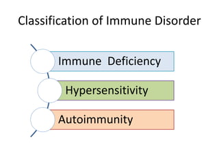
- 1. Immune Deficiency Hypersensitivity Autoimmunity Classification of Immune Disorder
- 2. Overview • Diseases of the immune system take many forms, including hypersensitivity reactions, autoimmune disorders and immunodeficiency states. • Autoimmune diseases are the result of a failure in the immune system to recognize self-antigens, resulting in the production of antibodies that react against the normal components of cells. • Immunodeficiency states can be hereditary or acquired. Severe combined immunodeficiency disorder (SCID) is a genetic disorder.
- 3. Immune Deficiency • Immune deficiency is a state in which immune system ability to fight infectious disease and cancer become compromised or entirely absent.
- 8. Hypersensitivity • A state of exaggerated or abnormal response to an antigen with resulting injury/damage to the host tissue instead of protection. Four Types of Hypersensitivity Reactions: • Type I (Anaphylactic) Reactions • Type II (Cytotoxic) Reactions • Type III (Immune Complex) Reactions • Type IV (Delayed or Cell-Mediated) Reactions
- 9. Type-I (Atopic or Anaphylactic) Reactions It occurs within minutes of exposure to antigen. Also known as IgE mediated or immediate hypersensitivity o Mechanism • First Exposure or Sensitization phase: The antigen is taken by antigen presenting cells (i.e. dendritic cells and macrophages) to the CD4+ (T helper) cells that differentiate to Th2 cells. • Th2 cells release IL-4 & IL-5. IL-4 activates B-cells to undergo class switching or isotype switching to produce IgE antibodies, while IL-5 stimulates production and activation of eosinophils. • Mast cells in the mucosal tissues or under the skin have high affinity to Fcε RI ( Fc epsilon region of immunoglobulin E) receptor which binds IgE even in absence of allergen. IgE are also called cytotropic antibodies since they can bind to cell surfaces. • IgE binds to mast cells.
- 10. Mechanism of Type I Cont… • Second Exposure/Activation Phase: Subsequent exposure to the same antigen results in binding of the antigen to IgE bound to mast cells. • Two or more antigens required to crosslink IgE antibodies, causing the mast cells to degranulate and thus release several pro-inflammatory molecules called mediators which cause the allergic effects. o Types of Mediators Released • Histamine: Causes vasodilation and increases vascular permeability (swelling & redness), increases mucus secretion (runny nose), attract neutrophils and eosinophils (chemotactic factor) contracts smooth muscles (in bronchi). • Prostaglandins: Cause vasodilation, contract bronchial smooth muscles and increase mucus secretion. • Leukotrienes: Cause bronchial spasms and attract more immune cells. • Proteases: Causes tissue damage (due to protein breakdown)
- 11. Mediators Involved Primary inflammatory mediators Histamine: Vascular permeability, smooth muscle contraction Serotonin: Neurotransmitter – smooth muscle contraction ECF-A: Eosinophil chemotactic factor of anaphylaxis causes Eosinophil chemotaxis. NCF-A: Neutrophil chemotactic factor of anaphylaxis causes Neutrophil chemotaxis. Proteases: mucus secretion, connective tissue degradation by protein breakdown Tryptase: It is an enzyme that is released, along with histamine and other chemicals, from mast cells when they are activated as part of a normal immune response as well as in allergic (hypersensitivity) responses. Serum tryptase concentration servers as a marker of mast cell activation. Heparin: Initiates bradykinin production causing swelling, anaphylaxis.
- 12. Mediators Involved Secondary inflammatory mediators Leukotrienes: Its release leads to Vascular permeability, smooth muscle contraction Prostaglandins: Causes vasodilation, smooth muscle contraction, platelet activation Bradykinin: Increases vascular permeability, smooth muscle contraction Cytokines: It signals the immune system to do its job. It causes activation of vascular endothelium, eosinophil recruitment and its activation
- 13. Mast Cells and the Allergic Response
- 14. Sequence of Events in Type-I Reaction 1- Early phase • It occurs within 5–30 minutes of re-exposure or 2nd exposure to an antigen. • Characterized by vasodilation, increased vascular permeability, edema, increased smooth muscles contraction, and urticarial (hives, is an outbreak of swollen, pale red bumps). • The early phase is due to binding of antigen to IgE bound to mast cells, with subsequent degranulation of the mast cells and release of chemical mediators mainly histamine. 2 - Late phase • It occurs in 24–48 hours after re-exposure to antigen. • Characterized by infiltration of neutrophils, basophils, eosinophils & monocytes and results in mucosal damage due to release of mediators such as leukotrienes and prostaglandins.
- 15. Forms of Type-I hypersensitivity Reactions Local anaphylaxis Skin Contact with allergen causes immediate redness, swelling and itching (urticaria). Allergen may come in contact with the skin directly, by injection (e.g. insect bite) and ingestion (e.g. food or drug allergies that produce skin reactions). Angioedema is characterized by laryngeal edema, edema of the eyelids, lips, tongue & trunk. Lungs Inhalation of allergens (e.g. pollens, dust particles) results in bronchial smooth muscles contraction, resulting in an acute airway obstruction and wheezing (e.g. allergic bronchial asthma).
- 16. Forms of Type-I hypersensitivity Reactions Local anaphylaxis Nose Inhalation of allergens (e.g. pollens, dust) leads to vasodilation and secretion of mucous, a condition known as hay fever and allergic rhinitis. Intestine Ingestion of allergens (e.g. nuts & sea foods) causes muscle contraction and fluid secretion that produce abdominal cramps and diarrhea, a condition called allergic gastroenteritis.
- 17. Forms of Type-I hypersensitivity Reactions Atopy • Genetic predisposition to allergic reactions is called atopy. • Atopy is a tendency of an individual to produce specific IgE antibodies after natural exposure to environmental allergens. • Atopic patients usually have markedly elevated total serum IgE levels. • The atopic trait is inherited in genes. • Examples include allergic rhinitis, asthma and atopic dermatitis (eczema) etc.
- 18. Forms of Type-I hypersensitivity Reactions Systemic Anaphylactic Reaction • It is a life-threatening systemic type-1 hypersensitivity reaction that typically results from injected allergens (e.g. penicillin antibiotics, local anesthetics, contrast dyes) and cutaneous allergens (e.g. bees stings). • Release of inflammatory mediators cause vasodilation & increased vascular permeability that leads to shock, allergic edema and may cause fatal asphyxia. • Asphyxia is generalized hypoxia that arises from abnormal breathing leading to unconsciousness and death.
- 19. Type-II Hypersensitivity Reaction Overview of General Mechanism • Antibodies (IgG and IgM) are directed against target antigens on cells or in extracellular matrix. • The target antigens may be endogenous (e.g. normal cell recognized by the immune system as foreign) or absorbed exogenous antigens (e.g. a drug). Specific Mechanisms • There are three specific mechanisms by which type-II hypersensitivity reactions occur. a) Complement-dependent reactions b) Antibody dependent cell-mediated cytotoxicity, and c) Antibody-mediated cellular dysfunction.
- 20. Type-II Hypersensitivity Reaction (a) Complement-dependent reactions Mechanism • Antibodies react with cell surface antigen, leading to activation of complement system that cause the direct lysis of the antigen-antibody complex containing the cell. • The complement proteins can also promote phagocytosis of the antigen by providing opsonization. Example-1: Reaction to Penicillin • The drug can bind to red blood cells, causing them to be recognized as foreign antigen. IgG & IgM antibodies bind to these antigens to form complexes that activate complement system to eliminate cells presenting foreign antigens.
- 21. Complement-dependent Reactions Example-2: Transfusion reactions • RBCs from an incompatible donor are destroyed by the antibodies normally present within the recipient. Such antibodies are directed against blood group antigens present on the cell membrane of RBCs.
- 22. Type-II Hypersensitivity Reaction (a) Complement-dependent reactions Contd.. Example-3: Good pasture's Syndrome Goodpasture syndrome, also known as anti-glomerular basement membrane disease, is a rare autoimmune disease in which antibodies attack the basement membrane in lungs and kidneys, leading to bleeding from the lungs, glomerulonephritis, and kidney failure. 1-Glomerulonephritis • Autoantibodies IgG are deposited along the capillaries of the glomeruli, where they react with glycoprotein present in the glomerular basement membrane. This results in a strong inflammatory reaction & complement fixation leading to necrosis of the glomerulus with impending renal failure. 2-Alveolar Basement membrane Damage • Auto-antibodies are produced against pulmonary alveolar basement membrane antigen, the resulting damage leads to bleeding from the lung.
- 23. Type-II Hypersensitivity Reaction Example 4: Hemolytic Disease of the Newborn • The Rh-negative mother is sensitized from Rh-positive baby during first pregnancy. • Rh-specific B-cells of the mother are sensitized which remain in the memory. • In the 2nd pregnancy, if the fetus is Rh-positive, memory B-cell will readily produce Rh-negative antibodies which can cross the placenta and cause the destruction of Rh- positive fetal red blood cells. • The resulting syndrome is called erythroblastosis fetalis or Hemolytic Disease of the Newborn.
- 25. Type-II Hypersensitivity Reaction (b) Antibody-dependent cell-mediated cytotoxicity (ADCC) Mechanism • This form of antibody-mediated cell injury does not involve fixation of the complement proteins but instead requires the cooperation of leukocytes. • Cell types that possess receptors for the Fc portion of IgG, such as neutrophils, eosinophils, macrophages and natural killer cells, mediate the destruction of antigen-antibody complex containing the target cell. Cellular lysis proceeds without phagocytosis. • ADCC may be relevant to the destruction of targets too large to be phagocytosed, such as parasites or tumor cells, and it may also play some role in graft rejection.
- 27. Type-II Hypersensitivity Reaction (c) Antibody-mediated cellular dysfunction (Non-cytotoxic) or Stimulatory Type II Hypersensitivity Also classified as Type V) Mechanism • Antibodies formed against cell surface receptors impair or dysregulate cell function without causing cell injury (i.e. may stimulate or inhibit cellular function). Example a) Stimulation Graves disease (autoimmune hyperthyroidism) is due to the production of autoantibodies that stimulate the thyroid- stimulating hormone (TSH) receptor, resulting in hyperthyroidism.
- 28. Type-II Hypersensitivity Reaction Antibody-mediated cellular dysfunction Example b) Inhibition Myasthenia Gravis is due to the production of auto- antibodies that block the acetylcholine receptors at the postsynaptic neuromuscular junction, inhibiting the excitatory effects of the neurotransmitter acetylcholine their receptors throughout neuromuscular junctions.
- 29. Effector Mechanisms of Antibody-Mediated Disease
- 30. Type-III (Immune Complex) Reaction • Mediated by immune complexes (antigen-antibody complexes) • Type-III reactions are caused by deposition of antigen-antibody complexes in the tissue & blood vessels. • Type-III reactions specifically occur when the antigen is in excess and small, immune complexes circulate in blood for longer periods and are slowly broken down. • The immune complex activates the complement system (family of complement proteins C1-C9) and attracts polymorpho-nuclear lymphocytes (Neutrophil, basophil, eosinophils) and macrophages to the site. • These attracted cells may release proteases, reactive oxygen species and inflammatory mediators that cause local tissue damage. Commonly occurs in kidneys lead to Glomerulonephritis & joints which lead to arthritis.
- 31. Immune Complex Mediated Hypersensitivity
- 32. Disorders of Type-III Reactions • Two types of immune complex injuries are resulted: 1) Systemic immune complex disease (serum sickness) 2) Local immune complex disease (Arthus reaction) • Serum Sickness Pathogenic immune complexes are formed in the circulation that are deposited in various tissues and cause inflammatory reactions there. Mechanism 1. Exposure to antigen leads to antibody formation 2. Antigen-antibody complexes are formed in the circulation.
- 33. Disorders of Type-III Reactions Mechanism Cont… 3. The complexes pass through the endothelial pores of small vessels and are deposited in the vessels wall where they activate complement system. This result in complement mediated necrosis and acute inflammation of the vessel wall, a condition called necrotizing vasculitis. Vasculitis may occur in a single organ (e.g. post streptococcus glomerulonephritis) or it may affect many organs (e.g. serum sickness due to injection of foreign serum (vaccines) or SLE)
- 34. Multiple Activities of the Complement System
- 35. Disorders of Type-III Reactions Arthus Reaction • This is localized immune complex injury in which tissue necrosis occurs at the site of entry of antigen. Mechanism • Repeated exposure to antigen results in high levels of antibody formation in the serum. • Subsequent exposure to the same antigen leads to the formation of antigen-antibody complexes that deposit locally in the small blood vessels. • The immune complexes activate complement & produce severe local acute inflammatory reaction with necrosis.
- 36. Disorders of Type-III Reactions Example of Arthus Reaction • A local type-III reaction can develop in the skin, for example, after vaccination. Hypersensitivity Pneumonitis • Repeated inhalation of small amounts of antigen results in the production of large amounts of IgG and immune complexes are formed that are deposited in the lung. • This results in cough, dyspnea and fever after 6-8 hours of exposure to antigen, a condition called Hypersensitivity Pneumonitis. • The lung disease caused by exposure to molds that grow on hay is known as farmer’s lung.
- 37. Type-IV (Cell-Mediated) Hypersensitivity • Mediated by sensitized T-cells rather than antibodies. • First contact sensitizes the person, subsequent contacts elicit a reaction. • Reactions are delayed and typically occur 48-72 hours after exposure to the antigen. • Delay is due to migration of macrophages and T-cells to the site of foreign antigens. • Type-IV reactions are categorized as (1) delayed hypersensitivity, (2) cell mediated cytotoxicity
- 38. Type-IV (Cell-Mediated) Hypersensitivity Delayed Type Hypersensitivity Reaction • First exposure leads to phagocytosis of the antigen by macrophages and presentation to T-cells forming memory T-cells. • On later exposure, memory T-cells (CD4+ helper T-cells) secrete interferon-γ, which activates macrophages. • Activated macrophages secrete IL-12, which causes differentiation of T-helper cells to TH1 cells that destroy the antigen. The activated macrophages also destroy the antigen intracellularly.
- 39. Type-IV (Cell-Mediated) Hypersensitivity Delayed Type Hypersensitivity Reaction • Microscopic morphology Stimulation of macrophages results in granulomas (i.e collections of epithelioid cells). • Inciting agents Mycobacteria, fungi, and parasites. Examples Tuberculin reaction Granulomatous inflammation
- 40. Overview of the DTH response
- 41. A prolonged DTH response can lead to formation of a granuloma
- 42. Type-IV (Cell-Mediated) Hypersensitivity T-cell Mediated Cytotoxicity • Sensitized CD8+ cells (cytotoxic T-cells) kill antigen- bearing cells. • The CD8+ cells accomplish this by releasing perforin that produces holes in the plasma membrane of cells, allowing granzyme to enter the cells. Granzyme then activates apoptosis. Example • Contact dermatitis
Editor's Notes
- Th2= T helper 2 cells
- Asphyxia= generalized hypoxia that arises from abnormal breathing leading to unconsciousness and death.
- Granulomas are masses of immune cells that form at the site of infection
- Epithelioid cells are activated macrophages that resembles epithelial cells