The hip joint is a ball and socket synovial joint that allows for movement in three planes. It is one of the most stable joints in the body. The hip joint is formed between the spherical head of the femur and the acetabulum of the pelvis. It is surrounded by strong ligaments and muscles that support the joint and allow for flexion, extension, abduction, adduction, and rotation. The hip joint receives its blood supply from various arteries and its nerve supply includes the femoral and obturator nerves.
Call Girls Rishikesh Just Call 9667172968 Top Class Call Girl Service Available
Hip_Joint_BMJ.ppt
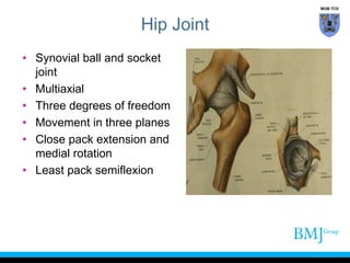
1. Hip Joint
• Synovial ball and socket
joint
• Multiaxial
• Three degrees of freedom
• Movement in three planes
• Close pack extension and
medial rotation
• Least pack semiflexion
MOB TCD
2. • One of most stable joints in
the body
• Articular surface of hip joint
are reciprocally curved
• Superior surface of femur and
acetabulum sustain greatest
pressure
Hip Joint
MOB TCD
3. Acetabulum
• Y-shaped epiphyseal cartilage
• Start to ossify at 12 years
• Fuse 16-17 years
• Acetabular notch is inferior
• Nonarticular fossa, thin related
medially to obturator internus
• Pad of fat, proprioceptive nerves
MOB TCD
4. Articular Surface of Hip Joint
• Semilunar articular surface
covered with hyaline
cartilage
• Deepened by acetabular
labrum
• Wedge shaped
fibrocartilage
MOB TCD
5. • Head of femur 2/3rd of sphere
• Pit for ligamentum teres
• Covered with articular cartilage
• Cartilage thicker posterior superior
• Epiphyseal line for head
intracapsular
Articular Surface
MOB TCD
6. Femur
• Trabeculae develop along lines
of stress
• Calcar femorale is the cortical
bone on inferior aspect of neck
• Neck is cancellous bone
MOB TCD
7. Capsule of Hip
• Proximally attached
• Margins of the acetabular
fossa
• Base of labrum
• Distally, anterior to the
intertrochanteric line
• Inferiorly, femoral neck close
to lesser trochanter
MOB TCD
8. • Posterior
• Free border, finger’s breath
from trochanteric crest due
to insertion of obturator
externus
• Into trochanteric fossa and
• Root greater trochanter
Capsule of Hip
MOB TCD
9. • Strongest superiorly
• Anteromedially, deep fibres
reflected head of rectus
femoris
• Iliopsoas is anterior
• Lateral deep fibres of gluteus
minimus
Capsule of Hip
MOB TCD
10. Retinacular Fibres
• Fibres of capsule reflected along
neck to articular margin called
retinacular fibres
• Blood supply to head run under
retinacular fibres
MOB TCD
11. Ligaments of Hip
• Acetabular labrum
• Transverse ligament
• Ligament of head
• Iliofemoral ligament
• Pubofemoral ligaments
• Ischiofemoral ligaments
• Zona orbicularis
MOB TCD
12. • Transverse ligament is part of
the labrum
• Ligamentum teres is
triangular, its base is attached
to transverse ligament, and
the apex to the pit on the
head of femur
• Blood supply to epiphysis
from obturator artery
• Only supplies a flake of bone
in elderly
Ligaments of Hip
MOB TCD
13. Iliofemoral Ligament
• Thickening of capsule
• Lower half of anterior
inferior iliac spine and
adjoining acetabulum
• Distally
• Upper and lower parts of
inter trochanteric line
MOB TCD
14. • One of strongest
ligaments in body
• Tightens in extension
• Helps maintain erect
posture
• Facet on anterior aspect
of neck
• Prevents hyperextension
• Fulcrum reducing hip
Iliofemoral Ligament
MOB TCD
15. Pubofemoral Ligament
• Superior pubic ramus
• Inferior part of inter
trochanteric line and upturned
part
• Relatively weak
• Prevents abduction
• Bursa between it and
iliofemoral
MOB TCD
16. Ischiofemoral Ligament
• Ischium to posterior part of
joint (weak)
• Circular fibres called zona
orbicularis
• Centre of gravity in front of
head
• Synovial under obturator
externus
MOB TCD
17. Synovial Membrane
• Lines inner portion of capsule
and non articular structures
• Ligament of head
• Fat in acetabular fossa
• May communicate with psoas
bursa
• Bursa under obturator
externus
MOB TCD
18. Bursa Under Gluteus Maximus
• Trochanteric bursa
• Posterolateral aspect of
greater trochanter
gluteofemoral
• Vastus lateralis ischial bursa
• Ischial tuberosity
MOB TCD
19. Blood Supply to Head of Femur
• Child, obturator artery via
ligamentum teres supplies
epiphysis
• Elderly, main supply via
retinacular vessels from
trochanteric and cruciate
anastamoses
• Medial and lateral circumflex
femoral vessels
MOB TCD
20. Blood Supply
• Superior gluteal supplies the upper
part of the acetabulum
• Inferior gluteal supplies the inferior
and posterior and the capsule
• Transverse and ascending
branches of lateral circumflex
femoral artery
• Transverse and ascending branch
of medial circumflex femoral
• Cruciate and trochanteric
anastomosis
MOB TCD
21. • Fractures of neck may cause
avascular necrosis, extra
capsular arteries enter the
trochanter at the base of neck
• Medial and lateral circumflex
femoral vessels and superior
gluteal
Blood Supply
MOB TCD
22. • Femoral nerve
• Obturator nerve
• Superior gluteal nerve
• Nerve to quadratus femoris
• Posterior dislocation may
damage sciatic
• Pain in hip referred to knee
Nerve Supply
MOB TCD
24. Inferior and Posterior Relations
• Obturator externus
• Passes inferior and then posterior
to joint
• Superior gluteal nerve
• Inferior gluteal nerve
• Sciatic nerve
• Posterior cutaneous nerve thigh
• Nerves to obturator internus and
quadratus femoris
• Pudendal nerve
MOB TCD
25. Lateral Relations
• Gluteus minimus
• Gluteus medius
• Superior gluteal vessels and
nerves between
• Iliotibial tract
• Superficial three quarters of
gluteus maximus
MOB TCD
26. Posterior Relations
• Piriformis
• Superior gemellus
• Obturator internus
• Inferior gemellus
• Quadratus femoris
• Adductor magnus
• Obturator externus
• Gluteus maximus
MOB TCD
28. Movements: Extension
• Hamstrings first 10°
• Long head of biceps
• Semitendinosus
• Semimembranosus
• 123, extended knee ++
• Adductor magnus
• Gluteus maximus most efficient when hip is
flexed 45°
MOB TCD
29. • Obturator nerve
• Adductor longus
• Adductor brevis
• Adductor magnus
• Can flex or extend depending
on position of hip
Movements: Adduction
MOB TCD
30. • Gluteus medius
• Gluteus minimus
• Standing on leg, gluteus medius and
minimus abduction
• By preventing adduction
Movements: Abduction
MOB TCD