SlideShare a Scribd company logo
1 of 18
2 2 N O V I E M B R E 2 0 1 0
Coroiditis Serpiginosa
Instituto de la visón
Servicio de retina
Epidemiologia
 Mayoría son Raza Blanca
 Hombres > Mujeres
 Negros, asiáticos y Hispanos
Presentación
 Inicialmente un ojo afectado
 PIO normal
 BMC “casi” siempre normal
 Celularidad de ¼ a 1/3 en el vitreo
Presentación Clínica
 La lesión Coroides Interna y/o EPR
 Color amarillo grisáceo y apariencia difusa
 Forma Geográfica, bordes bien definidos
 Las lesiones proceden del D.O
 Gris Pálido en pocas semanas en forma de serpentina
Presentación Clínica
 CV con lesiones/Escotomas/ Rejilla Amsler
 Pruebas electrofisiológicas
Diagnostico
 Evaluación Clínica
 FAG
 ICG
FAG
 Primera fase – Lesión aguda – Hipofluorecencia
 Etapas tardías – Fluoresce en zonas de lesión
 Lesiones agudas o crónicas hipofluorecen por la Coriocapilaris
 Coroides destruida
 Hiperfluorecencia tardía se observa en lesiones fibrosas cicatriciales
FAG
ICG
 Etapas Agudas – Hipofluorescencia en todas las
etapas
 Anormalidad de los vasos coroideos o resistencia
vascular coriocapilar
Diagnostico Diferencial
 Multifocal posterior placoid pigment epitheliopathy (AMPPPE)
 CNV
 Histoplasmosis ocular
 Degen. Macular
 Toxoplasmosis
 Escleritis Posterior
 Coroiditis multifocal
 Sarcoidosis
 Linfoma no Hodgking’s
 Tumor metatasico
 Osteoma Coroideo
Tratamiento
FAG
ICG
 PREGUNTAS Y COMENTARIOS

More Related Content

What's hot

CLASIFICACIÓN DEL AGUJERO MACULAR
CLASIFICACIÓN DEL AGUJERO MACULARCLASIFICACIÓN DEL AGUJERO MACULAR
CLASIFICACIÓN DEL AGUJERO MACULAR
ElviraLopezArroquia
 
Miopia degenerativa
Miopia degenerativaMiopia degenerativa
Miopia degenerativa
alejandra
 
Maculopatía miópica
Maculopatía miópicaMaculopatía miópica
Maculopatía miópica
zapatavictori
 
Alteraciones perifericas de la retina
Alteraciones perifericas de la retinaAlteraciones perifericas de la retina
Alteraciones perifericas de la retina
Juan Carlos Rivera
 

What's hot (20)

Membrana epiretiniana - Epiretinal membrane
Membrana epiretiniana - Epiretinal membraneMembrana epiretiniana - Epiretinal membrane
Membrana epiretiniana - Epiretinal membrane
 
Degeneración esferoidal
Degeneración esferoidalDegeneración esferoidal
Degeneración esferoidal
 
Glaucoma neovascular lml
Glaucoma neovascular lmlGlaucoma neovascular lml
Glaucoma neovascular lml
 
CLASIFICACIÓN DEL AGUJERO MACULAR
CLASIFICACIÓN DEL AGUJERO MACULARCLASIFICACIÓN DEL AGUJERO MACULAR
CLASIFICACIÓN DEL AGUJERO MACULAR
 
Sesioìn cliìnica queratocono
Sesioìn cliìnica queratoconoSesioìn cliìnica queratocono
Sesioìn cliìnica queratocono
 
Miopia degenerativa
Miopia degenerativaMiopia degenerativa
Miopia degenerativa
 
Retinopatía Diabética
Retinopatía DiabéticaRetinopatía Diabética
Retinopatía Diabética
 
Paquimetria y microscopia especular
Paquimetria y microscopia especularPaquimetria y microscopia especular
Paquimetria y microscopia especular
 
OCT: Interpretacion clínica
OCT: Interpretacion clínica OCT: Interpretacion clínica
OCT: Interpretacion clínica
 
Queratitis Por Herpes Simplex Virus (HSV)
Queratitis Por Herpes Simplex Virus (HSV)Queratitis Por Herpes Simplex Virus (HSV)
Queratitis Por Herpes Simplex Virus (HSV)
 
Oclusion venosa y arterial de retina
Oclusion venosa y arterial de retinaOclusion venosa y arterial de retina
Oclusion venosa y arterial de retina
 
Maculopatía miópica
Maculopatía miópicaMaculopatía miópica
Maculopatía miópica
 
Entendiendo angiografía con fluoresceína
Entendiendo angiografía con fluoresceínaEntendiendo angiografía con fluoresceína
Entendiendo angiografía con fluoresceína
 
Angiografia con Fluoresceina - Fluorescein angiography (FFA)
Angiografia con Fluoresceina - Fluorescein angiography (FFA)Angiografia con Fluoresceina - Fluorescein angiography (FFA)
Angiografia con Fluoresceina - Fluorescein angiography (FFA)
 
Degeneración macular relacionada con la edad, - Age related macular degenera...
Degeneración macular relacionada con la edad,  - Age related macular degenera...Degeneración macular relacionada con la edad,  - Age related macular degenera...
Degeneración macular relacionada con la edad, - Age related macular degenera...
 
Angiofluoresceinografía
AngiofluoresceinografíaAngiofluoresceinografía
Angiofluoresceinografía
 
Glaucoma
GlaucomaGlaucoma
Glaucoma
 
Idiopathic polypoidal choroidal vasculopathy
Idiopathic polypoidal choroidal vasculopathyIdiopathic polypoidal choroidal vasculopathy
Idiopathic polypoidal choroidal vasculopathy
 
Distrofias prueba 2
Distrofias prueba 2Distrofias prueba 2
Distrofias prueba 2
 
Alteraciones perifericas de la retina
Alteraciones perifericas de la retinaAlteraciones perifericas de la retina
Alteraciones perifericas de la retina
 

Viewers also liked (8)

Coroiditis multifocal con_panuveitis
Coroiditis multifocal con_panuveitisCoroiditis multifocal con_panuveitis
Coroiditis multifocal con_panuveitis
 
Estrias Angioides
Estrias AngioidesEstrias Angioides
Estrias Angioides
 
Estrias angioides
Estrias angioidesEstrias angioides
Estrias angioides
 
Estrias atróficas
Estrias atróficas  Estrias atróficas
Estrias atróficas
 
Toxina botulinica en oftalmologia
Toxina botulinica en oftalmologiaToxina botulinica en oftalmologia
Toxina botulinica en oftalmologia
 
Terapia Génica para Tratar Amaurosis Congénita de Leber (revisión)
Terapia Génica para Tratar Amaurosis Congénita de Leber (revisión)Terapia Génica para Tratar Amaurosis Congénita de Leber (revisión)
Terapia Génica para Tratar Amaurosis Congénita de Leber (revisión)
 
Cristalino
CristalinoCristalino
Cristalino
 
Patologia del cristalino
Patologia del cristalinoPatologia del cristalino
Patologia del cristalino
 

Similar to Coroiditis serpiginosa

Aproximación a La Angiografía
Aproximación a La AngiografíaAproximación a La Angiografía
Aproximación a La Angiografía
TElvira
 
3. enfermedad de stargardt (1)
3. enfermedad de stargardt (1)3. enfermedad de stargardt (1)
3. enfermedad de stargardt (1)
Marvin Barahona
 
Presión Intraocular y Glaucoma
Presión Intraocular y GlaucomaPresión Intraocular y Glaucoma
Presión Intraocular y Glaucoma
dra.martinez
 
ORBITAexposicionnnnnnnnnnnnnnnnnnnn.pptx
ORBITAexposicionnnnnnnnnnnnnnnnnnnn.pptxORBITAexposicionnnnnnnnnnnnnnnnnnnn.pptx
ORBITAexposicionnnnnnnnnnnnnnnnnnnn.pptx
DanielaG081
 
Clase 13 (patologías del polo posterior. criterios de derivación)
Clase 13 (patologías del polo posterior. criterios de derivación)Clase 13 (patologías del polo posterior. criterios de derivación)
Clase 13 (patologías del polo posterior. criterios de derivación)
OPTO2012
 

Similar to Coroiditis serpiginosa (20)

tumores uveovítreos correto 2.pptx
tumores uveovítreos correto 2.pptxtumores uveovítreos correto 2.pptx
tumores uveovítreos correto 2.pptx
 
Trastornos corneales degenerativos
Trastornos corneales degenerativosTrastornos corneales degenerativos
Trastornos corneales degenerativos
 
TCE, HTEC, Coma y Meningitis
TCE, HTEC, Coma y MeningitisTCE, HTEC, Coma y Meningitis
TCE, HTEC, Coma y Meningitis
 
Aproximación a La Angiografía
Aproximación a La AngiografíaAproximación a La Angiografía
Aproximación a La Angiografía
 
Urgencias Oftalmologicas
Urgencias OftalmologicasUrgencias Oftalmologicas
Urgencias Oftalmologicas
 
TRAUMA CRANEOENCEFALICO
TRAUMA CRANEOENCEFALICOTRAUMA CRANEOENCEFALICO
TRAUMA CRANEOENCEFALICO
 
3. enfermedad de stargardt (1)
3. enfermedad de stargardt (1)3. enfermedad de stargardt (1)
3. enfermedad de stargardt (1)
 
Comunicacion2
Comunicacion2Comunicacion2
Comunicacion2
 
Distrofias maculares
Distrofias macularesDistrofias maculares
Distrofias maculares
 
Orbita, patologia mas frecuente clinica del sol
Orbita, patologia mas frecuente clinica del solOrbita, patologia mas frecuente clinica del sol
Orbita, patologia mas frecuente clinica del sol
 
Oftalmololgía en neonatos
Oftalmololgía en neonatosOftalmololgía en neonatos
Oftalmololgía en neonatos
 
Presión Intraocular y Glaucoma
Presión Intraocular y GlaucomaPresión Intraocular y Glaucoma
Presión Intraocular y Glaucoma
 
tumores uveovítreos correto-1.pptx
tumores uveovítreos correto-1.pptxtumores uveovítreos correto-1.pptx
tumores uveovítreos correto-1.pptx
 
Uveítis, endoftalmitis en pediatría
Uveítis, endoftalmitis en pediatríaUveítis, endoftalmitis en pediatría
Uveítis, endoftalmitis en pediatría
 
RETINITIS PIGMENTOSA
RETINITIS PIGMENTOSARETINITIS PIGMENTOSA
RETINITIS PIGMENTOSA
 
ORBITAexposicionnnnnnnnnnnnnnnnnnnn.pptx
ORBITAexposicionnnnnnnnnnnnnnnnnnnn.pptxORBITAexposicionnnnnnnnnnnnnnnnnnnn.pptx
ORBITAexposicionnnnnnnnnnnnnnnnnnnn.pptx
 
Clase 13 (patologías del polo posterior. criterios de derivación)
Clase 13 (patologías del polo posterior. criterios de derivación)Clase 13 (patologías del polo posterior. criterios de derivación)
Clase 13 (patologías del polo posterior. criterios de derivación)
 
Glaucoma congenito
Glaucoma congenitoGlaucoma congenito
Glaucoma congenito
 
12. retina 2
12. retina 212. retina 2
12. retina 2
 
12. retina 2
12. retina 212. retina 2
12. retina 2
 

More from Montemorelos

More from Montemorelos (20)

Sospecha de glaucoma e hipertensión ocular
Sospecha de glaucoma e hipertensión ocularSospecha de glaucoma e hipertensión ocular
Sospecha de glaucoma e hipertensión ocular
 
Glaucoma inducido por esteroides
Glaucoma inducido por esteroidesGlaucoma inducido por esteroides
Glaucoma inducido por esteroides
 
Hipotonia
HipotoniaHipotonia
Hipotonia
 
Glaucomas de origen traumatico
Glaucomas de origen traumaticoGlaucomas de origen traumatico
Glaucomas de origen traumatico
 
Glaucoma neovascular
Glaucoma neovascularGlaucoma neovascular
Glaucoma neovascular
 
Queratitis acanthamoeba
Queratitis acanthamoebaQueratitis acanthamoeba
Queratitis acanthamoeba
 
Queratitis micotica
Queratitis micoticaQueratitis micotica
Queratitis micotica
 
Laser y procedimientos ciclodestructivos
Laser y procedimientos ciclodestructivosLaser y procedimientos ciclodestructivos
Laser y procedimientos ciclodestructivos
 
Exotropias
ExotropiasExotropias
Exotropias
 
Endotropias
EndotropiasEndotropias
Endotropias
 
Glaucoma primario de angulo cerrado
Glaucoma primario de angulo cerradoGlaucoma primario de angulo cerrado
Glaucoma primario de angulo cerrado
 
Terapeutica medica antiglaucomatosa
Terapeutica medica antiglaucomatosaTerapeutica medica antiglaucomatosa
Terapeutica medica antiglaucomatosa
 
Glaucoma y genetica
Glaucoma y geneticaGlaucoma y genetica
Glaucoma y genetica
 
Progresion del dano en el glaucoma y pio
Progresion del dano en el glaucoma y pioProgresion del dano en el glaucoma y pio
Progresion del dano en el glaucoma y pio
 
Enfermedad de coats
Enfermedad de coatsEnfermedad de coats
Enfermedad de coats
 
HIV OCULAR
HIV OCULARHIV OCULAR
HIV OCULAR
 
Agujero macular
Agujero macularAgujero macular
Agujero macular
 
Histoplasmosis ocular
Histoplasmosis ocularHistoplasmosis ocular
Histoplasmosis ocular
 
Eales disease
Eales diseaseEales disease
Eales disease
 
Membrana epiretinal
Membrana epiretinalMembrana epiretinal
Membrana epiretinal
 

Recently uploaded

Cuadro-comparativo-Aparato-Reproductor-Masculino-y-Femenino.pptx
Cuadro-comparativo-Aparato-Reproductor-Masculino-y-Femenino.pptxCuadro-comparativo-Aparato-Reproductor-Masculino-y-Femenino.pptx
Cuadro-comparativo-Aparato-Reproductor-Masculino-y-Femenino.pptx
guadalupedejesusrios
 
Dedo con deformidad en ojal o “boutonnière”
Dedo con deformidad en ojal o “boutonnière”Dedo con deformidad en ojal o “boutonnière”
Dedo con deformidad en ojal o “boutonnière”
AdyPunkiss1
 
Relacion final de ingresantes 23.11.2020 (2).pdf
Relacion final de ingresantes 23.11.2020 (2).pdfRelacion final de ingresantes 23.11.2020 (2).pdf
Relacion final de ingresantes 23.11.2020 (2).pdf
AlvaroLeiva18
 
11-incisiones-y-cierre-de-pared-abdominal.ppt
11-incisiones-y-cierre-de-pared-abdominal.ppt11-incisiones-y-cierre-de-pared-abdominal.ppt
11-incisiones-y-cierre-de-pared-abdominal.ppt
yuhelipm
 
SISTEMA OBLIGATORIO GARANTIA DE LA CALIDAD EN SALUD SOGCS.pdf
SISTEMA OBLIGATORIO GARANTIA DE LA CALIDAD EN SALUD SOGCS.pdfSISTEMA OBLIGATORIO GARANTIA DE LA CALIDAD EN SALUD SOGCS.pdf
SISTEMA OBLIGATORIO GARANTIA DE LA CALIDAD EN SALUD SOGCS.pdf
TruGaCshirley
 

Recently uploaded (20)

Cuadro-comparativo-Aparato-Reproductor-Masculino-y-Femenino.pptx
Cuadro-comparativo-Aparato-Reproductor-Masculino-y-Femenino.pptxCuadro-comparativo-Aparato-Reproductor-Masculino-y-Femenino.pptx
Cuadro-comparativo-Aparato-Reproductor-Masculino-y-Femenino.pptx
 
DETERMINISMO DEL TRABAJO DE PARTO-1.pptx
DETERMINISMO DEL TRABAJO DE PARTO-1.pptxDETERMINISMO DEL TRABAJO DE PARTO-1.pptx
DETERMINISMO DEL TRABAJO DE PARTO-1.pptx
 
Dedo con deformidad en ojal o “boutonnière”
Dedo con deformidad en ojal o “boutonnière”Dedo con deformidad en ojal o “boutonnière”
Dedo con deformidad en ojal o “boutonnière”
 
FISIOLOGIA BACTERIANA y mecanismos de acción (1).pptx
FISIOLOGIA BACTERIANA y mecanismos de acción (1).pptxFISIOLOGIA BACTERIANA y mecanismos de acción (1).pptx
FISIOLOGIA BACTERIANA y mecanismos de acción (1).pptx
 
Flashcard Anatomía del Craneo: Neurocráneo y Vicerocráneo.
Flashcard Anatomía del Craneo: Neurocráneo y Vicerocráneo.Flashcard Anatomía del Craneo: Neurocráneo y Vicerocráneo.
Flashcard Anatomía del Craneo: Neurocráneo y Vicerocráneo.
 
GENERALIDADES DEL SISTEMA HEMATOPOYETICO.pptx
GENERALIDADES DEL SISTEMA HEMATOPOYETICO.pptxGENERALIDADES DEL SISTEMA HEMATOPOYETICO.pptx
GENERALIDADES DEL SISTEMA HEMATOPOYETICO.pptx
 
Sangrado Uterino Anormal. Dr Carlos Quiroz_052747.pptx
Sangrado Uterino Anormal. Dr Carlos Quiroz_052747.pptxSangrado Uterino Anormal. Dr Carlos Quiroz_052747.pptx
Sangrado Uterino Anormal. Dr Carlos Quiroz_052747.pptx
 
Ovario. Ciclo ovárico o ciclo menstrual.pdf
Ovario. Ciclo ovárico o ciclo menstrual.pdfOvario. Ciclo ovárico o ciclo menstrual.pdf
Ovario. Ciclo ovárico o ciclo menstrual.pdf
 
AGENTES FÍSICOS EN FISIOTERAPIA (CFF OPHYSIO)
AGENTES FÍSICOS EN FISIOTERAPIA (CFF OPHYSIO)AGENTES FÍSICOS EN FISIOTERAPIA (CFF OPHYSIO)
AGENTES FÍSICOS EN FISIOTERAPIA (CFF OPHYSIO)
 
TRANSMISION DE LA INFORMACIÓN GENETICA - Clase 1.pptx
TRANSMISION DE LA INFORMACIÓN GENETICA - Clase 1.pptxTRANSMISION DE LA INFORMACIÓN GENETICA - Clase 1.pptx
TRANSMISION DE LA INFORMACIÓN GENETICA - Clase 1.pptx
 
6.METODOLOGIA ATENEA MICHAEL. ZAPATA.pdf
6.METODOLOGIA ATENEA MICHAEL. ZAPATA.pdf6.METODOLOGIA ATENEA MICHAEL. ZAPATA.pdf
6.METODOLOGIA ATENEA MICHAEL. ZAPATA.pdf
 
WE DO TRANSFORMATIONS DAY presentación .pptx
WE DO TRANSFORMATIONS DAY presentación   .pptxWE DO TRANSFORMATIONS DAY presentación   .pptx
WE DO TRANSFORMATIONS DAY presentación .pptx
 
Relacion final de ingresantes 23.11.2020 (2).pdf
Relacion final de ingresantes 23.11.2020 (2).pdfRelacion final de ingresantes 23.11.2020 (2).pdf
Relacion final de ingresantes 23.11.2020 (2).pdf
 
Psicología: Revista sobre las bases de la conducta humana.pdf
Psicología: Revista sobre las bases de la conducta humana.pdfPsicología: Revista sobre las bases de la conducta humana.pdf
Psicología: Revista sobre las bases de la conducta humana.pdf
 
OXIGENO TERAPIA: AEROSOLTERAPIA EN PACIENTES
OXIGENO TERAPIA: AEROSOLTERAPIA  EN PACIENTESOXIGENO TERAPIA: AEROSOLTERAPIA  EN PACIENTES
OXIGENO TERAPIA: AEROSOLTERAPIA EN PACIENTES
 
11-incisiones-y-cierre-de-pared-abdominal.ppt
11-incisiones-y-cierre-de-pared-abdominal.ppt11-incisiones-y-cierre-de-pared-abdominal.ppt
11-incisiones-y-cierre-de-pared-abdominal.ppt
 
Histologia del sistema respiratorio y sus funciones
Histologia del sistema respiratorio y sus funcionesHistologia del sistema respiratorio y sus funciones
Histologia del sistema respiratorio y sus funciones
 
Historia Clínica y Consentimiento Informado en Odontología
Historia Clínica y Consentimiento Informado en OdontologíaHistoria Clínica y Consentimiento Informado en Odontología
Historia Clínica y Consentimiento Informado en Odontología
 
SISTEMA OBLIGATORIO GARANTIA DE LA CALIDAD EN SALUD SOGCS.pdf
SISTEMA OBLIGATORIO GARANTIA DE LA CALIDAD EN SALUD SOGCS.pdfSISTEMA OBLIGATORIO GARANTIA DE LA CALIDAD EN SALUD SOGCS.pdf
SISTEMA OBLIGATORIO GARANTIA DE LA CALIDAD EN SALUD SOGCS.pdf
 
ACRONIMO TIMERS TRATAMIENTO DE HERIDAS AVANZADAS
ACRONIMO TIMERS TRATAMIENTO DE HERIDAS AVANZADASACRONIMO TIMERS TRATAMIENTO DE HERIDAS AVANZADAS
ACRONIMO TIMERS TRATAMIENTO DE HERIDAS AVANZADAS
 

Coroiditis serpiginosa

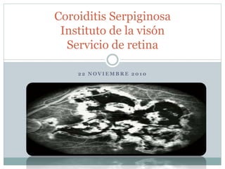
  • 1. 2 2 N O V I E M B R E 2 0 1 0 Coroiditis Serpiginosa Instituto de la visón Servicio de retina
  • 2. Epidemiologia  Mayoría son Raza Blanca  Hombres > Mujeres  Negros, asiáticos y Hispanos
  • 3. Presentación  Inicialmente un ojo afectado  PIO normal  BMC “casi” siempre normal  Celularidad de ¼ a 1/3 en el vitreo
  • 4. Presentación Clínica  La lesión Coroides Interna y/o EPR  Color amarillo grisáceo y apariencia difusa  Forma Geográfica, bordes bien definidos  Las lesiones proceden del D.O  Gris Pálido en pocas semanas en forma de serpentina
  • 5.
  • 6. Presentación Clínica  CV con lesiones/Escotomas/ Rejilla Amsler  Pruebas electrofisiológicas
  • 8. FAG  Primera fase – Lesión aguda – Hipofluorecencia  Etapas tardías – Fluoresce en zonas de lesión  Lesiones agudas o crónicas hipofluorecen por la Coriocapilaris  Coroides destruida  Hiperfluorecencia tardía se observa en lesiones fibrosas cicatriciales
  • 9. FAG
  • 10. ICG  Etapas Agudas – Hipofluorescencia en todas las etapas  Anormalidad de los vasos coroideos o resistencia vascular coriocapilar
  • 11.
  • 12. Diagnostico Diferencial  Multifocal posterior placoid pigment epitheliopathy (AMPPPE)  CNV  Histoplasmosis ocular  Degen. Macular  Toxoplasmosis  Escleritis Posterior  Coroiditis multifocal  Sarcoidosis  Linfoma no Hodgking’s  Tumor metatasico  Osteoma Coroideo
  • 13.
  • 15.
  • 16.
  • 18.  PREGUNTAS Y COMENTARIOS

Editor's Notes

  1. coroiditis serpiginosa, también llamada coroidopatía geográfica helicoidal peripapilar o coroiditis geográfica, es una enfermedad rara, generalmente bilateral, enfermedad inflamatoria crónica recurrente de causa desconocida coroides y la retina (EPR) Px. De Mediana Edad y Adultos pero tb se presenta en jovenes
  2. Negros, asiáticos y Hispanos sin manifestaciones sistémicas o asociación con las drogas, trauma o allergy No hay propensión familiar conocido
  3. A veces el paciente solo percibe una disminucion de la AV cuando involucra Macula y cuando se afecta el ojo contralateral Compromiso Foveal disminuye la AV reacción inflamatoria de camara anterior no granulomatosa
  4. Estas lesiones pueden crecer no sólo hacia el disco (fig. 105-2D), sino también peripherally.8 personas con coroiditis serpiginosa macular puede tener un peor pronóstico visual y diagnóstico preciso y el tratamiento rápido son primordiales. La neovascularización coroidea (NVC) puede ocurrir en asociación con coroiditis serpiginosa Un ojo puede tener una lesión macular aguda, mientras que el otro puede tener un peripapilar lesion.5 Del mismo modo, un ojo puede tener la CNV, mientras que el otro sólo puede tener cambios inflamatorios agudos
  5. FAG A, Red-free fundus photograph of the left macula of a 54-year-old white man. Note sensory detachment of the macula (arrows), subretinal hemorrhage centrally, and a geographic scar in the papillomacular bundle. B, Arteriovenous-phase fluorescein angiogram of the left macula. Note the patch of subretinal neovascularization in the central macula (arrows). The geographic lesion is in the papillomacular bundle. C, Late arteriovenous-phase fluorescein angiogram of the left macula. Note staining of the geographic lesion in the papillomacular bundle and fuzzy fluorescence of the subretinal neovascularization in the macula, which is leaking fluorescein under the sensory retinal detachment of the macula. D, Red-free fundus photograph of the right macula. The small, active inflammatory geographic lesion just temporal to the macula has "glowing" acute inflammatory edges (arrows).
  6. Los resultados de las pruebas electrofisiológicas son generalmente normales, aunque en casos muy graves en los que grandes áreas del fondo de ojo se ven afectadas, un electrorretinograma anormal o anormal oculogram electro-puede ser recorded.2
  7. Atrophic lesions show diffuse loss of pigment, choroidal vessels, and late staining on fluorescein angiography. Laboratory evaluation of these patients is invariably unrewarding. An association of serpiginous choroiditis and tuberculosis has been suggested. However, treatment with antimicrobial agents has made no difference to the course of the disease in these patients.[27]
  8. La fluorescencia por lo general comienza desde el borde de la lesión y se extiende hacia el interior. La angiografía es bastante típica, mostrando la imagen total de "dedos" del tejido afectado. Esto contrasta con la fluorescencia de la CNV, que muestra de encaje, irregular y nodular hiperfluorescencia en las fotos de fase temprana
  9. A, Red-free fundus photograph of the left macula of a 50-year-old white man. Note the acute geographic inflammatory lesion in the macula and papillomacular bundle (arrows). B, Early arteriovenous-phase fluorescein angiogram of the left macula. Note hypofluorescence of the acute inflammatory lesion. C, Late-phase fluorescein angiogram of the left macula. Note staining of the acute inflammatory lesion. D, Red-free fundus photograph of the left macula 4 months later. Note the scarring of the older geographic lesion in the macula and the fresh inflammatory lesion (arrow) extending superonasal from the macula in the upper part of the papillomacular bundle. E, Arteriovenous-phase fluorescein angiogram of the left macula. Note new acute lesions in the upper part of the papillomacular bundle. F, Late-phase fluorescein angiogram of the left macula. Note mild staining of the old inflammatory lesion in the macula and more intense fluorescent staining of the new inflammatory lesion in the upper part of the papillomacular bundle.
  10. También puede haber áreas localizadas de hiperfluorescencia fuera de estas zonas. Estas últimas lesiones no se corresponden con los cambios clínicamente visible de la retina, EPR, o de la coroides y podría representar áreas de la forma subclínica coroidea inflammation. Giovani y al. Sugirieron un sistema de clasificacion combinado de FAG y ICG
  11. FAG A, Red-free fundus photograph of the left macula of a 51-year-old white man. An extensive acute geographic inflammatory lesion extends from the optic nerve head and involves much of the central, lower, and temporal macula, as well as the upper part of the papillomacular bundle. The only area seemingly not involved is the upper macula. B, Early arteriovenous-phase fluorescein angiogram of the left macula. Note hypofluorescence of the acute inflammatory lesion in the macula and papillomacular bundle. C, Late-phase fluorescein angiogram of the left macula. Note staining of the geographic lesion in the macula. D, Note leakage of the retinal vein below the macula (phlebitis, arrow) overlying the acute inflammatory lesion. E, Red-free fundus photograph of the left macula 1 year later. The acute inflammatory geographic lesion in the upper part of the macula has extended to involve the entire macular area. Vision has been markedly reduced. F, Fluorescein angiogram of the left macula. There is extensive involvement of the entire macular and papillomacular bundle area.
  12. A number of inflammatory diseases can mimic serpiginous choroiditis.21 The condition that most resembles serpiginous choroiditis is acute multifocal posterior placoid pigment epitheliopathy (AMPPPE). Although AMPPPE can affect only one eye, with the fellow eye unaffected until a later date, in most instances both eyes are affected at the same time. In addition, AMPPPE does not usually recur; if it does, it is usually within the first few weeks or months of its initial presentation. Recurrences of AMPPPE do not happen over the course of years, as in serpiginous choroiditis. Finally, permanent vision loss in AMPPPE is unusual.
  13. A, Red-free fundus photograph of the left posterior pole of a 40-year-old white man. Large geographic areas of atrophy extend in a propeller-like fashion from the optic nerve head. The lesion just skips the left fovea (arrow). Note also the pigment epithelial hyperplasia. B, Early arteriovenous-phase fluorescein angiogram of the left posterior pole. Lesions show large choroidal vessels but no choriocapillaris; these are old lesions with absent choriocapillaris and pigment epithelium. C, Later arteriovenous-phase fluorescein angiogram of the left posterior pole. There is staining along the edges of the lesions. D, Late arteriovenous-phase fluorescein angiogram of the left nasal mid-periphery. Lesions extend out from the disc. Unattached lesions (arrows) show skipped areas.
  14. There are no reports of successful management of CNV associated with serpiginous choroiditis with anti-inflammatory agents. Thermal laser has been the main form of therapy for extrafoveal or juxtafoveal CNV.1,13,14 Recurrence after laser has been reported.11 Subfoveal CNV due to serpiginous choroiditis may be amenable to verteporfin photodynamic therapy but there are no reports describing results to date. Body_ID: P105028 Patients with serpiginous choroiditis are instructed to perform frequent Amsler grid testing to monitor for the presence of recurrent disease or CNV. Because similar central vision changes can occur with either, patients are instructed to return for immediate examination and angiography when symptoms occur.
  15. A, Red-free fundus photograph of the left macula of a 51-year-old white man. The atrophic geographic lesion extends from the optic nerve head and involves the central, temporal, and lower parts of the macula. B, Arteriovenous-phase fluorescein angiogram. The choriocapillaris and pigment epithelium are gone, as evidenced by the visibility of large choroidal vessels within the lesion. C, Late-phase fluorescein angiogram of the left macula. Note staining along the edges of the geographic lesion.
  16. The fellow eye of the patient shown in Figure 105-4. A, Red-free fundus photograph of the right posterior pole. Large lesions extend from the optic nerve head and involve the macula (arrow). This eye had a marked reduction in visual acuity. Note the scarring and pigment epithelial hyperplasia. B, Arteriovenous-phase fluorescein angiogram of the right posterior pole. Large choroidal vessels within lesions show that the choriocapillaries and pigment epithelium are gone. There is hyperfluorescence along the edges of the lesions. C, Late arteriovenous-phase fluorescein angiogram of the posterior pole, right fundus. There is early staining along the edges of the lesion and staining of the fibrous scar (arrow). D, Late arteriovenous-phase fluorescein angiogram of the right eye, superonasal area. Note the finger-like projections extending toward the mid-periphery.
  17. A, Clinical photograph demonstrating a creamy yellow lesion in the central macula, representing the macular variant of serpiginous choroidopathy. B, Early-phase fluorescein study demonstrating hypofluorescence of the acute lesion. The margins of the lesion are not well demarcated. C, Late-phase fluorescein study demonstrating intense hyperfluorescence and staining of the lesion. D, Early-phase indocyanine green (ICG) study demonstrating marked hypofluorescence of the lesion. Overlying retinal vessels are visualized, but all detail of the underlying choroidal vasculature is lost. E, Late-phase ICG study demonstrating persistent hypofluorescence and well-demarcated margins of the lesion. F, Very-late-phase ICG study demonstrating persistent hypofluorescence of the lesion and a halo of hyperfluorescence along its margin. Also note the focal, intense hyperfluorescent spots that may represent sites of subclinical choroidal inflammation.
  18. VISUAL PROGNOSIS Significant loss of central and even peripheral vision can occur as a result of inflammatory disease and its sequelae. Frequencies of central vision loss have been reported to be as high as 50% of involved eyes.7,8,32 This is especially true in patients with macular serpiginous choroiditis. In addition, CNV can occur in up to 25% of patients. Nevertheless, most patients with serpiginous choroiditis maintain central function in at least one eye.