More Related Content
Similar to Acs0527 Open Repair Of Abdominal Wall Hernia 2007 (20)
More from medbookonline (20)
Acs0527 Open Repair Of Abdominal Wall Hernia 2007
- 1. © 2007 WebMD, Inc. All rights reserved. ACS Surgery: Principles and Practice
5 GASTROINTESTINAL TRACT AND ABDOMEN 27 OPEN REPAIR OF ABDOMINAL WALL HERNIA — 1
27 OPEN REPAIR OF ABDOMINAL WALL
HERNIA
Robert J. Fitzgibbons, Jr., M.D., F.A.C.S., Alan T. Richards, M.D., F.A.C.S., and Thomas H. Quinn, Ph.D.
Abdominal wall hernias are so common that their management and in those who have previously undergone inguinal hernia
constitutes the largest part of the average general surgeon’s prac- repair.
tice. In the past, personal recollections and single-center series The prevalence of abdominal wall hernias is difficult to deter-
written by experts with a vested interest in publishing good results mine, as the wide range of published figures in the literature illus-
were the principal data sources that surgeons relied on in choosing trates.The major reasons for this difficulty are (1) the lack of stan-
the optimum treatment strategy for a patient. In recent years, for- dardization in how inguinal and ventral hernias are defined, (2) the
tunately, population-based studies have provided much better data inconsistency of the data sources used (which include self-report-
on the true failure rates associated with the various herniorrha- ing by patients, audits of routine physical examinations, and insur-
phies. In addition, trials designed to examine the natural history of ance company databases, among others), and (3) the subjectivity
hernias have shed some light on nonoperative treatment options. of physical examination, even when performed by trained sur-
In this chapter, we describe many different operations for geons. Most authorities, however, subscribe to the two-peak theo-
abdominal wall hernias. A well-known surgical dictum states that ry for inguinal hernias, which states that that a new diagnosis of an
when numerous different operations exist to treat the same dis- inguinal hernia is most likely in patients younger than 1 year and
ease, the perfect procedure does not exist. This dictum does not in patients older than 55 years. Clearly, though, hernias can be
hold true for abdominal wall herniorrhaphy, however. Because the diagnosed across any given age group.4 A 1996 analysis of a geo-
disease is so heterogeneous, many different procedures are needed graphically defined population in the United Kingdom estimated
to address individual patients’ needs; thus, it can be said that mul- that the lifetime risk of having to undergo an inguinal hernia repair
tiple perfect procedures exist. was 27% for men and 3% for women.5
The incidence of the most common type of ventral hernia, inci-
sional hernia, depends on how the condition is defined. The best
Epidemiology definition of incisional hernia is any abdominal wall gap, with or
In the United States, approximately 1,000,000 abdominal without a bulge, that is perceptible on clinical examination or diag-
wall herniorrhaphies are performed each year, of which 750,000 nostic imaging within 1 year after the index operation. A definition
are for inguinal hernias, 166,000 for umbilical hernias, 97,000 that requires the presence of a visible bulge will lead to underesti-
for incisional hernias, 25,000 for femoral hernias, and 76,000 for mation of the true incidence of the condition. The reported inci-
miscellaneous hernias.1 About 75% of all abdominal wall hernias dence of incisional hernia after a midline laparotomy ranges from
occur in the groin.Worldwide, some 20 million groin hernias are 3% and 20%, and it doubles if the index operation was associated
repaired each year.2 Inguinal hernias are more common on the with infection. Incisional hernias are most common after midline
right side than on the left. They occur seven times more fre- and transverse incisions, but they are also well documented after
quently in males than in females; only 8% of groin hernia repairs paramedian, subcostal, McBurney (gridiron), and Pfannenstiel
are performed in women. Femoral hernias account for fewer incisions.6 An analysis of 11 publications dealing with ventral her-
than 10% of all groin hernias; however, 40% present as emer- nia incidence after various types of incisions concluded that the
gencies (i.e., with incarceration or strangulation), and mortality risk was 10.5% for midline incisions, 7.5% for transverse incisions,
is higher for emergency repair than for elective repair. In male and 2.5% for paramedian incisions.7 Upper midline incisions are
patients, indirect inguinal hernias are the most common type, associated with the highest incidence of ventral hernia formation,
occurring approximately twice as frequently as direct inguinal transverse or oblique incisions with the lowest. Muscle-splitting
hernias; femoral hernias account for a much smaller percentage. incisions probably have a lower incidence of incisional hernias, but
In female patients, indirect inguinal hernias are also the most such incisions restrict access to the abdominal cavity. Most inci-
common type, but femoral hernias are seen more frequently sional hernias are detected within 1 year of surgery; the most com-
than direct hernias, which are rare in this population. Emergency mon cause is believed to be separation of aponeurotic edges in the
operations are more frequently required for female patients. In a early postoperative period. The male-to-female incidence ratio is
study from the Swedish Hernia Registry that analyzed 90,648 1:1, even though early evisceration is more common in males.
inguinal hernia operations (88,753 in men, 6,895 in women) At present, little information is available on the risk of major
between 1992 and 2003, emergency operations were more fre- complications arising from untreated abdominal wall hernias.
quently needed in women (16.9%) than in men (5.0%), leading The main reason for this scarcity of data is that surgeons are
to bowel resection in 16.6% and 5.6% of cases, respectively.3 taught, first, that all hernias, even asymptomatic ones, should be
Femoral recurrences were particularly common in women repaired at diagnosis to prevent potential strangulation or bowel
whose diagnosis at the time of the primary repair was direct or obstruction, and second, that herniorrhaphy becomes more diffi-
indirect hernia (41.6%, compared with 4.6% for men), a finding cult the longer repair is delayed. As a result, it is difficult to find
strongly suggesting that a hernia was missed at the original pro- a whole population in which at least some of the members do not
cedure. Femoral hernias are also more common in older patients routinely have their hernias repaired regardless of symptoms. In
- 2. © 2007 WebMD, Inc. All rights reserved. ACS Surgery: Principles and Practice
5 GASTROINTESTINAL TRACT AND ABDOMEN 27 OPEN REPAIR OF ABDOMINAL WALL HERNIA — 2
Table 1—Nyhus Classification System for Table 3—Classification System for Incisional Hernias
Groin Hernias
Parameter Categories
Type Description
Vertical
Midline, above or below umbilicus
1 Indirect hernia with normal internal abdominal ring. This type is
typically seen in infants, children, and small adults. Midline, including umbilicus
Paramedian
Indirect hernia in which internal ring is enlarged without impinge- Transverse
2 ment on the floor of the inguinal canal. Hernia does not extend Location Above or below umbilicus
to the scrotum.
Crosses midline
3A Direct hernia. Size is not taken into account. Oblique
Above or below umbilicus
Indirect hernia that has enlarged enough to encroach upon the
Combined
posterior inguinal wall. Indirect sliding or scrotal hernias are usu-
3B ally placed in this category because they are commonly associ-
< 5 cm
ated with extension to direct space. This type also includes
pantaloon hernias. Size* 5–10 cm
> 10 cm
3C Femoral hernia.
Primary
Recurrent hernia. Modifiers A, B, C, and D are sometimes added Recurrence Multiply recurrent
4 to type 4, corresponding to indirect, direct, femoral, and mixed,
Stratification for type of previous repair
respectively.
Yes
Obstruction
these circumstances, accurate estimates of the natural history of No obstruction
Reducibility
the disease are impossible. No
The natural history of an untreated, minimally symptomatic Obstruction
inguinal hernia was addressed in a randomized, controlled trial No obstruction
from 2006, in which 364 men were assigned to “watchful waiting” Asymptomatic
(WW), and 356 men underwent routine operation.8 Only two Symptoms
Symptomatic
patients in the WW group required emergency operations for
*Difficult to measure consistently.
strangulation over the follow-up period of 2 to 4.5 years. This
result translated into a rate of 1.8 per 1,000 patient-years (0.18%),
or about one fifth of 1% for each year that the hernia remains recovered uneventfully. The question that remained to be
unrepaired.The two patients who required emergency operations answered was, which group fared better overall, the WW group or
the group whose hernias were repaired immediately in accordance
with conventional teaching? The answer to this question was at
Table 2 Zollinger Classification System for variance with conventional assumptions. At the conclusion of the
Ventral Abdominal Wall Hernias study, functional status, as measured by quality-of-life instruments
and pain scales, was identical in the two groups. About one third
Type Examples of the patients in the WW group crossed over to undergo opera-
tive treatment, principally because of symptom progression.
Omphalocele
However, there appeared to be no penalty for delaying surgery.
Congenital Gastroschisis
Umbilical (infant)
Before this study, most surgeons assumed that a hernia would
become harder to repair the longer it remained (because of
Midline enlargement and buildup of scar tissue) and that patients whose
Diastasis recti operations were delayed would experience more complications.
Epigastric
The investigators found, however, that postoperative complication
Umbilical (adult, acquired, paraumbilical)
rates were the same in patients who underwent immediate surgery
Acquired Median
Supravesical (anterior, posterior, lateral)
as in those who were assigned to watchful waiting but had to cross
Paramedian over to surgical treatment.
Spigelian
Interparietal
Classification of Inguinal and Ventral Hernias
Midline
Numerous classification schemes for groin hernias have been
Paramedian
Incisional devised, usually bearing the name of the responsible investigator
Transverse
Special operative sites or investigators (e.g., Casten, Lichtenstein, Gilbert, Robbins and
Rutkow, Bendavid, Nyhus, Schumpelick, and Zollinger).The vari-
Penetrating, autopenetrating* ety of classifications in current use indicates that the perfect sys-
Blunt
tem has yet to be developed.9 The main problem in developing a
Focal, minimal injury
Traumatic single classification scheme suitable for wide application is that it
Moderate injury
Extensive force or shear
is impossible to eliminate subjective measurements so as to ensure
Destructive consistency from observer to observer.The advent of laparoscop-
ic herniorrhaphy has further complicated the issue in that some of
*Penetration from host tissue such as bone. the measurements needed cannot be obtained via a laparoscopic
- 3. © 2007 WebMD, Inc. All rights reserved. ACS Surgery: Principles and Practice
5 GASTROINTESTINAL TRACT AND ABDOMEN 27 OPEN REPAIR OF ABDOMINAL WALL HERNIA — 3
approach. At present, the Nyhus system enjoys the greatest degree and enter the skin through the subcutaneous tissue.
of acceptance [see Table 1]. The first layers encountered beneath the skin are Camper’s and
A classification system for abdominal wall hernias outside the Scarpa’s fasciae in the subcutaneous tissue. The only significance
groin has been proposed by Zollinger [see Table 2].10 Ventral inci- of these layers is that when sufficiently developed, they can be reap-
sional hernias are common enough to warrant their own discrete proximated to provide another layer between a repaired abdominal
classification system.The scheme most often used for categorizing wall and the outside. The major blood vessels of this superficial
incisional hernias [see Table 3] was the result of a 1998 consensus fatty layer are the superficial inferior and superior epigastric ves-
conference held in conjunction with the European Hernia sels, the intercostal vessels, and the superficial circumflex iliac ves-
Society’s annual congress.11 This system is important in that it sels (which are branches of the femoral vessels).
affords investigators a reliable means of comparing results between The external oblique muscle is the most superficial of the great
one procedure and another or between one center and another. flat muscles of the abdominal wall [see Figure 1].This muscle aris-
es from the posterior aspects of the lower eight ribs and interdigi-
tates with both the serratus anterior and the latissimus dorsi at its
Abdominal Wall Anatomy origin.The posterior portion of the external oblique muscle is ori-
The skin of the lower anterior abdominal wall is innervated ented vertically and inserts on the crest of the ilium. The anterior
by anterior and lateral cutaneous branches of the ventral rami portion of the muscle courses inferiorly and obliquely toward the
of the seventh through 12th intercostal nerves and by the ven- midline and the pubis. The muscle fibers give way to form its
tral rami of the first and second lumbar nerves. These nerves aponeurosis, which occurs well above the inguinal region. The
course between the lateral flat muscles of the abdominal wall obliquely arranged anterior inferior fibers of the aponeurosis of the
Linea Alba
Anterior
Rectus
Sheath
Semilunar Rectus
Line Abdominis
Internal
Oblique Muscle
Transversus
Abdominis
External
Oblique Muscle
Posterior
Rectus
Inguinal Sheath
Ligament
Arcuate Line
External
Oblique
Muscle Aponeurosis of
Internal Internal Oblique
Oblique Muscle (Fused with
Muscle Anterior Rectus
Sheath)
Transversus
Abdominis
Muscle and External Ring
Aponeurosis
Medial Crus
Spermatic Cord
Inferior
Reflected Epigastric
External Vessels
Oblique
Aponeurosis
Superficial
Transversalis Inguinal
Fascia Ring
Spermatic Cord
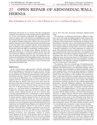
Figure 1 Shown are the great flat muscles of the abdominal wall. The insert depicts the relationship of the
great muscles to the groin.
- 4. © 2007 WebMD, Inc. All rights reserved. ACS Surgery: Principles and Practice
5 GASTROINTESTINAL TRACT AND ABDOMEN 27 OPEN REPAIR OF ABDOMINAL WALL HERNIA — 4
external oblique muscle fold back upon themselves to form the face of the muscle, leaving only transversalis fascia to cover the pos-
inguinal ligament, which attaches laterally to the anterior superior terior surface of the rectus abdominis.
iliac spine. In most persons, the medial insertion of the inguinal lig- The innervation of the anterior wall muscles is multifaceted.
ament is dual: one portion of the ligament inserts on the pubic The seventh through 12th intercostal nerves and the first and sec-
tubercle and the pubic bone, whereas the other portion is fan- ond lumbar nerves provide most of the innervation of the lateral
shaped and spans the distance between the inguinal ligament prop- muscles, as well as of the rectus abdominis and the overlying skin.
er and the pectineal line of the pubis. This fan-shaped portion of The nerves pass anteriorly in a plane between the internal oblique
the inguinal ligament is called the lacunar ligament. It blends lat- muscle and the transversus abdominis, eventually piercing the lat-
erally with Cooper’s ligament (or, to be anatomically correct, the eral aspect of the rectus sheath to innervate the muscle therein.The
pectineal ligament). The more medial fibers of the aponeurosis of external oblique muscle receives branches of the intercostal nerves,
the external oblique muscle divide into a medial crus and a lateral which penetrate the internal oblique muscle to reach it. The ante-
crus to form the external or superficial inguinal ring, through rior ends of the nerves form part of the cutaneous innervation of
which the spermatic cord (in females, the round ligament) and the abdominal wall. The first lumbar nerve divides into the ilioin-
branches of the ilioinguinal and genitofemoral nerves pass.The rest guinal nerve and the iliohypogastric nerve [see Figure 2]. These
of the medial fibers insert into the linea alba after contributing to important nerves lie in the space between the internal oblique
the anterior portion of the rectus sheath. muscle and the external oblique aponeurosis. They may divide
Beneath the external oblique muscle is the internal oblique within the psoas major or between the internal oblique muscle and
muscle.The fibers of the internal oblique muscle fan out following the transversus abdominis. The ilioinguinal nerve may communi-
the shape of the iliac crest, so that the superior fibers course cate with the iliohypogastric nerve before innervating the internal
obliquely upward toward the distal ends of the lower three or four oblique muscle. The ilioinguinal nerve then passes through the
ribs while the lower fibers orient themselves inferomedially toward external inguinal ring to run parallel to the spermatic cord, while
the pubis to run parallel to the external oblique aponeurotic fibers. the iliohypogastric nerve pierces the external oblique muscle to
These fibers arch over the round ligament or the spermatic cord, innervate the skin above the pubis. The cremaster muscle fibers,
forming the superficial part of the internal (deep) inguinal ring. which are derived from the internal oblique muscle, are innervat-
Beneath the internal oblique muscle is the transversus abdo- ed by the genitofemoral nerve.There can be considerable variabil-
minis.This muscle arises from the inguinal ligament, the inner side ity and overlap.
of the iliac crest, the endoabdominal fascia, and the lower six costal The blood supply of the lateral muscles of the anterior wall
cartilages and ribs, where it interdigitates with the lateral diaphrag- comes primarily from the lower three or four intercostal arteries,
matic fibers. The medial aponeurotic fibers of the transversus the deep circumflex iliac artery, and the lumbar arteries. The rec-
abdominis contribute to the rectus sheath and insert on the pecten tus abdominis has a complicated blood supply that derives from
ossis pubis and the crest of the pubis, forming the falx inguinalis. the superior epigastric artery (a terminal branch of the internal
Infrequently, these fibers are joined by a portion of the internal thoracic [internal mammary] artery), the inferior epigastric artery
oblique aponeurosis; only when this occurs is a true conjoined ten- (a branch of the external iliac artery), and the lower intercostal
don formed.12
Aponeurotic fibers of the transversus abdominis also form the
Quadratus
structure known as the aponeurotic arch. It is theorized that con- Lumborum
traction of the transversus abdominis causes the arch to move
downward toward the inguinal ligament, thereby constituting a
form of shutter mechanism that reinforces the weakest area of the
L3
groin when intra-abdominal pressure is raised. The area beneath
Iliohypogastric
the arch varies. Many authorities believe that a high arch, resulting Nerve Genitofemoral
in a larger area from which the transversus abdominis is by defin- Nerve
ition absent, is a predisposing factor for a direct inguinal hernia. Ilioinguinal
The transverse aponeurotic arch is also important because the Nerve
term is used by many authors to describe the medial structure that Sympathetic Trunk
is sewn to the inguinal ligament in many of the older inguinal her-
nia repairs. Psoas Muscle
The rectus abdominis forms the central anchoring muscle mass
of the anterior abdomen. It arises from the fifth through seventh Genital Branch
costal cartilages and inserts on the pubic symphysis and the pubic of Genitofemoral
Nerve
crest. It is innervated by the seventh through 12th intercostal
nerves, which laterally pierce the aponeurotic sheath of the muscle.
The semilunar line is the slight depression in the aponeurotic fibers Iliohypogastric
coursing towards the muscle. In a minority of persons, the small Nerve
pyramidalis muscle accompanies the rectus abdominis at its inser- Ilioinguinal
tion.This muscle arises from the pubic symphysis. It lies within the Nerve
rectus sheath and tapers to attach to the linea alba, which repre- Lateral Femoral
External
sents the conjunction of the two rectus sheaths and is the major site Cutaneous Nerve
Spermatic Nerve
of insertion for three aponeuroses from all three lateral muscle lay- Femoral Branch
ers.The line of Douglas (i.e., the arcuate line of the rectus sheath) of Genitofemoral
is formed at a variable distance between the umbilicus and the Nerve
inguinal space because the fasciae of the large flat muscles of the Figure 2 Shown are the important nerves of the lower abdomi-
abdominal wall contribute their aponeuroses to the anterior sur- nal wall.
- 5. © 2007 WebMD, Inc. All rights reserved. ACS Surgery: Principles and Practice
5 GASTROINTESTINAL TRACT AND ABDOMEN 27 OPEN REPAIR OF ABDOMINAL WALL HERNIA — 5
arteries.The lower intercostal arteries enter the sides of the muscle
after traveling between the oblique muscles; the superior and the Table 4—Commercially Available Synthetic
inferior epigastric arteries enter the rectus sheath and anastomose Prostheses for Abdominal Wall Hernia Repair
near the umbilicus.
The endoabdominal fascia is the deep fascia covering the inter- Polypropylene/polyester
nal surface of the transversus abdominis, the iliacus, the psoas Bard Composix E/X Mesh (PPL + ePTFE)
major and minor, the obturator internus, and portions of the Bard Dulex Mesh (dual-sided) (PPL + ePTFE)
periosteum. It is a continuous sheet that extends throughout the Bard Kugel Hernia Patch (PPL + ePTFE + PPL ring)
extraperitoneal space and is sometimes referred to as the wallpaper Bard Ventralex (PPL + ePTFE + PPL tail)
Sofradim Parietene (PPL + hydrophilic collagen)
of the abdominal cavity. Commonly, the endoabdominal fascia is
Sofradim Parietex (PPL + hydrophilic collagen)
subclassified according to the muscle being covered (e.g., iliac fas- Genzyme Sepramesh (PPL + Seprafilm)
cia or obturator fascia). Ethicon Prolene Soft Mesh (PPL)
The transversalis fascia is particularly important for inguinal Ethicon Proceed (PPL + PDS + ORC)
hernia repair because it forms anatomic landmarks known as ana- Ethicon Ultrapro (PPL + poliglecaprone 25)
logues or derivatives. The most significant of these analogues for Ethicon Vicryl Knitted Mesh
groin hernia surgeons are the iliopectineal arch, the iliopubic tract, Gore-Tex Soft Tissue Patch (ePTFE)
the crura of the deep inguinal ring, and Cooper’s ligament (i.e., the Gore-Tex DualMesh (ePTFE)
pectineal ligament). The superior and inferior crura form a Gore-Tex DualMesh Plus (ePTFE + silver + chlorhexidine)
“monk’s hood”–shaped sling around the deep inguinal ring. This Gore-Tex MycroMesh (ePTFE)
sling has functional significance, in that as the crura of the ring are PPL—polypropylene ePTFE—expanded polytetrafluoroethylene ORC—oxidized
pulled upward and laterally by the contraction of the transversus regenerated cellulose PPL—polypropylene
abdominis, a valvular action is generated that helps preclude indi-
rect hernia formation.The iliopubic tract is the thickened band of
the transversalis fascia that courses parallel to the more superfi- of Bogros. The preperitoneal space is of particular importance for
cially located inguinal ligament. It is attached to the iliac crest lat- surgeons because many of the inguinal hernia repairs (see below)
erally and inserts on the pubic tubercle medially. The insertion are performed in this area.The inferior epigastric vessels, the deep
curves inferolaterally for 1 to 2 cm along the pectineal line of the inferior epigastric vein, the iliopubic vein, the rectusial vein, the
pubis to blend with Cooper’s ligament, ending at about the mid- retropubic vein, the communicating rectusioepigastric vein, the
portion of the superior pubic ramus. Cooper’s ligament is actually internal spermatic vessels, and the vas deferens are all encountered
a condensation of the periosteum and is not a true analogue of the in this space.13
transversalis fascia.
Hesselbach’s inguinal triangle is the site of direct inguinal her-
nias. As viewed from the anterior aspect, the inguinal ligament Choice of Prosthetic Material
forms the base of the triangle, the edge of the rectus abdominis For most abdominal wall hernias, the procedure of choice
forms the medial border, and the inferior epigastric vessels form includes the use of a prosthesis. A detailed discussion comparing
the superolateral border. (It should be noted, however, that Hessel- and contrasting various prosthetic materials is beyond the scope of
bach actually described Cooper’s ligament as the base.) this chapter; however, some general statements may be made. As a
Below the iliopubic tract are the critical anatomic elements from rule, North American surgeons tend to consider polypropylene
which a femoral hernia may develop. The iliopectineal arch sepa- mesh the favored prosthetic material, whereas European surgeons
rates the vascular compartment that contains the femoral vessels are more likely to employ polyester mesh. Of course, the use of
from the neuromuscular compartment that contains the iliopsoas mesh presupposes a situation in which the prosthesis can be iso-
muscle, the femoral nerve, and the lateral femoral cutaneous nerve. lated from contact with intra-abdominal viscera by one or more
The vascular compartment is invested by the femoral sheath, layers of human tissue (e.g., peritoneum). In situations where con-
which has three subcompartments: (1) the lateral, containing the tact with intra-abdominal viscera cannot be avoided, a standard
femoral artery and the femoral branch of the genitofemoral nerve; mesh prosthesis should not be used. Either the prosthesis should
(2) the middle, containing the femoral vein; and (3) the medial, be composed of a nonmesh material, such as expanded polytetra-
which is the cone-shaped cul-de-sac known as the femoral canal. fluoroethylene (ePTFE), or a dual-layer prosthesis should be used,
The femoral canal is normally a 1 to 2 cm blind pouch that begins with a standard plastic mesh on the side facing the abdominal wall
at the femoral ring and extends to the level of the fossa ovalis.The (to encourage an intense fibroplastic response) and an adhesion
femoral ring is bordered by the superior pubic ramus inferiorly, the barrier of some type coating the peritoneal side. Numerous dual-
femoral vein laterally, and the iliopubic tract (with its curved inser- sided prosthetics, incorporating a variety of adhesion barriers, are
tion onto the pubic ramus) anteriorly and medially. The femoral now available [see Table 4]. It has consistently been shown that
canal normally contains preperitoneal fat, connective tissue, and when these materials are used, adhesions are not only less common
lymph nodes (including Cloquet’s node at the femoral ring), which but also less tenacious than when mesh alone is used. Often, bowel
collectively make up the femoral pad.This pad acts as a cushion for adhesions can be literally wiped from the peritoneal surface of a
the femoral vein, allowing expansion such as might occur during a dual-layer prosthesis with gentle blunt traction, in sharp contrast to
Valsalva maneuver, and serves as a plug to prevent abdominal con- the typically tedious and sometimes impossible dissection of bowel
tents from entering the thigh. A femoral hernia exists when the loops from a mesh prosthesis. Although all of the dual-layer pros-
blind end of the femoral canal becomes an opening (the femoral theses currently on the market are approved for decreasing adhe-
orifice) through which a peritoneal sac can protrude. sions to the adhesion barrier side, no manufacturer has sought
Between the transversalis fascia and the peritoneum is the approval for complete prevention of adhesions. Consequently, the
preperitoneal space. In the midline behind the pubis, this space is long-term effects of these less severe (but still present) adhesions
known as the space of Retzius; laterally, it is referred to as the space are unknown; further study is required to address this issue.
- 6. © 2007 WebMD, Inc. All rights reserved. ACS Surgery: Principles and Practice
5 GASTROINTESTINAL TRACT AND ABDOMEN 27 OPEN REPAIR OF ABDOMINAL WALL HERNIA — 6
Table 5—Commercially Available Biologic space, the posterior space, or both and (2) whether a prosthesis is
Prostheses for Abdominal Wall Hernia Repair included or omitted [see Table 6]. In reality, most of the numerous
eponyms used to name inguinal herniorrhaphies refer not to fun-
Approximate Price
damentally distinct operations but, rather, to relatively minor mod-
Prosthesis ($/cm2) ifications of standard hernia procedures [see Table 6]. Accordingly,
rather than address every known variant, we describe only the
Cook Surgisis Freeze-Dried Soft Tissue Graft (porcine 3.40 major repairs on which these variants are based.
small intestine)
The most important consideration in choosing an inguinal her-
LifeCell AlloDerm (human cadaver skin) 26.08 nia procedure is the experience of the surgeon. Knowing the ideal
operation for a given clinical scenario is of no significance if the sur-
Tissue Science Laboratory Permacol (porcine dermis) 8.33
geon is not skilled in performing it. On the assumption that the sur-
TEI Bioscience SurgiMend (fetal calf) 22.00 geon’s expertise is equal to the task, the next consideration should
Synovis Surgical Veritas (bovine pericardium) 8.60
be to tailor the operation to the patient’s particular hernia. For
example, a simple Marcy repair would be completely adequate for
Tutogen Tutopatch (bovine pericardium) — a pediatric patient with a Nyhus type 1 hernia [see Table 1] but not
Bard Tutomesh/Allomax (human dermis) 26.00 for an elderly patient who has an indirect hernia in conjunction
with extensive destruction of the inguinal floor. The conventional
Bard Collamend (porcine dermis) 16.00 anterior prosthetic repairs are particularly useful in high-risk
patients because they can easily be performed with local anesthe-
sia.19 On the other hand, giant prosthetic reinforcement of the vis-
A number of so-called biologic prostheses have been developed ceral sac (GPRVS), especially when bilateral, necessitates general
that are designed to promote vessel ingrowth and eventual remod- or regional anesthesia and thus is best for patients with bilateral
eling of tissue to resemble the native type [see Table 5]. Although direct or recurrent hernias or, perhaps, for patients with connective
biologic prostheses are much more expensive than synthetic pros- tissue disorders that appear to be associated with their hernia. If
theses, they may be the better choice when the operative field is surgery has previously been done in either the anterior or the
contaminated or when an abdominal wall defect is so large that the preperitoneal space, the surgeon should choose a procedure that
prosthesis cannot be covered by skin. Clearly, more study is uses the undissected space. If local or systemic infection is present,
required before their exact place in the armamentarium of the a nonprosthetic repair is usually considered preferable, though the
abdominal wall hernia surgeon can be determined. newer biologic prostheses now being evaluated may eventually
At present, there is some controversy regarding the weight of the change this view. Uncorrected coagulopathy is a contraindication
polypropylene mesh used in abdominal wall hernia repairs. (The to elective repair.
controversy almost certainly applies to the other types of mesh
prosthesis as well.) Data from randomized studies indicate that use
of a lightweight mesh results in less long-term pain than use of a Inguinal Hernia Repair: Operative Technique
normal mesh, without having any negative effect on the recurrence
rate.14,15 Lighter-weight mesh also addresses the theoretical con- ANTERIOR HERNIORRHAPHY
cern about the possible carcinogenic effects of polypropylene, as
has been suggested by experimental studies in rats, though it Steps Common to Prosthetic and Nonprosthetic Repairs
should be kept in mind that there has never been a documented The various anterior herniorrhaphies have a number of initial
case of a sarcoma developing in a human being as a result of an technical steps in common; they differ primarily with respect to the
inguinal hernia prosthesis.16 To illustrate the difference between a specific details of floor reconstruction.
lightweight mesh and a normal one, a 7.5 × 15 cm piece of
polypropylene mesh (Prolene; Ethicon, Inc., Somerville, New Step 1: choice of anesthetic Local anesthesia is entirely ade-
Jersey) weighs about 80 g/m2, whereas a similarly sized piece of a quate, especially when combined with intravenous sedation. In
polypropylene–poliglecaprone 25 (Monocryl; Ethicon, Inc., specialty hernia clinics, it is the approach most commonly
Somerville, New Jersey) lightweight mesh (UltraPro; Ethicon, Inc., employed. In general practice, however, general anesthesia is the
Somerville, New Jersey) weighs less than 30 g/m2 after absorption rule. This approach is reasonable in fit patients but is associated
of the poliglecaprone 25 component. North American surgeons with a higher incidence of postoperative urinary retention.20 If gen-
have been slow to accept the use of lightweight mesh for inguinal eral anesthesia is used, a local anesthetic should be given at the end
hernia repair, fearing a higher recurrence rate (as was suggested by of the procedure as an adjuvant to reduce immediate postoperative
one of the earlier randomized trials).17 Many also have some con- pain. Regional (spinal or epidural) anesthesia can also be used, but
cerns about possible bias in the data, noting that the research sup- it is less popular, having the highest incidence of systemic side
porting the use of lightweight mesh has been almost exclusively effects (primarily cardiovascular).19
funded by industry. Nevertheless, the randomized trials mentioned We prefer local anesthesia combined with I.V. infusion of a
earlier cannot be entirely discounted. rapid-acting, short-lasting, amnesic, and anxiolytic agent (e.g.,
propofol).This technique affords the patient all the benefits of gen-
eral anesthesia in terms of comfort, without the higher incidence
Inguinal Hernia Repair: Choice of Procedure of urinary retention seen with regional or general endotracheal
Practical considerations do not allow a description of every sin- anesthesia. An added benefit is that the patient can be aroused
gle named inguinal hernia repair in the literature. The nonpros- from sedation periodically to perform Valsalva maneuvers to test
thetic named repairs alone number more than 70.18 For the pur- the repair.
poses of this chapter, inguinal hernia repairs may be grouped The techniques and drug dosages employed by different experts
according to (1) whether the operation makes use of the anterior vary considerably. Compounding factors include the age of the
- 7. © 2007 WebMD, Inc. All rights reserved. ACS Surgery: Principles and Practice
5 GASTROINTESTINAL TRACT AND ABDOMEN 27 OPEN REPAIR OF ABDOMINAL WALL HERNIA — 7
patient and the amount of I.V. sedation used. Our preference is to sistent correlation with postoperative groin pain either way. The
use a solution containing 50 ml of 0.5% lidocaine with epineph- ilioinguinal and genitofemoral nerves are usually left with the cord
rine and 50 ml of 0.25% bupivacaine with epinephrine. The epi- structures. The genitofemoral nerve cannot always be identified
nephrine is optional and may be omitted if the patient has a histo- with certainty. It will be sacrificed in those procedures that include
ry of coronary artery disease or if there is concern about delayed division of the cremaster muscle (e.g., Shouldice repair).
bleeding. In an adult of normal size, 70 ml of this solution is inject-
ed before preparation and draping: 10 ml is placed 1 cm medial Step 4: mobilization of cord structures The cord struc-
and 1 cm inferior to the anterior superior iliac spine in an attempt tures are bluntly dissected away from the inferior flap of the exter-
to block the major nerves innervating the groin area [see nal oblique aponeurosis to expose the inguinal ligament (shelving
Abdominal Wall Anatomy, above], and the other 60 ml is used as a edge) and the iliopubic tract.This dissection is continued over the
field block along the orientation of the eventual incision in the sub- pubic tubercle and onto the anterior rectus sheath for at least 2 cm,
cutaneous and deeper tissues. Care is taken to ensure that some of defining the point where the most medial edge of a prosthesis will
the material is injected into the areas of the pubic tubercle and eventually be sutured if a Lichtenstein prosthetic repair is being
Cooper’s ligament, which are easily identified by tactile sensation performed. This measure facilitates en masse lifting of the cord
(except in very obese patients). The remaining 30 ml of the solu- structures with the fingers of one hand at the pubic tubercle so that
tion is reserved for discretionary use during the procedure. the index finger can be passed underneath to meet the ipsilateral
thumb or the fingers of the other hand. Mobilization of the cord
Step 2: initial incision Traditionally, the skin is opened by structures is completed by means of blunt dissection, and a
making an oblique incision between the anterior superior iliac Penrose drain is placed around them so that they can be retracted
spine and the pubic tubercle. For cosmetic reasons, however, many during the procedure.
surgeons now prefer a more horizontal skin incision placed in the
natural skin lines. In either case, the incision is deepened through Step 5: division of cremaster muscle For decades, com-
Scarpa’s fasciae and the subcutaneous tissue to expose the exter- plete division of the cremaster muscle with concomitant sacrifice
nal oblique aponeurosis. The external oblique aponeurosis is then of the genitofemoral nerve was common practice, especially with
opened through the external inguinal ring. If a prosthesis is to be indirect hernias. The purpose of this step was to facilitate identifi-
used, a large space is created beneath the external oblique aponeu- cation of the sac and to lengthen the cord for better visualization of
rosis from the anterior rectus sheath medially to the anterior supe- the inguinal floor. It is clear, however, that adequate exposure can
rior iliac spine laterally to prepare for the eventual placement. almost always be obtained by opening the muscle longitudinally,
which reduces the chances of damage to the cord and prevents tes-
Step 3: care of the sensory nerves The iliohypogastric ticular descent. Accordingly, the latter approach should be consid-
nerve is identified; it can be either left in situ or freed from the sur- ered best practice unless circumstances argue for division of the
rounding tissue and isolated from the operative field by passing a muscle.
hemostat under the nerve and grasping the upper flap of the exter-
nal oblique aponeurosis. Routine division of the iliohypogastric Step 6: management of hernial sac The term high ligation
nerve along with the ilioinguinal nerve is practiced by some but is of the sac is used frequently in discussing inguinal hernia repair; its
not advised by most, though there does not seem to be any con- historical significance has ingrained it in the descriptions of most
of the older operations. For our purposes in this chapter, high lig-
ation of the sac should be considered equivalent to reduction of the
Table 6—Selected Major Inguinal sac into the preperitoneal space without excision. The two meth-
Herniorrhaphy Techniques* ods work equally well and are highly effective. Proponents of sac
inversion believe that this measure results in less pain (because the
Category Anterior Repairs Posterior Repairs richly innervated peritoneum is not incised) and may be less likely
Marcy to cause adhesive complications.To date, however, no randomized
Bassini trials have been done to determine whether this is so.21 Sac ever-
Nonprosthetic Nyhus-Condon (iliopubic sion in lieu of excision does protect intra-abdominal viscera in
(pure tissue) Maloney darn tract repair)
Shouldice cases of unrecognized incarcerated sac contents or sliding hernia.
McVay Cooper’s ligament Many surgeons (especially urologists) believe that complete
excision of all indirect inguinal hernial sacs, even when inguinal-
Anterior approach
Read-Rives
scrotal, is important for preventing excessive postoperative hydro-
Posterior approach cele formation.The downside of this practice is that the incidence
GPRVS of ischemic orchitis from excessive trauma to the cord rises sub-
Prosthetic Lichtenstein tension-free stantially.The logical sequela of ischemic orchitis is testicular atro-
Modified Nyhus-Condon
(tension-free hernioplasty
repair) Mesh plug-and-patch
Kugel-Ugahary phy, though this presumed relationship has not been conclusively
Laparoscopic proved. In our view, it is better to divide an indirect inguinal her-
TAPP nial sac in the midportion of the inguinal canal once it is clear that
TEP the hernia is not sliding and no abdominal contents are present.
IPOM
The distal sac is not removed, but its anterior wall is opened as far
Bilayer prosthetic repair† distally as is convenient. We have not observed an increased inci-
dence of hydroceles with this approach.
*Many other named repairs have been described. For the most part, however, these other Direct hernial sacs are separated from the cord and other sur-
named repairs are relatively minor modifications of procedures listed in this table.
†Both the anterior space and the posterior space are used.
rounding structures and reduced into the preperitoneal space.
GPRVS—giant prosthetic reinforcement of the visceral sac IPOM—intraperitoneal onlay Dividing the superficial layers of the neck of the sac circumferen-
mesh TAPP—transabdominal preperitoneal TEP—totally extraperitoneal tially—thereby, in effect, opening the inguinal floor—usually facil-
- 8. © 2007 WebMD, Inc. All rights reserved. ACS Surgery: Principles and Practice
5 GASTROINTESTINAL TRACT AND ABDOMEN 27 OPEN REPAIR OF ABDOMINAL WALL HERNIA — 8
Nonprosthetic Repairs
Marcy repair The Marcy repair is the simplest nonprosthet-
ic repair performed today. Its main indication is for treatment of
Nyhus type 1 hernias (i.e., indirect inguinal hernias in which the
internal ring is normal). It is appropriate for children and young
adults in whom there is concern about the long-term effects of
prosthetic material. The essential features of the Marcy repair are
high ligation of the sac and narrowing of the internal ring.
Displacing the cord structures laterally allows the placement of
sutures through the muscular and fascial layers [see Figure 3].
Bassini repair Edoardo Bassini (1844–1924) is considered
the father of modern inguinal hernia surgery. It was during the
19th century that many of the great anatomists—Scarpa, Cooper,
Hesselbach, Bogros, Retzius, Cloquet, Gay, and others—made
their discoveries. By combining high ligation of the hernial sac with
reconstruction of the inguinal floor (based on the principles for-
mulated by the 19th-century anatomists), as well as taking advan-
tage of the developing disciplines of antisepsis and anesthesia,
Bassini was able to reduce recurrence and morbidity substantially.
Before Bassini’s achievements, elective herniorrhaphy was almost
never recommended, because the results were so bad. Bassini’s
operation, known as the radical cure, became the gold standard for
Figure 3 Marcy repair. The deep inguinal ring is narrowed inguinal hernia repair for most of the 20th century.
medially with several sutures that approximate the trans- The initial steps in the procedure are as previously described [see
verse aponeurotic arch to the iliopubic tract.
Steps Common to Prosthetic and Nonprosthetic Repairs, above].
Bassini felt that the incision in the external oblique aponeurosis
should be as superior as possible while still allowing the superficial
itates reduction and helps to maintain it while the prosthesis is external ring to be opened, so that the reapproximation suture line
being placed.The opening in the inguinal floor also allows the sur- created later in the operation would not be directly over the suture
geon to palpate for a femoral hernia. Sutures can be used to main- line of the inguinal floor reconstruction.22 Whether this technical
tain reduction of the sac, but they have no real strength in this set- point is significant is debatable. Bassini also felt that lengthwise
ting; their main purpose is to allow the repair to proceed without division of the cremaster muscle followed by resection was impor-
being hindered by continual extrusion of the sac into the field, tant for ensuring that an indirect hernial sac could not be missed
especially when the patient strains. and for achieving adequate exposure of the inguinal floor.
After performing the initial dissection and the reduction or liga-
Step 7: repair of inguinal floor Methods of repairing the tion of the sac, Bassini began the reconstruction of the inguinal
inguinal floor differ significantly among the various anterior floor by opening the transversalis fascia from the internal inguinal
herniorrhaphies and thus are described separately under the rele- ring to the pubic tubercle, thereby exposing the preperitoneal fat,
vant headings [see Nonprosthetic Repairs and Prosthetic Repairs, which he then bluntly dissected away from the undersurface of the
below]. superior flap of the transversalis fascia [see Figure 4a]. This step
allowed him to properly prepare the deepest structure in his
Step 8: relaxing incision A relaxing incision is employed famous “triple layer” (comprising the transversalis fascia, the trans-
only if a nonprosthetic repair is being performed. The incision is versus abdominis, and the internal oblique muscle).
made through the anterior rectus sheath and down to the rectus The first stitch in Bassini’s repair includes the triple layer supe-
abdominis, extending superiorly from the pubic tubercle for a riorly and the periosteum of the medial side of the pubic tubercle,
variable distance, as determined by the degree of tension present. along with the rectus sheath. In current practice, however, most
A so-called hockey-stick incision oriented laterally at the superior surgeons try to avoid the periosteum of the pubic tubercle so as to
end is a common choice. The posterior rectus sheath is strong decrease the incidence of osteitis pubis. The repair is then contin-
enough to prevent future incisional herniation. The relaxing inci- ued laterally, and the triple layer is secured to the reflected inguinal
sion works because as the anterior rectus sheath separates, the ligament (Poupart’s ligament) with nonabsorbable sutures. The
various components of the abdominal wall are displaced laterally sutures are continued until the internal ring is closed on its medi-
and inferiorly. al side [see Figure 4b]. A relaxing incision was not part of Bassini’s
original description but now is commonly added.
Step 9: closure Closure of the external oblique fascia serves Concerns about injuries to neurovascular structures in the
to reconstruct the superficial (external) ring. The external ring preperitoneal space and to the bladder led many surgeons, espe-
must be loose enough to prevent strangulation of the cord struc- cially in North America, to abandon the opening of the transver-
tures yet tight enough to ensure that an inexperienced examiner salis fascia. The unfortunate consequence of this decision is that
will not confuse a dilated ring with a recurrence. A dilated external the proper development of the triple layer is severely compromised.
ring is sometimes referred to as an industrial hernia, because over In lieu of opening the floor, a forceps (e.g., an Allis clamp) is used
the years it has occasionally been a problem during preemploy- to grasp tissue blindly in the hope of including the transversalis fas-
ment physical examinations. Scarpa’s fascia and the skin are closed cia and the transversus abdominis.The layer is then sutured, along
to complete the operation. with the internal oblique muscle, to the reflected inguinal ligament
- 9. © 2007 WebMD, Inc. All rights reserved. ACS Surgery: Principles and Practice
5 GASTROINTESTINAL TRACT AND ABDOMEN 27 OPEN REPAIR OF ABDOMINAL WALL HERNIA — 9
Transversus
Abdominis Internal Oblique
a Muscle b
Transversalis
Fascia
Figure 4 Bassini repair. (a) The transversalis fascia has been opened and the preperitoneal fat stripped away to
prepare the deepest structure in Bassini’s triple layer (comprising the transversalis fascia, the transversus abdominis,
and the internal oblique muscle). (b) The triple layer superiorly is approximated to the inguinal ligament, beginning
medially at the pubic tubercle and extending laterally until the deep inguinal ring is sufficiently narrowed.
as in the classic Bassini repair.The structure grasped in this mod- steps of the procedure.24 A continuous nonabsorbable suture (typ-
ified procedure is sometimes referred to as the conjoined tendon, ically of monofilament steel wire) is used to repair the floor. The
but this term is not accurate, because of the variability in what is Shouldice surgeons believe that a continuous suture distributes
actually grasped in the clamp. This imprecise “good stuff to good tension evenly and prevents potential defects between interrupted
stuff” approach almost certainly accounts for the inferior results sutures that could lead to recurrence.
achieved with the Bassini procedure in the United States.
Maloney darn The Maloney darn derives its name from the
way in which a long nylon suture is repeatedly passed between the
tissues to create a weave that one might consider similar to a mesh.
After initial preparation of the groin (see above), a continuous
nylon suture is used to oppose the transversus abdominis, the rec-
tus abdominis, the internal oblique muscle, and the transversalis
fascia medially to Poupart’s ligament laterally. The suture is con-
tinued into the muscle around the cord and is woven in and out to
form a reinforcement around the cord [see Figure 5]. On the later-
al side of the cord, it is sutured to the inguinal ligament and tied.
The darn is a second layer. The sutures are placed either parallel
or in a criss-cross fashion and are plicated well into the inguinal lig-
ament below.The darn must be carried well over the medial edge
of the inguinal canal. Once the darn is complete, the external
oblique fascia is closed over the cord structures.The Maloney darn
can be considered a forerunner of the mesh repairs, in that the pur-
pose of the darn is to provide a scaffold for tissue ingrowth.23
Shouldice repair Steps 1 through 6 of this repair are per-
formed essentially as previously described [see Steps Common to
Prosthetic and Nonprosthetic Repairs, above]. Particular impor-
tance is placed on freeing of the cord from its surrounding adhe- Figure 5 Maloney darn. The weave is made from a continuous
sions, resection of the cremaster muscle, high dissection of the her- nylon suture and is considered by many to be the precursor of
nial sac, and division of the transversalis fascia during the initial the mesh repairs.
- 10. © 2007 WebMD, Inc. All rights reserved. ACS Surgery: Principles and Practice
5 GASTROINTESTINAL TRACT AND ABDOMEN 27 OPEN REPAIR OF ABDOMINAL WALL HERNIA — 10
a b
Figure 6 Shouldice repair. (a) The first suture line starts at the pubic tubercle by approximating the iliopubic
tract laterally to the undersurface of the lateral edge of the rectus abdominis. The suture is continued laterally,
approximating the iliopubic tract to the medial flap (made up of the transversalis fascia, the internal oblique mus-
cle, and the transversus abdominis). (b) The second suture line begins after the stump of the divided cremaster
muscle has been picked up. The direction of the suture is reversed back toward the pubic tubercle, approximating
the medial edges of the internal oblique muscle and the transversus abdominis to Poupart’s ligament. Two more
suture lines will be constructed by approximating the internal oblique muscle and the transversus abdominis to a
band of the inferior flap of the external oblique aponeurosis superficial and parallel to Poupart’s ligament—in
effect, creating a second and a third artificial Poupart’s ligament.
The repair is started at the pubic tubercle by approximating the and continue to be so today. For a time, the Shouldice repair was
iliopubic tract laterally to the undersurface of the lateral edge of the the gold standard against which all newer procedures were com-
rectus abdominis [see Figure 6a]. The suture is continued laterally, pared. The major criticism of this operation is that it is difficult to
approximating the iliopubic tract to the medial flap, which is made teach because surgeons have problems understanding what is real-
up of the transversalis fascia, the internal oblique muscle, and the ly being sewn to what. Unless one is specifically trained at the
transversus abdominis. Eventually, four suture lines are developed Shouldice clinic and has the opportunity to work with the surgeons
from the medial flap. The continuous suture is extended to the there, one may find it hard to identify the various layers in the
internal ring, where the lateral stump of the cremaster muscle is medial flap reliably and reproducibly—a step that is crucial for
picked up to form a new internal ring. Next, the direction of the developing the multiple suture lines. To compound the difficulty,
suture is reversed back toward the pubic tubercle, approximating modifications developed outside the Shouldice clinic have given
the medial edges of the internal oblique muscle and the transver- rise to different versions of the procedure. For example, some sur-
sus abdominis to Poupart’s ligament, and the wire is tied to itself geons use three continuous layers instead of four for reconstruc-
and then the first knot [see Figure 6b]. Thus, two suture lines are tion of the inguinal floor.
formed by the first suture.
A second wire suture is started near the internal ring, approx- McVay Cooper’s ligament repair This operation is similar
imating the internal oblique muscle and the transversus abdo- to the Bassini repair, except that it uses Cooper’s ligament instead
minis to a band of external oblique aponeurosis superficial and of the inguinal ligament for the medial portion of the repair.
parallel to Poupart’s ligament—in effect, creating a second, arti- Interrupted sutures are placed from the pubic tubercle laterally
ficial Poupart’s ligament. This third suture line ends at the pubic along Cooper’s ligament, progressively narrowing the femoral ring;
crest.The suture is then reversed, and a fourth suture line is con- this constitutes the most common application of the repair—
structed in a similar manner, superficial to the third line. At the namely, treatment of a femoral hernia [see Figure 7].The last stitch
Shouldice clinic, the cribriform fascia is always incised in the in Cooper’s ligament is known as a transition stitch and includes
thigh, parallel to the inguinal ligament, to make the inner side of the inguinal ligament.This stitch has two purposes: (1) to complete
the lower flap of the external oblique aponeurosis available for the narrowing of the femoral ring by approximating the inguinal
these multiple layers. In general practice, however, this step is ligament to Cooper’s ligament, as well as to the medial tissue, and
commonly omitted. (2) to provide a smooth transition to the inguinal ligament over the
The results at the Shouldice clinic have been truly outstanding femoral vessel so that the repair can be continued laterally (as in a
- 11. © 2007 WebMD, Inc. All rights reserved. ACS Surgery: Principles and Practice
5 GASTROINTESTINAL TRACT AND ABDOMEN 27 OPEN REPAIR OF ABDOMINAL WALL HERNIA — 11
Bassini repair). Given the considerable tension required to bridge
such a large distance, a relaxing incision should always be used. In
the view of many authorities, this tension results in more pain than
is noted with other herniorrhaphies and predisposes to recurrence.
For this reason, the McVay repair is rarely chosen today, the main
exception being for treatment of a patient with a femoral hernia or
a patient with specific contraindications to mesh repair.
Prosthetic Repairs
Lichtenstein repair This operation is now considered the
gold standard for inguinal herniorrhaphy.The initial preparation of
the inguinal floor does not differ substantially from that carried out
in a nonprosthetic repair.The transversalis fascia is not opened—a
practice that has occasionally been criticized on the grounds that it
might cause an occult femoral hernia to be missed. To date, how-
ever, an excessive incidence of missed femoral hernias has not been
reported in men. The situation may be different in women: evi-
dence from the large population-based Swedish study cited earlier
suggests that femoral recurrence is much more common than one
might assume when the entire myopectineal orifice is not
addressed (as is the case with a McVay procedure or any of the
preperitoneal operations).3
The key to the operation is the placement of a large prosthesis
(at least 15 × 10 cm for an adult) extending from a point 2 cm
Figure 8 Lichtenstein repair. A mesh prosthesis is positioned
medial to the pubic tubercle (to prevent the pubic tubercle recur- over the inguinal floor and secured to the rectus sheath with a
rences all too commonly seen with other operations) to the ante- continuous suture. A slit is made in the mesh to accommodate the
rior superior iliac spine laterally. The medial end is rounded to cord structures, and the two tails are secured to each other and to
correspond to the patient’s particular anatomy, and a continuous the shelving edge of the inguinal ligament with a single interrupt-
suture of either nonabsorbable or long-lasting absorbable mater- ed suture. The superior and medial aspects of the prosthesis are
ial is begun between the prosthesis and the anterior rectus sheath secured to the internal oblique muscle and the rectus fascia with a
2 cm medial to the pubic tubercle [see Figure 8]. The suture is few interrupted sutures.
continued laterally in a locking fashion, securing the prosthesis to
either side of the pubic tubercle (not into it) and then to the above and a narrower one below. The tails are positioned around
shelving edge of the inguinal ligament. The suture is tied at the the cord structures and placed beneath the external oblique
internal ring. aponeurosis laterally to about the anterior superior iliac spine, with
A slit is made on the lateral side of the prosthesis to create two the upper tail placed on top of the lower. A single interrupted
tails, a wider one (approximately two thirds of the total height) suture is placed to secure the lower edge of the superior tail to the
lower edge of the inferior tail and the inguinal ligament—thereby,
in effect, creating a shutter valve.This step is considered crucial for
preventing the indirect recurrences occasionally seen when the tails
are simply reapproximated. The maneuver provides a cradling
effect as well, preventing direct contact between the cut edges of
the prosthesis and the cord structures, which could result in dam-
age when linear approximation is used. The suture also incorpo-
rates the shelving edge of the inguinal ligament so as to create a
domelike buckling effect over the direct space, thereby ensuring
that there is no tension, especially when the patient assumes an
upright position. The Lichtenstein group has developed a cus-
tomized prosthesis with a built-in domelike configuration, which,
in their view, makes suturing the approximated tails to the inguinal
ligament unnecessary.
A few interrupted sutures are placed to attach the superior and
medial aspects of the prostheses to the underlying internal oblique
muscle and rectus fascia. Care is taken to tie these loosely (with an
“air knot”) and to avoid placing them laterally so as to minimize
the risk of damaging the intramuscular and therefore invisible por-
tions of the important nerves. On occasion, the iliohypogastric
nerve, which courses on top of the internal oblique muscle, pene-
trates the medial flap of the external oblique aponeurosis. In this
situation, the prosthesis should be slit to accommodate the nerve.
The prosthesis can be trimmed in situ, but enough laxity must be
Figure 7 McVay Cooper’s ligament repair. The lateral stitch is the maintained to allow for the difference between the supine and
transition stitch to the femoral sheath and the inguinal ligament. upright positions, as well as for possible shrinkage of the mesh.
- 12. © 2007 WebMD, Inc. All rights reserved. ACS Surgery: Principles and Practice
5 GASTROINTESTINAL TRACT AND ABDOMEN 27 OPEN REPAIR OF ABDOMINAL WALL HERNIA — 12
bulk. Many surgeons consider this step important for preventing
erosion into surrounding structures (e.g., the bladder); indeed,
such complications have been reported, albeit rarely.
Millikan further modified the procedure by recommending that
the inside petals be sewn to the ring of the defect. For an indirect
hernia, the inside pedals are sewn to the internal oblique portion
of the internal ring; this forces the outside of the prosthesis under-
neath the inner side of the defect and makes it act like a preperi-
toneal underlay. For direct hernias, the inside petals are sewn to
Cooper’s ligament and the shelving edge of the inguinal ligament,
as well as to the conjoined tendon; this, again, forces the outside of
the prosthesis to act as an underlay.
The patch portion of the procedure is optional and involves
placing a flat piece of polypropylene in the conventional inguinal
space so that it widely overlaps the plug, much as in a Lichtenstein
repair.The difference with a plug-and-patch repair is that only one
or two sutures—or, sometimes, no sutures—are used to secure the
flat prosthesis to the underlying inguinal floor. Some surgeons,
however, place so many sutures that they have in effect performed
a Lichtenstein operation on top of the plug (sometimes referred to
as a “plugstenstein” repair).
To the credit of its proponents, the plug-and-patch repair, in all
of its varieties, has been skillfully presented and has rapidly taken a
Figure 9 Gilbert plug-and-patch repair. Depicted is the mesh
significant share of the overall inguinal hernia market. It is not only
plug technique for repair of an inguinal hernia. A flat sheet of fast but also extremely easy to teach, which has made it popular in
polypropylene mesh is rolled up like a cigarette or formed into a both private and academic centers. A randomized, controlled trial
cone (as shown here), inserted into the defect, and secured to has shown it to be equivalent to the Lichtenstein repair in terms of
either the internal ring (for an indirect hernia) or the neck of the recurrence and morbidity.28 However, numerous case reports in
defect (for a direct hernia) with interrupted sutures. the literature have described removal of plugs for pain, migration,
Prefabricated mesh plugs are now available. or erosion, and as a result, the plug-and-patch repair has been the
focus of considerable medicolegal scrutiny.
If a femoral hernia is recognized, the transversalis fascia is POSTERIOR (PREPERITONEAL) HERNIORRHAPHY
opened and the hernia reduced to expose Cooper’s ligament. The
Lichtenstein group’s approach is still to suture the inferior edge of Nonprosthetic Repairs
the prosthesis to the inguinal ligament. The femoral space is then A key technical issue in a preperitoneal hernia repair is how the
addressed by suturing the posterior surface of the prosthesis to surgeon chooses to enter the preperitoneal space. In fact, within this
Cooper’s ligament, thereby covering the entire myopectineal ori- general class of repair, it is the method of entry into this space that
fice, and finally by completing the superior and lateral sutures.We constitutes the major difference between the various procedures.
prefer to forgo the approximation of the inferior edge of the pros- Many approaches to the preperitoneal space have been
thesis to the inguinal ligament in favor of using interrupted sutures described. For example, the space can be entered either anteriorly
between that edge and Cooper’s ligament, much as in a McVay or posteriorly. If an anterior technique is to be used, the initial steps
repair (the “Fitztenstein” technique). A transition stitch is required of the operation are similar to those of a conventional anterior
between the inferior edge of the prosthesis, Cooper’s ligament, and herniorrhaphy. If a posterior technique is to be used, any of sever-
the inguinal ligament on the medial side of the femoral vein. This al incisions (lower midline, paramedian, or Pfannenstiel) will allow
stitch closes the femoral canal and sets the stage for the lateral side an extraperitoneal dissection. The preperitoneal space can also be
of the prosthesis to be sutured to the inguinal ligament.The rest of entered transabdominally.This approach is useful when the patient
the operation then proceeds in the same manner as a classic is undergoing a laparotomy for some other condition and the her-
Lichtenstein repair. nia is to be repaired incidentally. Of course, the transabdominal
preperitoneal laparoscopic repair described elsewhere [see 5:28
Plug-and-patch repair The mesh plug technique was first Laparoscopic Hernia Repair], by definition, enters the preperitoneal
developed by Gilbert and subsequently modified by Rutkow and space from the abdomen.
Robbins, Millikan, and others [see Figure 9].25-27 The groin is
entered via a standard anterior approach. The hernial sac is dis-
sected away from surrounding structures and reduced into the
preperitoneal space. A flat sheet of polypropylene mesh is rolled up Table 7—Contraindications to Use of
like a cigarette, tied, inserted in the defect, and secured with inter- Prosthesis for Herniorrhaphy
rupted sutures to either the internal ring (for an indirect hernia) or
the neck of the defect (for a direct hernia). Local infection* Allergy
A prefabricated prosthesis that has the configuration of a flower Systemic infection Patient preference (phobia)
Economic constraints
is commercially available and is recommended by Rutkow and
Robbins. This prosthesis is tailored to each patient’s particular *The newer biologic prostheses may be acceptable.
anatomy by removing some of the “petals” to avoid unnecessary
- 13. © 2007 WebMD, Inc. All rights reserved. ACS Surgery: Principles and Practice
5 GASTROINTESTINAL TRACT AND ABDOMEN 27 OPEN REPAIR OF ABDOMINAL WALL HERNIA — 13
Reed credits Annandale as being the first surgeon to describe
the anterior method of gaining access to the preperitoneal space.29
Bassini’s operation, as classically performed, is technically an ante-
rior preperitoneal operation, but it is never discussed in this group,
because in the American variant of the procedure, the preperi-
toneal space is not entered. Cheatle suggested the posterior
approach to the preperitoneal space for repair of an inguinal her-
nia but used a laparotomy to do it.30 Cheatle and Henry subse-
quently modified the operation so as to render it entirely extraperi-
toneal (the Cheatle-Henry approach), which made the procedure
more acceptable to surgeons.31
The preperitoneal nonprosthetic method remained popular
into the second half of the 20th century, championed by propo-
nents such as Nyhus and Condon, who emphasized the impor-
tance of the iliopubic tract as the inferior border in primary closure
of direct or indirect hernia defects.32 Today, however, these opera-
tions are of little more than historical significance, because it is
now universally agreed that better results are obtained in this space
when a prosthesis is used. Indeed, after 1975, Nyhus and Condon
began routinely placing a 6 × 14 cm piece of polypropylene mesh
to buttress the primary repair for all recurrent hernias.33 When
contraindications to a prosthesis are present [see Table 7], most sur-
geons would opt for a conventional anterior herniorrhaphy (e.g., a
Bassini or Shouldice repair) rather than a preperitoneal nonpros-
thetic herniorrhaphy.
Prosthetic Repairs
The most important step in any preperitoneal prosthetic repair
Figure 10 Depicted is the myopectineal orifice of Fruchaud.
is the placement of a large prothesis in the preperitoneal space on
The area is bounded superiorly by the internal oblique muscle
the abdominal side of the defect.The theoretical advantage of this and the transversus abdominis, medially by the rectus abdominis
measure is that whereas in a conventional repair, abdominal pres- and the rectus sheath, laterally by the iliopsoas muscle, and infe-
sure might contribute to recurrence, in a preperitoneal repair, the riorly by Cooper’s ligament. Critical anatomic landmarks (e.g.,
abdominal pressure would actually help fix the mesh material the inguinal ligament, the spermatic cord, and the femoral ves-
against the abdominal wall, thereby adding strength to the repair. sels) are contained within this structure.
The hernia defect itself may or may not be closed, depending on
the preference of the surgeon. The strength of the repair depends
on the prosthesis rather than on closure of the defect; however, Instead of subdividing hernias into direct, indirect, and femoral
such closure may decrease the seroma formation that inevitably and then examining their specific causes, he emphasized that the
occurs at the site of the undisturbed residual sac. Although these common cause of all inguinal hernias was the failure of the trans-
seromas almost always are self-limiting and disappear with time, versalis fascia to retain the peritoneum.This concept led Stoppa to
they can be confused with recurrences by both patients and refer- develop GPRVS, which reestablishes the integrity of the peritoneal
ring physicians. Accordingly, some surgeons prefer to take every sac by inserting a large permanent prosthesis that entirely replaces
step possible to prevent them. the transversalis fascia over the myopectineal orifice of Fruchaud
with wide overlapping of surrounding tissue. With GPRVS, the
Read-Rives repair The posterior space is accessed directly exact type of hernia present (direct, indirect, or femoral) is unim-
through the groin, and thus, the initial part of a Read-Rives repair, portant, because the abdominal wall defect is not addressed.
including the opening of the inguinal floor, is much like that of a
classic Bassini repair. The inferior epigastric vessels are identified Step 1: skin incision. A lower midline, inguinal, or Pfannenstiel
and the preperitoneal space completely dissected. The spermatic incision may be used, depending on the surgeon’s preference.The
cord is parietalized by separating the ductus deferens from the inguinal incision is placed 2 to 3 cm below the level of the anteri-
spermatic vessels. A 12 × 16 cm piece of mesh is positioned in the or superior iliac spine but above the internal ring; it is begun at the
preperitoneal space deep to the inferior epigastric vessels and midline and extended laterally for 8 to 9 cm.35
secured with three sutures placed in the pubic tubercle, in
Cooper’s ligament, and in the psoas muscle laterally.The transver- Step 2: preperitoneal dissection. The fascia overlying the space of
salis fascia is closed over the prosthesis and the cord structures Retzius is opened without violation of the peritoneum. A combi-
replaced.The rest of the closure is accomplished much as in a con- nation of blunt and sharp dissection is continued laterally posteri-
ventional anterior prosthetic repair. or to the rectus abdominis and the inferior epigastric vessels. The
preperitoneal space is completely dissected to a point lateral to the
Stoppa-Rignault-Wantz repair (GPRVS) GPRVS has its anterior superior iliac spine [see Figure 11]. The symphysis pubis,
roots in the important contribution that Henri Fruchaud made to Cooper’s ligament, and the iliopubic tract are identified. Inferiorly,
herniology. In describing the myopectineal orifice that bears his the peritoneum is generously dissected away from the vas deferens
name [see Figure 10], Fruchaud, who was Stoppa’s mentor, popu- and the internal spermatic vessels to create a large pocket, which
larized a different viewpoint on the etiology of inguinal hernias.34 will eventually accommodate a prosthesis without the possibility of