BIOMIMESYS® Hydroscaffold for 3D cell culture: automation of the processes, from cell seeding to imaging

The method of 2D cell culture is not a true reflection of the physiological cell environment where cells connect to other cells and, also to the extracellular matrix (ECM). Therefore some of the processes studied in 2D culture such as gene expression, apoptosis and importantly drug uptake and toxicity may not be directly transferable to in vivo experiments. HCS Pharma has developed a physiological and natural hydroscaffold, based on Hyaluronic Acid (HA) biofunctionalized with ECM components as collagens I, IV or VI, fibronectin and other molecules of interest. We will present the use of this scaffold on an automated platform, with the development of a 3D cell model for steatosis and phospholipidosis assessment as an example. Automated HepG2 cells seeding, medium renewal, spheroids treatment, fixation, staining and imaging will be discussed. BIOMIMESYS® represents a new generation of mimetic hydroscaffold for 3D cell culture. Available in a ready-to-use format it enables the culture of cells under physiological conditions that are representative of the microenvironment found in whole tissues. The highly porous nature of the hydroscaffold allows the rapid uptake of nutrients, oxygen, etc. into the cells to create a reproducible study model for all downstream analytical techniques used with 3D cell culture. Biomimesys® scaffolds technology is an innovative tool, closely mimeting the ECM matrix for relevant 3D cell culture allowing advanced drug discovery, ADME-Tox profiling and disease modelling. It is physiological, easy to use and compatible with all standard analysis methods (fluorescent plate reader, microscopy, Immunofluorescence, direct cell lysis for nucleic acids and protein extraction, histology).
Recommended

Recommended
More Related Content
More from HCS Pharma
More from HCS Pharma (20)
Recently uploaded
Recently uploaded (20)
BIOMIMESYS® Hydroscaffold for 3D cell culture: automation of the processes, from cell seeding to imaging
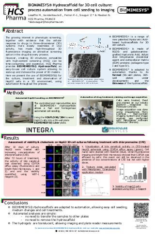
- 1. BIOMIMESYS® Hydroscaffold for 3D cell culture: process automation from cell seeding to imaging Lesaffre M., Vandenhaute E., Ferron P.-J., Souguir Z.* & Maubon N. HCS Pharma, FRANCE *zied.souguir@hcs-pharma.com Abstract Conclusions To get this poster, please flash the QR- code You can use the I-NIGMA application from your store The growing interest in phenotypic screening, together with evidence that the cellular response to drugs in three-dimensional (3D) systems more closely resembles in vivo activity, has made high-throughput 3D fluorescence imaging an attractive screening option in the drug discovery process. However, creating 3D structures compatible with high-content screening (HCS) can be time-consuming and expensive. HCS Pharma provides BIOMIMESYS® Hydroscaffold, an easy-to-use cell culture system allowing to scale up and increase the power of 3D models. Here we present the use of BIOMIMESYS® for the culture, treatment and observation of HepG2 cells in a 3D environment, using automation throughout the process. BIOMIMESYS® is a range of new patented Hyaluronic Acid- based hydroscaffolds for 3D cell culture. BIOMIMESYS® is made of RGDS- and galactosamine- grafted Hyaluronic Acid, Adipic Acid Dihydrazide crosslinker agent and extracellular matrix (ECM) proteins (collagens type I and type IV). BIOMIMESYS® is adapted to automation thanks to its format (96-well plates, 384- well plates under development) and the thinness of the hydroscaffold. 200 µm 10µm 20µm The controlled and reproductible size of BIOMIMESYS® Hydroscaffolds allows a fast and homogenous automated cell seeding. Using the VIAFLO 96/384 to seed HepG2 cells into a 96-well plate BIOMIMESYS® Liver plate. 0 µM Hoechst CDFDA Phase contrast Merged 1 µM 10 µM 25 µM 50 µM 100 µM 1. Vizualization of bile canaliculi activity in CPZ-treated HepG2 spheroids using CDFDA efflux assay (green). Cell nuclei were stained with Hoechst (blue). Direct fluorescence imaging and bright field microscopy showed that CDFDA was effluxed by cells: this event can still be observed in the presence of low concentrations of CPZ but not with higher doses. 2. Texture index calculated with MetaXpress ‘Granularity’ application module After 10 days of culture, HepG2 were treated with increasing concentrations of chlorpromazine (CPZ). After 72 hours of treatment, the activity of bile canaliculi was observed using CDFDA efflux assay (fluorescence and bright field microscopy, 1 and 2) and and the viability quantified using WST-1 reagent (3). BIOMIMESYS® Hydroscaffolds are adapted to automation, allowing easy cell seeding, medium changes and cell treatments. Automated analyses are simple: - no need to transfer the samples to other plates - no need to remove the hydroscaffold The hydrogels are translucent, allowing imaging and plate reader measurements Methods Cells were treated, fixed and stained on our robotic platform integrated by Molecular Devices. Image acquisition was done using ImageXpress Micro Confocal High-Content Imaging System and image analysis was performed with MetaXpress software (Molecular Devices). Automated HepG2 handling on BIOMIMESYS® Automation of drug treatment, staining and image acquisition Results Assessment of viability & cholestasis on HepG2 3D cell cultures following treatment with chlorpromazine (CPZ) Scanning electron microscopy observation of BIOMIMESYS® Hydroscaffold. The porosity ranges from 100 to 200 μm. 3. Viability of HepG2 cells assessed using WST-1 reagent To know more about BIOMIMESYS® Hydroscaffold, visit our website: www.biomimesys.com +33(0) 769 999 137 hello@biomimesys.com