ACC 2015 by Krittanawong et al - All Rights Reserved
•
0 likes•249 views

Recommended

Recommended
More Related Content
What's hot
What's hot (17)
Similar to ACC 2015 by Krittanawong et al - All Rights Reserved
Similar to ACC 2015 by Krittanawong et al - All Rights Reserved (20)
ACC 2015 by Krittanawong et al - All Rights Reserved
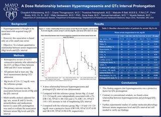
- 1. Table 2: Baseline characteristics of patients by serum Mg levels A Dose Relationship between Hypermagnesemia and QTc Interval Prolongation © 2014 Mayo Foundation for Medical Education and Research Chayakrit Krittanawong, M.D.1, Charat Thongprayoon, M.D.2, Thoetchai Peeraphatdit, M.D.1, Malcolm R Bell, M.B.B.S., F.RA.C.P1, Peter Brady, M.B. Ch. B., M.D1, Vitaly Herasevich, M.D.2, PhD., Suraj Kapa, M.D.1, Samuel Asirvatham, M.D.1,3, Niyada Naksuk, M.D.1 1Department of Cardiovascular disease, 2Department of Anesthesiology, 3Department of Pediatrics and Adolescent Medicine, Mayo Clinic, Rochester, MN, USA • Hypomagnesemia is thought to be associated with acquired long QT syndrome • However, this association is based only on a few small case series • Objective: To evaluate quantitative relationship between serum magnesium (Mg) levels and QTc interval Background • A dose relationship between hypermagnesemia and prolonged QTc interval was demonstrated • Compared with the reference group, Serum Mg ≥2.4 and 2.2-<2.4 mg/dL were independently associated with a 2% (OR 1.02, 95%CI 1.00-1.02) and a 1% (OR 1.01, 95%CI 1.01-1.03) increase in risk of lengthening QTc interval • Compared with the reference group, Mg <1.8 and 1.8-<2.0 mg/dL were a protective factor (OR 0.98, 95%CI 0.97-0.98 and OR 0.99, 95%CI 0.98-0.99, respectively) Mean serum magnesium levels, mg/dL < 1.8 1.8-<2.0 2.0-<2.2 2.2-<2.4 ≥ 𝟐𝟐. 𝟒𝟒 P-value Age, mean + SD, y 67+16 67+15 67+15 68+15 70+16 < 0.0001 Male, % 54 62 66 68 67 < 0.0001 White, % 86 85 84 84 84 0.44 Diabetes mellitus, % 32 27 26 28 35 < 0.0001 Hypertension, % 66 62 61 64 63 0.01 CKD, % 19 17 18 23 36 < 0.0001 CVA , % 7 7 6 5 6 0.54 AMI, % 50 55 51 48 35 < 0.0001 Cardiac arrest, % 8 10 9 7 10 0.08 Cardiogenic shock, % 7 9 9 9 12 0.01 ADHF, % 37 39 43 48 63 < 0.0001 K, mean + SD , mEq/L 4.1+0.4 4.1+0.4 4.2+0.4 4.3+0.4 4.4+0.6 < 0.0001 Ca, mean + SD, mg/dL 4.7+0.4 4.7+0.3 4.8+0.3 4.8+0.3 4.7+0.4 < 0.0001 Antiarrhythmic, % 22 27 30 29 33 < 0.0001 Beta-blockers, No. (%) 69 74 72 70 62 <0.001 Prolonged QTc meds, % 51 50 49 48 50 0.44 • Retrospective review of 8,512 consecutive patients who admitted to the CCU at Mayo Clinic between 2004 and 2013 were included • All patients had at least one Mg level measurement during CCU admission • Mg levels of 2.0-<2.2 mg/dL were the reference groups • The primary outcome was the association between levels of Mg and the QTc interval • Multivariable analysis adjusted for age, sex, serum K and Ca levels, antiarrhythmic and medications known to cause QTc prolongation was used to evaluate the association between serum Mg levels and QTc interval Results Methods Figure 1: A dose relationship between mean serum Mg levels (mg/dL), mean serum K levels (mg/dL), mean serum Ca levels (mg/dL) and mean QTc interval (ms) Conclusions • This finding suggests that hypomagnesemia was a protective factor for QTc prolongation • Contrary to conventional wisdom, we found a dose relationship between hypermagnesemia and prolonged QTc interval • Further experimental studies of cardiac molecular physiology between serum magnesium level and QTc interval are still needed to verify our findings Table 2: Baseline characteristics of patients by serum Mg levels