Case Review #1: 42 year old make with Adult Scoliosis Harrington Rod Removal
•
2 likes•2,400 views

A 42 year old male, post status seven fusions for scoliosis. Dr. Pashman performed revision surgery T2-Pelvis to correct 70 degree+ curvature, remove harrington rods and correct flatback. KIM/SRP Classification 3.

Recommended
Recommended
More Related Content
More from Robert Pashman
More from Robert Pashman (20)
Recently uploaded
Recently uploaded (20)
Case Review #1: 42 year old make with Adult Scoliosis Harrington Rod Removal
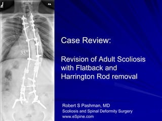
- 1. Case Review: 35° Revision of Adult Scoliosis with Flatback and Harrington Rod removal Robert S Pashman, MD Scoliosis and Spinal Deformity Surgery www.eSpine.com
- 2. Patient History 42 year old male, seven posterior instrumented fusions for massive scoliosis. 25 years ago had a posterior instrument fusion for severe thoracic scoliosis. This included Harrington compression and distraction rods along the selected curvature. Revision surgery adding on below the 70° instrumentation which ends at T12 or so, with significant curvature in both frontal and sagittal planes from the T9 through L2 area. Plate pseudoarthrosis at approximately T10 or so. Flat back deformity has been formed by the distraction instrumentation, although his head plumbs over the mid sacrum, he still has significant thoracolumbar kyphosis. Increasing pain at the thoracolumbar junction adjacent to the fusion and was told variably that he needed osteotomies, posterior spinal fusion versus nothing at all. He was told that he may be paralyzed if he does nothing.
- 3. Indications for Surgery Adult/adolescent idiopathic scoliosis excess 70 degrees. Now status post posterior spinal fusion x 7. Increased lumbar curve "adding on" with fractional curve below fusion causing #5 sagittal and coronal plane decompensation. Hardware failure. Now with unremitting low back pain due to collapse and cosmetic deformity.
- 4. Surgical Strategy 1. Segmental spinal instrumentation, T2 to pelvis. 16 levels using CD Legacy stainless steel screw/rod construct. 2. Posterior spinal fusion, T2 to pelvis using locally harvested autogenous bone. 3. Spinal osteotomy, Smith-Peterson osteotomy for correction of sagittal and coronal plane deformity, L1-T12. 4. T4-5 osteotomy for removal of proximal hardware hook. 5. T11-12 osteotomy for removal of distal hardware hook. 6. Re-exploration laminectomy, T12, L1, L2, L3-4 correction of sagittal plane deformity in anticipation of kyphectomy, Smith-Peterson osteotomy. 7. Laminotomy fusion mass, T3-4, T4-5 and T5-6 for placement of proximal thoracic pedicle screws through large fusion mass. 8. Pelvic instrumentation, bilateral iliac crest exploration through separate incisions for placement of screws.
- 5. Post-op x-rays Balance has been restored in both sagittal and frontal planes. 35°
- 6. Post-op x-ray Comparison The patient’s curvature was reduced by 50%, from 70° to 35°. He is well balanced 70° 35° in both planes.
- 7. Post-op x-rays Comparison The patient’s posture has improved dramatically. His films look good, and he is doing very well post- operatively.