2. 1 | INTRODUCTION
The panel adopted the following scheme, adapted from the American
Heart Association,1
to rate the strength of the recommendations in these
guidelines. The recommendation class designation appears with each rec-
ommendation, along with a second term that independently rates the
quality of the evidence upon which the recommendation was based.
2 | CLASSIFICATION OF
RECOMMENDATIONS
Class I recommendations are strong, reflecting the panel's conviction
that the recommended action appears to have definite benefit for
most patients, outweighing the risk to most patients.
Class I recommendations can be summarized as “benefit >>> risk”.
Class IIa recommendations are moderately strong, reflecting the
panel's belief that the recommended action should benefit most
patients, probably outweighing the risk to most patients.
Class IIa recommendations can be summarized as “benefit >> risk”.
Class IIb recommendations are weak, reflecting the belief that the
recommended action possibly benefits some patients and may out-
weigh the risk of taking the proposed action in most patients.
Class IIb recommendations can be summarized as “benefit > risk”.
Class III is used to describe recommendations in which the panel
believes that the potential risk and benefit of the proposed action are
essentially equal, such that these actions should probably not be pur-
sued under most circumstances.
Class III recommendations can be summarized as “benefit = risk”.
Class IV recommendations indicate the panel's belief that the pro-
posed action is more likely to cause harm than benefit to most
patients, such that Class IV designates actions that the panel believes
are contraindicated under most circumstances.
Class IV recommendations can be summarized as “risk >> benefit”.
The recommendation classification and level of evidence (LOE)
upon which that recommendation was based were independently
determined by the panel (ie, any class of recommendation may be
paired with any LOE).
The panel acknowledges that future evidence may change the
strength of any of these recommendations, as well as the quality of the
evidence on which they are based. Many important clinical questions
addressed in the guidelines have not yet been adequately addressed by
clinical trials. At times, the panel has made strong recommendations
based on more than the available evidence—thus weak or even absent
evidence does not necessarily accompany a weak recommendation.
Although randomized clinical trial evidence may be unavailable, a clear
clinical consensus that a particular test or treatment is useful may exist.
3 | LEVELS OF EVIDENCE
The methods of assessing the quality of the scientific evidence that
supports clinical decision making are evolving. The panel chose to
use a hybrid of the American Heart Association and Veterinary
Emergency Critical Care RECOVER evidence grading criteria, as out-
lined below.1,2
3.1 | Strong
High-quality evidence is from ≥1 randomized controlled trial, or a
moderate-quality randomized controlled trial, corroborated by a high-
quality observational study or other moderate-quality trials. These
prospective clinical studies were performed in dogs and either ran-
domly allocated subjects to an intervention or control group or used
concurrent controls (ie, controls recruited at the same time as the
experimental subjects) without randomization. Strong evidence also
could have been obtained from prospectively enrolled, controlled,
observational clinical studies in dogs with spontaneously occurring
mitral valve disease. These studies asked clinically relevant questions,
were considered to be adequately powered, and did not experience
excessive loss of subjects to follow up in any group, generating a clear
and statistically valid result.
3.2 | Moderate
Moderate-quality evidence is from ≥1 well-designed, well-executed
nonrandomized studies, observational studies, or registry studies, or
meta-analyses of such studies. Evidence rated as “moderate” by the
panel was generated from controlled, retrospective studies in dogs
(ie, studies in which dogs with mitral valve disease [or appropriate
controls] were selected from a previous period in time). Moderate
evidence also could have been generated from blinded and con-
trolled laboratory studies that were performed in experimental dogs.
3.3 | Weak
Weak-quality evidence is from randomized or nonrandomized obser-
vational or registry studies with limitations of design or execution,
but performed in dogs with clinical myxomatous mitral valve disease
(MMVD), or physiological, mechanistic, or experimental studies per-
formed in research dogs. Evidence rated as “weak” by the panel was
generated from uncontrolled clinical case reports or case series in
dogs with mitral valve disease, as well as by experimental or clinical
studies that were not performed in dogs with MMVD. These could
include experimental models of mitral valve disease in other species,
as well as high-quality studies in humans (such as meta-analyses, ran-
domized controlled trials, and clinical studies with concurrent con-
trols, including observational studies) with spontaneous mitral valve
disease.
3.4 | Expert opinion
Expert opinion based on clinical experience, common sense, or physi-
ologic or mechanistic studies performed in species other than dogs is
considered the weakest LOE.
2 KEENE ET AL.
3. 4 | INCIDENCE, PATHOLOGY, AND
PATHOGENESIS OF MMVD
It is estimated that approximately 10% of dogs presented to primary
care veterinary practices have heart disease, and MMVD is the most
common heart disease of dogs in many parts of the world, accounting
for approximately 75% of heart disease cases seen in dogs by veteri-
nary practices in North America.
The pathology of MMVD has been relatively recently reviewed,3
and some progress in understanding the genetics and pathophysiology
of the disease has been reported.4,5
Myxomatous mitral valve disease
most commonly affects the left atrioventricular or mitral valve, although
in at least 30% of cases, the right atrioventricular (tricuspid) valve also is
involved.6
The disease is approximately 1.5 times more common in
males than in females. Prevalence is also higher in smaller (<20 kg) dogs,
although large breeds sometimes are affected, and larger dogs also
often experience faster disease progression with more apparent myo-
cardial dysfunction, and have a more guarded prognosis.7
In small breed
dogs, the disease generally is slowly but at times unpredictably progres-
sive. Most dogs experience the onset of a recognizable murmur of
mitral valve regurgitation years before the clinical onset of heart failure.
Cavalier King Charles Spaniels notably are predisposed to developing
MMVD at a relatively young age, although the time course of their dis-
ease progression to heart failure does not appear to be markedly differ-
ent from that of other small breed dogs.8,9
Although the cause of MMVD remains unknown, the disease has an
inherited component in some breeds,5,10
and the severity of the disease
may have a genetic component in other breeds. The disease consistently
is characterized by changes in the cellular constituents as well as the
intercellular matrix of the valve apparatus (including the valve leaflets
and chordae tendineae).11,12
These changes involve both the collagen
content and the alignment of collagen fibrils within the valve. Expansion
of the spongiosa layer is characterized by changes in the proteoglycan
content of this layer. Dysregulation of the extracellular matrix appears to
be central to these changes. Valvular interstitial cells, possibly with post-
transcriptional regulation from microsatellite ribonucleic acids, acquire
properties of activated myofibroblasts, and activated myofibroblasts
increase proteolytic enzymes, including matrix metalloproteinases, which
degrade collagen and elastin faster than they can be produced by
unactivated valvular interstitial cells.13–15
Endothelial cell changes and subendothelial thickening also
occur,16–18
but these changes do not appear to put dogs with MMVD
at increased risk for either arterial thromboembolism or infective
endocarditis. Mitral valve prolapse is a common finding in dogs
with myxomatous valve degeneration and represents a prominent
echocardiographic feature of MMVD in some breeds.10,19,20
Pro-
gressive deformation of the valve structure eventually prevents
effective coaptation, allowing regurgitation (valve leakage). Pro-
gressive valvular regurgitation increases cardiac work, leading to
ventricular remodeling (eccentric hypertrophy of both the atrium
and ventricle, and intercellular matrix changes), and eventually to
ventricular dysfunction.
It has been hypothesized that abnormal numbers or types of mito-
gen receptors (ie, any of the subtypes of serotonin, endothelin, or angio-
tensin receptors) on fibroblast cell membranes in the valves of affected
dogs play a role in the pathophysiology of acquired valvular lesions.21–23
Systemic or local metabolic, neurohormonal, or inflammatory mediators
(eg, endogenous catecholamines, inflammatory cytokines) also may
influence progression of the valve lesion or the subsequent myocardial
remodeling and ventricular dysfunction that accompany long-standing,
hemodynamically important valvular regurgitation. The interactions of
these factors, as well as the impact of changes in mitral valve annular
geometry and mechanical stress on the pathogenesis and progression
of MMVD, are incompletely understood.11–13,24
The prevalence of MMVD increases markedly with age in small
breed dogs, with up to 85% showing evidence of the valve lesion by
13 years of age.25
The presence of the pathologic lesion of MMVD in
an individual does not necessarily identify a dog that will develop clini-
cally relevant valve regurgitation or signs of heart failure. Depending
on the rate of progression of the individual's valvular disease relative
to other common pathologic conditions that occur late in life and
often prove fatal, the presence of MMVD in the absence of clinical
signs may or may not influence the course of the affected dog's life.
It has become clear that age, progressive heart enlargement
(of the left atrium [LA] and ventricle), increased transmitral E wave
blood flow velocities, increased serum N-terminal pro-B-type natri-
uretic peptide (NT-proBNP) concentrations, and increases in resting
heart rate are at least moderately predictive of the rate of progression
of MMVD and can help identify dogs at risk for impending heart
failure.26–29
The rate of change of echocardiographic and radiographic
variables also may identify animals at increased risk of heart failure or death
from cardiac cause.30,31
Development of truly predictive (sensitive and spe-
cific) risk stratification schemes, however, awaits further refinement.
5 | CLASSIFICATION OF HEART DISEASE
AND HEART FAILURE
The term heart disease is used synonymously with cardiac pathology—
in this case, myxomatous degenerative changes of the mitral valve.
Heart disease, depending on its nature, rate of progression, and patient
age and condition may or may not lead to heart failure. The term “heart
failure” refers to clinical signs caused by heart dysfunction. Heart failure
is caused by heart disease that affects heart function such that either
venous pressures increase so severely that fluid accumulates in the lungs
or a body cavity (congestive heart failure [CHF], sometimes called “back-
ward heart failure because the heart fails to drain the veins adequately), or
the heart's pumping ability is compromised such that it cannot meet the
body's needs either during exercise or at rest, in the face of either normal
or increased venous pressures (sometimes called “forward heart failure”).
In 2009, the consensus panel adapted a staging system for heart
disease and heart failure, and sought to link the severity of morpho-
logic changes and clinical signs to appropriate treatments at each
stage.32
According to this approach, patients are expected to advance
from 1 stage to the next stage, unless progression of the disease is
KEENE ET AL. 3
4. altered by corrective treatment (such as surgery). This staging system,
applied to dogs with MMVD, remains useful, although recent clinical
trial results necessitate a more critical clinical evaluation of dogs in
Stage B to facilitate sound therapeutic decision making.
This staging system for MMVD describes 4 basic stages of heart
disease and heart failure:
• Stage A identifies dogs at high risk for developing heart disease but
that currently have no identifiable structural disorder of the heart
(eg, every Cavalier King Charles Spaniel or other predisposed breed
without a heart murmur).
• Stage B identifies dogs with structural heart disease (eg, the typical
murmur of mitral valve regurgitation, accompanied by some typical
valve pathology, is present), but that have never developed clinical
signs caused by heart failure. In a change from the 2009 recommenda-
tions, strong evidence now supports initiating treatment to delay the
onset of clinical signs of heart failure in a subset of stage B patients
with more advanced cardiac morphologic changes (outlined below).
Stage B1 describes asymptomatic dogs that have no radio-
graphic or echocardiographic evidence of cardiac remodeling in
response to their MMVD, as well as those in which remodeling
changes are present, but not severe enough to meet current
clinical trial criteria that have been used to determine that initi-
ating treatment is warranted (see specific criteria below).
Stage B2 refers to asymptomatic dogs that have more advanced
mitral valve regurgitation that is hemodynamically severe and
long-standing enough to have caused radiographic and echocar-
diographic findings of left atrial and ventricular enlargement that
meet clinical trial criteria used to identify dogs that clearly
should benefit from initiating pharmacologic treatment to delay
the onset of heart failure (specific criteria detailed below).
• Stage C denotes dogs with either current or past clinical signs of
heart failure caused by MMVD. Because of important treatment dif-
ferences between dogs with acute heart failure requiring hospital
care and those in which heart failure can be treated on an outpatient
basis, these issues have been addressed separately by the panel. It is
important to note that some dogs presented with heart failure for
the first time may have severe clinical signs requiring aggressive
treatment (eg, with additional afterload reducers or temporary venti-
latory assistance) that more typically would be reserved for those
patients refractory to standard treatment (see Stage D below).
• Stage D refers to dogs with end-stage MMVD, in which clinical signs
of heart failure are refractory to standard treatment (defined later in
this consensus statement). Such patients require advanced or spe-
cialized treatment strategies to remain clinically comfortable with
their disease, and at some point, treatment efforts become futile
without surgical repair of the valve. As with Stage C, the panel has
distinguished between dogs in Stage D that require acute, hospital-
based treatment and those that can be managed as outpatients.
This staging system emphasizes that there are known risk factors
and structural prerequisites for the development of heart failure caused
by MMVD. Accordingly, the classification system is designed to aid in:
• developing screening programs for the presence of MMVD in dogs
known to be at risk;
• implementing interventions that may (now and in the future)
decrease the risk of disease development or progression;
• identifying asymptomatic dogs with MMVD early in the course of
their disease, comparable to in situ cancer, so they can be more
effectively managed medically as chronic disease patients, or possi-
bly be treated surgically;
• identifying symptomatic dogs with MMVD so that these patients
can be managed medically as chronic disease patients or possibly
treated surgically; and
• identifying symptomatic dogs with advanced heart failure caused by
MMVD refractory to conventional medical treatment. These patients
require aggressive or new treatment strategies, possibly including
surgery, or potentially palliative or hospice-type end-of-life care.
6 | GUIDELINES FOR DIAGNOSIS AND
TREATMENT OF MMVD
6.1 | Stage A
Dogs at higher than average risk for developing heart failure but with-
out any apparent structural abnormality (ie, no audible heart murmur)
at the time of examination.
6.1.1 | Recommendations for diagnosis of Stage A
(unchanged from 2009)
• Small breed dogs, including breeds with known predisposition to
develop MMVD (eg, Cavalier King Charles Spaniels, Dachshunds,
Miniature, and Toy Poodles) should undergo regular evaluations
(yearly auscultation by the family veterinarian) as part of routine
health care. (Class I, LOE: expert opinion)
• Owners of breeding dogs or those at especially high risk, such as
Cavalier King Charles Spaniels, may choose to participate in yearly
screening events at dog shows or other events sponsored by their
breed association or kennel club and conducted by board-certified
cardiologists participating in an ACVIM-approved disease registry.
(Class I, LOE: expert opinion)
6.1.2 | Recommendations for treatment of Stage A
(unchanged from 2009)
• No drug treatment recommended for any patient. (Class I, LOE:
expert opinion)
• No dietary treatment recommended for any patient. (Class I, LOE:
expert opinion)
• Potential breeding animals should no longer be bred if a murmur or
echocardiographic evidence of mitral regurgitation (MR) is identi-
fied early, during the normal breeding age range (6-8 years of
age). (Class I, LOE: moderate)33,34
4 KEENE ET AL.
5. 6.2 | Stage B
Dogs in Stage B have a structural abnormality (eg, the presence of
MMVD) but have never had clinical signs of heart failure associated
with their disease.
6.2.1 | Recommendations for diagnosis and further
categorization of Stage B
• Myxomatous mitral valve disease typically is recognized during a
screening or routine health examination by auscultation of a heart
murmur typical of mitral valve regurgitation.
• Thoracic radiography is recommended in all patients to assess the
hemodynamic relevance of the valve disease and to obtain baseline
thoracic radiographs at a time when the patient is asymptomatic for
MMVD. Patients with MMVD frequently have concurrent tracheal
or bronchial diseases and having baseline radiographs at a time when
the dog is asymptomatic can enhance the ability to radiographically
differentiate cardiac from noncardiac causes of cough in the face of
future clinical signs. (Class I, LOE: expert opinion).
• Blood pressure measurement is recommended for all patients to
identify or rule out concurrent systemic hypertension and to estab-
lish baseline blood pressure. (Class I, LOE: expert opinion)
• Echocardiography, performed by an experienced operator, is rec-
ommended to definitively identify the cause of the murmur, answer
specific questions regarding the severity of cardiac chamber enlarge-
ment, and identify comorbidities. A specialist's examination might
identify hemodynamic abnormalities including pulmonary hyperten-
sion or increased left atrial pressure. Echocardiographic identification
of mild left atrial or ventricular enlargement can be challenging, and
comparisons to breed-specific normal values may be required (Class I,
LOE: Moderate).35–41
In addition to short axis basilar views, recently
described 2-dimensional, long-axis echocardiographic ratios (left ven-
tricle (LV)/aorta (Ao), LA/Ao, and LA/LV) have proven to be effective
for identifying left atrial and ventricular enlargement in dogs with
MMVD.42
(Class I, LOE: strong)
• The panel recognizes that it is sometimes necessary to use thoracic
radiography in the absence of echocardiography to further refine
Stage B. Under these circumstances, the clinician must be cautious
because of marked variation in thoracic conformation and breed
differences in normal vertebral heart scales; the use of the verte-
bral left atrial size (VLAS) (details below) may be beneficial. (Class I,
LOE: moderate)
6.3 | Stage B1: Asymptomatic dogs with mitral valve
regurgitation caused by MMVD that is not severe
enough to meet criteria used to trigger the use of
medical treatment to delay the onset of heart failure.
Stage B1 dogs are characterized by a spectrum of imaging findings
ranging from those with radiographically and echocardiographically
normal left atrial [LA] and ventricular [LV] dimensions with normal LV
systolic function and normal radiographic vertebral heart or VLAS to
those with echocardiographic or radiographic evidence of left atrial
and ventricular enlargement that does not meet specific criteria out-
lined below.
6.3.1 | Recommendations for treatment and
monitoring of Stage B1 (both pharmacologic and
dietary, small and large breed dogs) remain unchanged
from the 2009 recommendations
Treatment is not recommended in these dogs because at this early
stage of disease, progression to heart failure is uncertain, unlikely to
occur within the recommended evaluation interval, and there is no
evidence that medication is effective at this stage. To summarize,
• No drug or dietary treatment is recommended (Class I, LOE: expert
opinion)
• Reevaluation by echocardiography is suggested (or radiography if
echocardiography is unavailable) in 6-12 months, depending on
the imaging results (some panelists recommend more frequent
follow-up in large dogs). (Class I, LOE: expert opinion)
6.4 | Stage B2: Asymptomatic MMVD causing MR
severe enough to result in cardiac remodeling (LA and
LV enlargement) sufficient to recommend treatment
before the onset of clinical signs based on the results
of a clinical trial.43,44
Dogs in this category should
meet the current criteria outlined below.
• Stage B2 criteria for heart enlargement identify dogs that are likely
to benefit substantially from treatment before the onset of clinical
signs of heart failure. (Class I, LOE: Strong):
murmur intensity ≥3/6;
echocardiographic LA : Ao ratio in the right-sided short axis view
in early diastole ≥1.6 (Figure 1)45
;
Left ventricular internal diameter in diastole, normalized for body
weight (LVIDDN) ≥1.7 (Table 1)46
;
breed-adjusted47–53
radiographic vertebral heart score (VHS)
10.5.
• Ideally, all these criteria should be met before initiating treatment,
because treatment represents a lifelong commitment. Of these
criteria, echocardiographic evidence of left atrial and ventricular
enlargement meeting or exceeding these criteria is considered to
be the most reliable way to identify dogs expected to benefit from
treatment.
• Although studies to identify reliable radiographic markers of Stage
B2 cardiac remodeling and enlargement in MMVD are underway,
definitive criteria for the radiographic identification of this stage
are currently not available. In the absence of echocardiographic mea-
surements, clear radiographic evidence of cardiomegaly (eg, a “gen-
eral breed” VHS ≥11.5, or a comparable “breed-adjusted” VHS in
cases where breed-specific VHS normal values are available) or evi-
dence of accelerating (increasing) interval change in radiographic or
KEENE ET AL. 5
6. echocardiographic cardiac enlargement patterns42
can substitute for
quantitative echocardiography to identify Stage B2. (Class I, LOE:
expert opinion)
• A newer index of radiographic left atrial enlargement, the VLAS,
provides a quantitative method of estimating left atrial size. Mea-
sured on either the right or left lateral radiograph by drawing a line
from the center of the most ventral aspect of the carina to the
most caudal aspect of the LA where it intersects with the dorsal
border of the caudal vena cava, that line then is transposed to the
cranial edge of the 4th thoracic vertebral body.54
Studies are ongo-
ing to determine a VLAS value that accurately predicts B2 remo-
deling, but in the absence of echocardiography, VLAS values of ≥3
likely identify Stage B2 MMVD. (Class 1, LOE: moderate)
6.4.1 | Recommendations for treatment of Stage B2
• Pimobendan is recommended at a dosage of 0.25-0.3 mg/kg PO
q12h.44,55
(Class I, LOE: strong)
• Dietary treatment is recommended. Principles guiding dietary treat-
ment at this stage include mild dietary sodium restriction and provi-
sion of a highly palatable diet with adequate protein and calories for
maintaining optimal body condition.56
(Class IIa, LOE: weak)
• Angiotensin converting enzyme inhibitors (ACEI): For patients in
Stage B2 on either initial examination, or in which the LA has
increased markedly in size on successive monitoring examinations,
5 (of 10) panelists recommend treatment with ACEI.57–59
(Class IIa in
geographic regions where ACEI are low cost, LOE: weak)
Clinical trials addressing the efficacy of ACEI for treatment of
dogs in Stage B have shown mixed results.
• Beta blockade is not recommended routinely to delay the onset of
heart failure in dogs in Stage B2, regardless of heart enlargement.
Clinical trials addressing the efficacy of beta blockers for the treat-
ment of dogs in Stage B2 have shown no benefit to date. (Class III,
LOE: weak)
• Spironolactone also is not recommended for routine use to delay
the onset of heart failure in dogs. Clinical trials addressing the effi-
cacy of spironolactone for the treatment of dogs in Stage B2 have
not been published as of this writing (2019), although a pilot study
suggests this approach should be used60
(Class IIb, LOE: expert
opinion).
• No other pharmacologic treatments for Stage B were recommended
by a majority of panelists. A few panelists considered the use of the
following medications for patients in advanced Stage B2 under spe-
cific circumstances: beta blockers, amlodipine. These treatment strat-
egies require further investigation to assess their efficacy and safety
in this patient population before definitive recommendations can be
made. (Class III, LOE: expert opinion)
FIGURE 1 One of the 4 criteria that identify advanced Stage B2
in dogs is that the echocardiographic LA : Ao ratio as measured in the
right-sided short-axis view in early diastole is ≥1.6. The measurement
is illustrated. The blue arrow illustrates the measurement of the aortic
dimension at the level of the aortic valve, and the orange arrow
illustrates measurement of the left atrial dimension TABLE 1 One of the 4 criteria that identify advanced Stage B2 in
dogs is an increase in their left ventricular chamber size, such that
normalized to their body weight (BW), it is ≥1.7
BW (kg) LVIDD (cm)
1 1.7
2 2.1
3 2.4
4 2.6
5 2.7
6 2.9
7 3.0
8 3.1
9 3.2
10 3.3
11 3.4
12 3.5
13 3.6
14 3.7
15 3.8
16 3.8
17 3.9
18 4.0
19 4.0
20 4.1
For dogs weighing between 1 and 20 kg (BW, left hand column), the left
ventricular end diastolic diameter (LVIDD) meeting this criterion must be
more than or equal to the dimension (in cm) in the right hand column. In the
referenced clinical trial, the end-diastolic LV dimension was obtained from a
2D short-axis-guided M-mode echocardiogram of the chamber.55
The
formula for normalizing LVIDD to BW is LVIDdN = LVIDd (cm)/weight
(kg)0294
.
6 KEENE ET AL.
7. • Some panelists find the use of cough suppressants useful in occa-
sional patients in advanced Stage B2 when their cough is thought
to be the result of pressure from cardiac enlargement (without
pulmonary edema) on adjacent bronchi. (Class IIa, LOE: expert
opinion)
• Surgical intervention in advanced Stage B2 is possible and rec-
ommended by some panelists for clients who can afford and access
mitral valve repair at the few centers demonstrating evidence of
acceptably low complication rates and effective, durable results.61–63
(Class IIa, LOE: moderate)
6.5 | Stage C
Stage C dogs have MMVD severe enough to cause current or past
clinical signs of heart failure. Stage C includes all dogs with MMVD
that have experienced an episode of clinical heart failure and that are
not refractory to standard heart failure treatment (standard treatment
is defined below). These patients continue to be categorized as Stage
C even after improvement or complete resolution of their clinical signs
with standard treatment. In exceptional cases that undergo successful
surgical mitral valve repair, reclassification to Stage B is warranted.
Guidelines for standard pharmacologic treatment are provided for
both in-hospital (acute) management of heart failure and for home
care (chronic) management of heart failure, as well as recommenda-
tions for chronic dietary management. Some patients in Stage C may
have life-threatening clinical signs and require more extensive acute
treatment than is considered standard. These acute care patients tem-
porarily may share medical management strategies with dogs that
have progressed to Stage D (refractory heart failure, see below).
For both Stages C and D (MMVD patients with symptomatic heart
failure), the acute care of heart failure is focused on regulating the
patient's hemodynamic status and tissue oxygen delivery. This is accom-
plished by monitoring (as much as possible, under existing clinical cir-
cumstances) and optimizing the patient's preload, afterload, heart rate,
contractility, and oxygenation, while decreasing oxygen demand. The
ultimate goals include improving cardiac output, decreasing mitral valve
regurgitation, and relieving clinical signs associated with either low car-
diac output or excessively increased venous pressures (congestion),
especially pulmonary dysfunction.
The broad goals of chronic management (in clinical settings where
surgery to effectively repair the mitral valve is not possible) are focused
on maintaining hemodynamic improvements while providing additional
treatments aimed at slowing the progression of disease, prolonging sur-
vival, decreasing the clinical signs of CHF, enhancing exercise capacity,
maintaining body weight (BW), and improving quality of life.
6.5.1 | Recommendations for diagnosis of Stage C
The signalment, history, and physical examination can be helpful in
determining the pretest probability of heart failure as a cause of clinical
signs in patients with MMVD. For example, obese dogs with no history
of weight loss are less likely to be in heart failure secondary to MMVD;
dogs with marked sinus arrhythmia and relatively slow heart rates also
are less likely to have clinical signs attributable to MMVD than are those
with similar clinical signs (eg, cough, dyspnea) with sinus rhythm or sinus
tachycardia. (Class I, LOE: expert opinion)
• The typical dog in Stage C from MMVD presents with clinical signs
of left-sided CHF and a history that can include tachypnea, restless-
ness, respiratory distress, or cough. Because of the relatively high
prevalence of chronic tracheobronchial disease in the population
most at risk for MMVD, the presence of a typical left apical reg-
urgitant quality murmur in a coughing dog does not necessarily
mean that clinical signs are the result of CHF. A clinical database
(including thoracic radiographs and ideally an echocardiogram)
should be obtained. Additionally, basic laboratory tests, including at
a minimum PCV as well as serum total protein, creatinine, urea nitro-
gen and electrolyte concentrations, and urine specific gravity) should
be obtained as soon as practical in dogs with heart failure. Impaired
renal function in particular represents an important comorbidity in
dogs with heart failure. (Class I, LOE: expert opinion)
• Echocardiography with Doppler studies also is useful in the diagno-
sis of dogs with MMVD that have advanced to Stages C and
D. Cardiac ultrasound examination can confirm the presence of
MMVD, quantify chamber enlargements and cardiac function, pro-
vide general estimates of LV filling pressures, and identify com-
orbidities and complications of chronic MR. These might include
pulmonary hypertension, acquired atrial septal defect, and pericardial
effusion from an atrial tear or unrelated cardiac tumor. As an exam-
ple, a pretreatment finding of a low-velocity E-wave on pulsed-wave
Doppler strongly argues against a diagnosis of left-sided heart fail-
ure. Conversely, most dogs in Stages C and D have high-velocity
early filling waves. In dogs with evidence of symptomatic pulmonary
hypertension (eg, exertional fatigue, collapse or syncope, ascites
from right-sided CHF), spectral Doppler findings can substantiate
the diagnosis and help guide therapeutic decision-making.
• Serum NT-proBNP concentrations (obtained using a commercially
available test) can add useful adjunct evidence when determining
the cause of clinical signs in dogs with MMVD, especially when the
NT-proBNP concentration is normal or nearly normal in a symp-
tomatic animal. As a group, dogs with clinical signs caused by heart
failure have higher serum NT-proBNP concentrations than do dogs
in which clinical signs are caused by primary pulmonary disease,
although the positive predictive value of any single specific NT-
proBNP concentration has not been adequately characterized. A
normal or near normal NT-proBNP concentration in a dog with
clinical signs of cough, dyspnea, or exercise intolerance strongly
suggests that heart failure is not the cause of the clinical signs.64,65
(Class I, LOE: moderate)
• Most symptomatic dogs with MMVD are middle-aged or older, and
it is prudent to complete the clinical database with a blood pres-
sure assessment, CBC, serum biochemical profile, and urinalysis,
especially if treatment for CHF is anticipated. (Class I, LOE: expert
opinion)
KEENE ET AL. 7
8. 6.5.2 | Recommendations for acute (hospital-based)
treatment of Stage C
• Furosemide 2 mg/kg administered IV (or intramuscularly [IM]),
followed by 2 mg/kg IV or IM hourly until the patient's respiratory
signs are substantially improved (ie, respiratory rate and effort are
decreased) or a total dosage of 8 mg/kg has been reached over
4 hours. (Class I, LOE: expert opinion)
• For life-threatening pulmonary edema (ie, expectoration of froth
associated with severe dyspnea, radiographic white-out lung, poor
initial response to furosemide bolus with failure of respiratory effort
and rate to improve over 2 hours), furosemide also may be adminis-
tered as a constant rate infusion (CRI) at a dosage of 0.66-1 mg/kg/
hour after the initial bolus.66,67
(Class IIa, LOE: weak)
• Allow the patient free access to water once diuresis has begun.
(Class I, LOE: expert opinion; humane considerations apply)
• Pimobendan, 0.25-0.3 mg/kg administered PO q12h. Although the
clinical trial evidence supporting the chronic use of pimobendan in
the management of Stage C heart failure from MMVD is stronger
than for the acute presentation, the recommendation to use
pimobendan in acute heart failure treatment is strongly supported
by hemodynamic and experimental evidence68
as well as the anec-
dotal experience of the panelists. In many countries outside of the
United States, pimobendan for IV administration is available. (Class I,
LOE: weak)
• Oxygen supplementation, if needed, can be administered via a
humidity and temperature-controlled oxygen cage or incubator or
via a nasal oxygen cannula. (Class I, LOE: expert opinion)
• Mechanical treatments (eg, abdominal paracentesis, thoracentesis)
are recommended to relieve effusions judged sufficient to impair
ventilation or cause respiratory distress. (Class I, LOE: expert
opinion)
• Sedation-anxiety associated with dyspnea should be treated. Nar-
cotics, or a narcotic combined with an anxiolytic agent, most often
are used by panelists. Care must be taken to monitor the blood
pressure and respiratory response to narcotics and tranquilizers in
the setting of acute heart failure. No specific treatment or dosage
regimen was used by all panelists. Butorphanol 0.2 to 0.25 mg/kg
administered IM or IV was the narcotic most often utilized for this
purpose; combinations of buprenorphine (0.0075-0.01 mg/kg) and
acepromazine (0.01-0.03 mg/kg IV, IM, or SC) as well as other nar-
cotics, including morphine and hydrocodone, also were suggested.
(Class I, LOE: expert opinion)
• Provide optimal nursing care, including maintenance of appropriate
environmental temperature and humidity, increase of the head on
pillows, and placement of sedated patients in sternal posture.
(Class I, LOE: expert opinion)
• Dobutamine (2.5-10 μg/kg/min as a CRI, starting at 2.5 μg/kg/min
and increasing the dosage incrementally) may be used in addition
to the above treatments to improve the left ventricular function in
patients that fail to respond adequately to diuretics, pimobendan,
sedation, oxygen, and comfort care measures. Continuous ECG
monitoring is recommended where available during dobutamine
infusion, with dosage reduction indicated if tachycardia or ectopic
beats occur. (Class I, LOE: expert opinion)
• Constant IV infusion of sodium nitroprusside at dosages ranging
from 1 to 15 μg/kg/min) for up to 48 hours often is useful for life-
threatening, poorly responsive pulmonary edema69
; this medication
is currently (2018) expensive in the United States. The use and PO
titration of additional arterial dilators (eg, hydralazine or amlodipine,
specific dosing recommendations also in Class D below) also may be
useful in patients when administration of nitroprusside is not feasi-
ble. (Class I, LOE: weak)
• ACEI, for example, enalapril or benazepril, 0.5 mg/kg PO q12h.
Although treatment with an ACEI is a Class I recommendation for
chronic Stage C heart failure (see below) and some panelists also
treat acute heart failure with ACEI, the evidence supporting ACEI
efficacy and safety in acute treatment, when combined with furo-
semide and pimobendan, is less clear. There is, however, clear evi-
dence that the acute administration of enalapril plus furosemide in
acute heart failure results in significant improvement in pulmonary
capillary wedge pressure when compared with the administration
of furosemide alone.70
(Class IIb, LOE: weak)
• Nitroglycerin ointment, approximately half an inch paste/10 kg
BW, applied to an unhaired or shaved area of skin, can be used for
the first 24 to 36 hours of hospitalization.71,72
Some panelists rec-
ommend administering the ointment at intervals (12 hours on,
12 hours off). Other panelists do not use nitroglycerin in this set-
ting. (Class IIb, LOE: weak)
6.5.3 | Recommendations for chronic (home-based)
treatment of Stage C
• Continue PO furosemide administration to effect, commonly at a
dosage of 2 mg/kg administered q12h, or as needed to maintain
patient comfort. Some panelists now choose to substitute torsemide
for furosemide at 1/10-1/20 or approximately 5% to 10% of the furo-
semide dosage, or approximately 0.1-0.3 mg/kg q24h73
for home
care in animals in which hospitalized CHF management using furo-
semide was difficult or met with limited success. (Class I, LOE:
moderate)
• Chronic PO furosemide dosages ≥8 mg/kg q24h in any dosing regi-
men (or the equipotent torsemide dosage) needed to maintain
patient comfort in the face of appropriate dosages of pimobendan,
an ACEI, and spironolactone indicate disease progression to Stage
D. Consideration of known causes of diuretic resistance, including
noncompliance (ie, not receiving the drug), high sodium intake,
slow absorption (eg, gut edema), impaired secretion into the renal
tubular lumen (eg, chronic kidney disease, advanced age, concur-
rent nonsteroidal anti-inflammatory drug use), hypoproteinemia,
hypotension, nephron remodeling, and neurohormonal activation is
warranted. (Class I, LOE: weak)
• Measurement of serum creatinine, blood urea nitrogen, and elec-
trolyte concentrations 3-14 days after initiating furosemide
8 KEENE ET AL.
9. treatment is recommended for animals with Stage C heart failure.
(Class I, LOE: weak)
• Continue or start ACEI (eg, enalapril or benazepril, 0.5 mg/kg PO
q12h) or an equivalent dosage of another ACEI, if approved for this
use. Measurement of serum creatinine and electrolyte concentra-
tions 3-14 days after beginning an ACEI is recommended for ani-
mals with Stage C heart failure. Concern for development of acute
kidney injury is warranted should serum creatinine concentrations
increase by ≥30% of the baseline concentration. (Class I, LOE: weak)
• Spironolactone (2.0 mg/kg PO q12 - 24 h) is recommended as an
adjunct for chronic treatment of dogs in Stage C heart failure. The
primary benefit of spironolactone in this situation is thought to be
aldosterone antagonism.74,75
(Class I, LOE: moderate)
• Continue pimobendan, 0.25-0.3 mg/kg PO q12h.76,77
(Class I, LOE:
strong)
• Panelists recommend against starting a beta blocker in the face of
active clinical signs of CHF (eg, cardiogenic pulmonary edema) cau-
sed by MMVD. (Class IV, LOE: weak)
• None of the panelists routinely use nitroglycerin in the chronic
treatment of Stage C heart failure. (Class III, LOE: expert opinion)
• Participation in a structured, home-based extended care program
to promote ideal BW, appetite, respiratory and heart rate monitor-
ing while providing client support to enhance medication regimen
adherence and dosage adjustments in patients with heart failure is
encouraged. (Class I, LOE: expert opinion)
Of these variables, identification of increases in resting respi-
ratory rate above normal baseline has the best predictive value for
impending clinical decompensation.78,79
(Class I, LOE: moderate).
• In centers with low complication rates, Stage C patients benefit
from surgical intervention to repair their mitral valve appara-
tus.61,63
(Class I, LOE: moderate)
• In cases complicated by atrial fibrillation, diltiazem, often in combina-
tion with digoxin (see below), is recommended to control ventricular
rate. Multiple preparations of diltiazem are available; treatment
should be started at a modest dosage for the preparation chosen
and titrated to achieve heart rate control. Ideally, mean heart rate as
measured by Holter monitoring in dogs with well-controlled signs of
CHF receiving stable drug dosage regimens should be close to nor-
mal or at least 125 beats per minute.80,81
(Class I, LOE: moderate)
• Digoxin 0.0025-0.005 mg/kg, administered PO q12h to achieve a
target steady-state plasma concentration (approximately 8 hours
post-pill) of 0.8-1.5 ng/mL. For the chronic management of Stage C
heart failure, panelists recommended the addition of digoxin only in
cases complicated by persistent atrial fibrillation to slow the ventric-
ular response rate. In these cases, digoxin generally is used in combi-
nation with diltiazem. Digoxin may not be tolerated in patients with
factors known to put animals at risk for adverse effects or toxicity
(eg, increases of serum creatinine concentration above normal, ven-
tricular ectopy, concerns over owner adherence, or chronic gastroin-
testinal disease resulting in frequent or unpredictable bouts of
vomiting or diarrhea).82
(Class IIb, LOE: moderate)
• In patients receiving a beta blocker before the onset of Stage C
heart failure, the majority of panelists continue beta blockade; some
panelists consider dosage reduction if needed clinically because of
clinical signs of low cardiac output, hypothermia, or bradycardia.
(Class IIB, LOE: expert opinion)
• Some panelists find the use of cough suppressants useful in occa-
sional patients in Stage C heart failure from MMVD. (Class IIa,
LOE: expert opinion)
• Some panelists find the use of bronchodilators useful in occasional
patients in Stage C MMVD patients. (Class IIb, LOE: expert opinion)
6.5.4 | Recommendations for dietary treatment for
Stage C
• Cardiac cachexia is defined as a loss of muscle or lean body mass
associated with heart failure, with or without clinically relevant
accompanying weight loss. Cachexia has substantial negative prog-
nostic implications and is much easier to prevent than to treat.83,84
(Class I, LOE: moderate)
• Maintain adequate calorie intake (maintenance calorie intake in
Stage C should be approximately 60 kcal/kg BW) to minimize
weight loss that often occurs in CHF.85,86
Simple culinary strate-
gies to improve appetite may be beneficial in accomplishing this
goal (eg, warming food, mixing wet food with dry food, offering a
variety of foods). (Class I, LOE: moderate)
• Specifically address and inquire about the occurrence of anorexia
and make efforts to treat any drug-induced or other identifiable
causes of anorexia that occur. (Class I, LOE: expert opinion)
• Record body condition score and accurate weight of the patient at
every clinic visit and investigate the cause of clinically relevant changes
in body condition, weight gain or loss. (Class I, LOE: expert opinion)
• Ensure adequate protein intake and avoid low-protein diets designed
to treat chronic kidney disease, unless severe concurrent renal failure
is present.83
(Class I, LOE: moderate)
• Modestly restrict sodium intake, taking into consideration sodium
from all dietary sources (including dog food, treats, table food, and
foods used to administer medications) and avoid any processed or
other salted foods.87,88
(Class I, LOE: moderate)
• Monitor serum electrolyte concentrations and supplement the diet
with potassium from either natural or commercial sources only if
hypokalemia is identified. The panel's anecdotal experience is that
hypokalemia is much more common in animals receiving torsemide.
(Class I, LOE: expert opinion)
• Hyperkalemia is relatively rare in patients treated for CHF with
diuretics, even in those concurrently receiving ACEI in combination
with spironolactone. Diets and foods with high potassium content
should be avoided when hyperkalemia is present. (Class I, LOE:
expert opinion)
• Consider monitoring serum magnesium concentrations, especially
as heart failure progresses and in dogs with arrhythmias. Supple-
ment with magnesium in cases in which hypomagnesemia is identi-
fied. (Class IIa, LOE: expert opinion)
KEENE ET AL. 9
10. • Consider supplementing with omega-3 fatty acids, especially in
dogs with decreased appetite, muscle loss, or arrhythmia.86
(Class
IIa, LOE: moderate)
6.6 | Stage D
Patients have clinical signs of failure refractory to standard treatment
for Stage C heart failure from MMVD. Stage D dogs thus require more
than a total daily dosage of 8 mg/kg of furosemide or the equivalent
dosage of torsemide, administered concurrently with standard doses
of the other medications thought to control the clinical signs of heart
failure (eg, pimobendan, 0.25-0.3 mg/kg PO q12h, a standard dosage
of approved ACEI, and 2.0 mg/kg of spironolactone daily). When
needed, antiarrhythmic medication to maintain sinus rhythm or regu-
late the ventricular response to atrial fibrillation (mean daily heart rate
125/minute)81
should be in use before a patient is considered to be
refractory to standard treatment.
Few clinical trials have addressed drug efficacy and safety in this
patient population. This deficiency leaves cardiologists treating heart
failure refractory to conventional medical treatment with a perplexing
variety of treatment options. Because of the relative lack of clinical trial
evidence and the diverse clinical presentations of patients with end-
stage heart failure, development of meaningful consensus guidelines
regarding the timing and implementation of individual pharmacologic
and dietary treatment strategies for Stage D patients proved difficult.
Surgical intervention to repair the mitral valve at Stage D is possible
and indicated where feasible, although it is associated with higher peri-
operative mortality and decreased overall survival in studies reported to
date.61
As with Stage C, guidelines for pharmacologic treatment are pro-
vided for both in-hospital (acute) and at-home care (chronic) manage-
ment of heart failure, as well as recommendations for chronic dietary
management.
6.6.1 | Recommendations for the diagnosis of Stage
D (refractory heart failure)
• Because Stage D heart failure patients are, by definition, refractory
to the standard treatments for Stage C patients, defining refractory
CHF involves the same diagnostic steps outlined for Stage C plus the
finding of failure to respond to treatments outlined in the Stage C
guidelines.
6.6.2 | Recommendations for acute (hospital-based)
treatment of Stage D
• In the absence of severe renal insufficiency (eg, serum creatinine
concentration 3 mg/dL), additional furosemide can be administered
to dyspneic patients diagnosed with refractory heart failure as an ini-
tial 2 mg/kg IV bolus followed by either additional bolus doses or a
furosemide CRI at a dosage of 0.66-1 mg/kg/h, until respiratory
distress (rate and effort) has decreased, or for a maximum of
4 hours. (Class I, LOE: expert opinion)
• Torsemide, a potent long-acting loop diuretic may be used to treat
dogs no longer adequately responsive to furosemide (0.1-0.2 mg/kg
q12h-q24h or approximately 5%-10% of the current furosemide
dosage to deliver a furosemide-equivalent dose).28
It appears that
the diuresis induced by torsemide produces less renin-angiotensin-
aldosterone system activation than more frequent doses of furose-
mide, similar to what has been shown in dogs and horses with the
diuresis induced by furosemide CRI.89,90
Clinicians should continue
to allow patients free access to water, once diuresis has begun.
(Class I, LOE: expert opinion)
• Cavitary centesis (abdominal paracentesis, thoracentesis), as needed
to relieve respiratory distress or discomfort. (Class I, LOE: expert
opinion)
In addition to oxygen supplementation as in Stage C (above),
mechanical ventilatory assistance may be useful in making the
patient comfortable, in allowing time for medications to have an
effect, and in providing time for left atrial dilatation to accommo-
date sudden increases in mitral valve regurgitant volume in
patients with acute exacerbation of MMVD (eg, chordae ten-
dineae rupture with severe cardiogenic pulmonary edema) and
impending respiratory failure.91
(Class I, LOE: weak)
• In patients that can tolerate it, more vigorous afterload reduction
(arterial vasodilation) is recommended, with close monitoring of arte-
rial blood pressure. In cases in which mechanical ventilation and IV
vasodilator or inotropic support is needed, arterial pressure monitor-
ing via peripheral arterial catheterization is preferred over noninva-
sive blood pressure monitoring when possible. In dogs judged to be
too sick to wait for the effects of PO afterload reduction or inotropic
support (eg, pimobendan with or without hydralazine or amlodipine),
the administration of a CRI IV of sodium nitroprusside (for
afterload reduction) or dobutamine (for inotropic support, especially
in hypotensive patients) or both is recommended by a majority of
panelists.69
Both are started at dosages of 1.0 μg/kg/min and
up-titrated every 15-30 minutes to a maximum of approximately
10-15 μg/kg/min. These rates may be used for 12-48 hours to
improve hemodynamic status and control refractory cardio-
genic pulmonary edema. Continuous ECG and blood pressure
monitoring are recommended to minimize the potential risks of
this treatment. (Class IIa, LOE: weak)
• Potentially beneficial PO drugs that decrease afterload in this situ-
ation include hydralazine (0.5-2.0 mg/kg PO, starting at a low dos-
age and titrating to effect as described above with nitroprusside,
but with hourly dosage increases or amlodipine (approximately
0.05-0.1 mg/kg PO, also to effect, although maximal drug effect
does not occur for approximately 3 hours, mandating a slower
titration). (Class I, LOE: expert opinion)
These drugs are recommended in addition to an ACEI and
pimobendan. Vigilance is needed to avoid serious, prolonged hypo-
tension (monitor blood pressure closely, maintaining arterial systolic
blood pressure 85 mm Hg, or mean arterial blood pressure 60 mm
10 KEENE ET AL.
11. Hg). Serum creatinine concentration should be reevaluated no more
than 24 to 72 hours after initiating these drugs.
The panel emphasized that because afterload reduction may
increase cardiac output substantially in the setting of severe MR
and heart failure, administration of an effective arterial dilator drug
in this setting does not necessarily compromise blood pressure.
(Class IIa, LOE: expert opinion).
• Sildenafil, (starting at 1-2 mg/kg PO q8h, and titrating if needed) is
used by panelists to treat Stage D heart failure from MMVD that is
complicated by clinically relevant estimated pulmonary hypertension.
Pulmonary hypertension is recognized as an increasingly frequent
complication of MMVD, either as a direct consequence of severe
mitral valve regurgitation or as an independent comorbidity that can
be responsible for clinical signs including syncope, cough, and short-
ness of breath (dyspnea), and sometimes radiographically evident
pulmonary infiltrates.92,93
(Class I, LOE: moderate) The occurrence of
ascites or jugular distension in patients with primarily left-sided heart
disease is suggestive of pulmonary hypertension and should prompt
an attempt to conclusively diagnose and identify patients that may
benefit from sildenafil. (Class IIa, LOE: weak)
• Pimobendan dosage may be increased (off-label use) to include a
third 0.3 mg/kg daily PO dose (ie, 0.3 mg/kg PO q8h); some panel-
ists administer an additional dose of pimobendan on admission to
Stage D patients with acute pulmonary edema regardless of the
timing of the last dose given at home. This dosage recommenda-
tion is outside the US Food and Drug Administration–approved
labeling for pimobendan (off-label use), and this use of the drug
should be explained to and approved by the client. (Class IIa, LOE:
expert opinion)
• Some panelists recommend adjunctive treatment with bronchodi-
lators in treating cardiogenic pulmonary edema in hospitalized
patients. (Class IIb, LOE: expert opinion)
6.6.3 | Recommendations for chronic (home-based)
Stage D treatment
• Furosemide (or torsemide) dosage should be increased as needed to
decrease the accumulation of pulmonary edema or body cavity
effusions, if not limited by renal dysfunction (indicators of which
generally should be monitored 12-48 hours after dosage increases).
Inappetence may increase the risk of development of azotemia
associated with medications for heart failure. The specific strategy
and magnitude of dosage increase (eg, same dosage divided q8h
instead of 2 higher doses, substituting 1 SC dose for a PO dose
q4h, or flexible SC dose supplementation, based on BW or girth
measurements) varied widely among the panelists. See Stage C rec-
ommendations (above) for a brief discussion of diuretic resistance.
(Class IIa, LOE: expert opinion)
• Torsemide, a potent and longer-acting loop diuretic, may be used
to treat dogs no longer adequately responsive to furosemide
(torsemide beginning dosage of 0.1-0.2 mg/kg PO, or approximately
5%-10% of the current furosemide dosage, up to approximately
0.6 mg/kg, divided q12h if necessary).94
(Class I, LOE: moderate)
• Spironolactone, if not already started as recommended in Stage C,
is indicated for chronic treatment of Stage D patients.74
(Class I,
LOE: moderate)
• Beta blockers generally should not be initiated at this stage, unless
they are being used as an adjunct to control heart rate in atrial
fibrillation. (Class IV, LOE: expert opinion)
• Hydrochlorothiazide was recommended by several panelists as
adjunctive treatment to furosemide or torsemide, utilizing various
dosing schedules (including intermittent use every 2nd-4th day).
Some panelists warned of the risk of acute kidney insufficiency
and marked electrolyte disturbances, based on personal experi-
ence. (Class IIb, LOE: expert opinion)
• Pimobendan dosage is increased by some panelists to include a
third 0.3 mg/kg daily dose (off-label use; routine explanations and
cautions to the owner apply as in hospital care described above) or
an even higher dosage when repeated rescue is necessary. (Class
IIa, LOE: expert opinion)
• Additional afterload reduction, using either amlodipine or hydral-
azine (see dosages and cautions above), may provide additional
hemodynamic benefit and decrease cough frequency.
• Digoxin, at the same (relatively low) dosages recommended by
some panelists for Stage C heart failure with atrial fibrillation, is
recommended for the treatment of atrial fibrillation in Stage D
patients lacking a concrete contraindication.82
(Class IIb, LOE:
moderate)
• Digoxin, at the same (relatively low) dosages recommended by
some panelists for Stage C heart failure with atrial fibrillation, also
is recommended by some panelists for all Stage D patients, includ-
ing those in sinus rhythm, lacking a concrete contraindication.
(Class IIb, LOE: expert opinion)
• Sildenafil (1-2 mg/kg PO q8h) may be useful in the management of
patients with clinical signs related to exertion and in management
of ascites when there is echocardiographic evidence of moderate
to severe pulmonary hypertension.95
(Class IIa, LOE: weak)
• Beta blockade may be useful in decreasing the ventricular response
rate in atrial fibrillation after stabilization and digitalization, but
caution should be used because of the negative inotropic effects
of beta blockers. (Class IIb, LOE: expert opinion)
• The majority of panelists felt that beta blockade initiated previ-
ously should not be stopped, but that dosage reduction may be
needed if shortness of breath cannot be controlled by the addition
of other medications or if bradycardia, hypotension, or both were
present. (Class IIb, LOE: expert opinion)
• Cough suppressants are recommended to treat chronic, intractable
cough in Stage D home care patients by some panelists. (Class IIa,
LOE: expert opinion)
• Bronchodilators are recommended to treat chronic, intractable
coughing in Stage D home care patients by some panelists. (Class
IIb, LOE: expert opinion)
KEENE ET AL. 11
12. 6.6.4 | Recommendation for chronic (home-based)
dietary treatment for Stage D
• All of the dietary considerations for Stage C (above) apply.
• In patients with refractory fluid accumulations, attempts should be
made to further decrease dietary sodium intake if it can be done
without compromising appetite or renal function. (Class IIa, LOE:
expert opinion)
ACKNOWLEDGMENT
This work was presented in part at the 2017 ACVIM Forum, Seattle,
WA, in the plenary program.
CONFLICT OF INTEREST DECLARATION
Bruce W. Keene—Consulted for Boehringer Ingelheim and CEVA Ani-
mal Health.
Clarke E. Atkins—Consulted for Boehringer Ingelheim and CEVA
Animal Health.
John D. Bonagura —Consulted for Boehringer Ingelheim, IDEXX
and CEVA Animal Health.
Philip R. Fox—Consulted for Boehringer Ingelheim, IDEXX and
CEVA Animal Health.
Jens Häggström—Consulted for Boehringer Ingelheim, IDEXX and
CEVA Animal Health.
Virginia Luis Fuentes—Consulted for Boehringer Ingelheim and
CEVA Animal Health.
Mark A. Oyama—Consulted for Boehringer Ingelheim, CEVA Ani-
mal Health, and IDEXX.
John E. Rush—Consulted for Boehringer Ingelheim and IDEXX.
Rebecca Stepien—Consulted for Boehringer Ingelheim and IDEXX.
Masami Uechi—Consulted for Boehringer Ingelheim and TERUMO
Corporation.
OFF-LABEL ANTIMICROBIAL DECLARATION
Authors declare no off-label use of antimicrobials.
INSTITUTIONAL ANIMAL CARE AND USE COMMITTEE
(IACUC) OR OTHER APPROVAL DECLARATION
Authors declare no IACUC or other approval was needed.
HUMAN ETHICS APPROVAL DECLARATION
Authors declare human ethics approval was not needed for this study.
ORCID
Bruce W. Keene https://orcid.org/0000-0002-4758-5654
Philip R. Fox https://orcid.org/0000-0003-4089-0573
Jens Häggström https://orcid.org/0000-0003-3402-023X
John E. Rush https://orcid.org/0000-0002-8277-8996
REFERENCES
1. Yancy CW, Jessup M, Bozkurt B, et al. 2017 ACC/AHA/HFSA focused
update of the 2013 ACCF/AHA guideline for the management of heart
failure: a report of the American College of Cardiology/American Heart
Association task force on clinical practice guidelines and the Heart Fail-
ure Society of America. J Am Coll Cardiol. 2017;70:776-803.
2. Boller M, Fletcher DJ. RECOVER evidence and knowledge gap analy-
sis on veterinary CPR. Part 1: evidence analysis and consensus pro-
cess: collaborative path toward small animal CPR guidelines. J Vet
Emerg Crit Care. 2012;22(Suppl.1):S4-S12.
3. Fox PR. Pathology of myxomatous mitral valve disease in the dog.
J Vet Cardiol. 2012;14:103-126.
4. Meurs KM, Friedenberg SG, Williams B, et al. Evaluation of genes
associated with human myxomatous mitral valve disease in dogs with
familial myxomatous mitral valve degeneration. Vet J. 2018;232:
16-19.
5. Madsen MB, Olsen LH, Häggström J, et al. Identification of 2 loci
associated with development of myxomatous mitral valve disease in
cavalier king charles spaniels. J Hered. 2011;102:S62-S67.
6. Borgarelli M, Buchanan JW. Historical review, epidemiology and natu-
ral history of degenerative mitral valve disease. J Vet Cardiol. 2012;
14:93-101.
7. Borgarelli M, Zini E, D'Agnolo G, et al. Comparison of primary mitral
valve disease in German shepherd dogs and in small breeds. J Vet Car-
diol. 2004;6:27-34.
8. Borgarelli M, Häggström J. Canine degenerative myxomatous mitral
valve disease: natural history, clinical presentation and therapy. Vet
Clin North Am Small Anim Pract. 2010;40:651-663.
9. Häggström J, Höglund K, Borgarelli M. An update on treatment and
prognostic indicators in canine myxomatous mitral valve disease.
J Small Anim Pract. 2009;5:25-33.
10. Olsen LH, Fredholm M, Pedersen HD. Epidemiology and inheritance
of mitral valve prolapse in dachshunds. J Vet Intern Med. 1999;13:
448-456.
11. Aupperle H, Disatian S. Pathology, protein expression and signaling in
myxomatous mitral valve degeneration: comparison of dogs and
humans. J Vet Cardiol. 2012;14(1):59-71.
12. Han RI, Black A, Culshaw GJ, French AT, Else RW, Corcoran BM. Dis-
tribution of myofibroblasts, smooth muscle-like cells, macrophages,
and mast cells in mitral valve leaflets of dogs with myxomatous mitral
valve disease. Am J Vet Res. 2008;69:763-769.
13. Yang VK, Loughran KA, Meola DM, et al. Circulating exosome micro-
RNA associated with heart failure secondary to myxomatous mitral
valve disease in a naturally occurring canine model. J Extracell Vesicles.
2017;6(1):1350088.
14. Markby G, Summers KM, MacRae VE, et al. Myxomatous degenera-
tion of the canine mitral valve: from gross changes to molecular
events. J Comp Pathol. 2017;156;156:371-383.
15. Markby G, Summers K, MacRae V, et al. Comparative transcriptomic
profiling and gene expression for myxomatous mitral valve disease in
the dog and human. Vet Sci. 2017;4:3,E34.
16. Corcoran BM, Black A, Anderson H, et al. Identification of surface
morphologic changes in the mitral valve leaflets and chordae ten-
dineae of dogs with myxomatous degeneration. Am J Vet Res. 2004;
65:198-206.
17. Han RI, Black A, Culshaw G, French AT, Corcoran BM. Structural and
cellular changes in canine myxomatous mitral valve disease: an image
analysis study. J Heart Valve Dis. 2010;19:60-70.
12 KEENE ET AL.
13. 18. Hadian M, Corcoran BM, Bradshaw JP. Molecular changes in fibrillar
collagen in myxomatous mitral valve disease. Cardiovasc Pathol. 2010;
19:e141-e148.
19. Sargent J, Connolly DJ, Watts V, et al. Assessment of mitral regurgita-
tion in dogs: comparison of results of echocardiography with mag-
netic resonance imaging. J Small Anim Pract. 2015;56:641-650.
20. Pedersen HD, Lorentzen KA, Kristensen KB. Echocardiographic mitral
valve prolapse in Cavalier King Charles Spaniels: epidemiology and
prognostic significance for regurgitation. Vet Rec. 1999;144:315-320.
21. Mow T, Pedersen HD. Increased endothelin-receptor density in myx-
omatous canine mitral valve leaflets. J Cardiovasc Pharmacol. 1999;
34:254-260.
22. Cremer SE, Moesgaard SG, Rasmussen CE, et al. Alpha-smooth mus-
cle actin and serotonin receptors 2A and 2B in dogs with myxoma-
tous mitral valve disease. Res Vet Sci. 2015;100:197-206.
23. Oyama M a, Levy RJ. Insights into serotonin signaling mechanisms
associated with canine degenerative mitral valve disease. J Vet Intern
Med. 2010;24:27-36.
24. Menciotti G, Borgarelli M, Aherne M, et al. Mitral valve morphology
assessed by three-dimensional transthoracic echocardiography in healthy
dogs and dogs with myxomatous mitral valve disease. J Vet Cardiol. 2017;
19(2):113-123.
25. Buchanan JW. Chronic valvular disease (endocardiosis) in dogs. Adv
Vet Sci Comp Med. 1977;21:75-106.
26. Sargent J, Muzzi R, Mukherjee R, et al. Echocardiographic predictors
of survival in dogs with myxomatous mitral valve disease. J Vet Car-
diol. 2015;17:1-12.
27. Hezzell MJ, Falk T, Olsen LH, Boswood A, Elliott J. Associations
between N-terminal procollagen type III, fibrosis and echocardio-
graphic indices in dogs that died due to myxomatous mitral valve dis-
ease. J Vet Cardiol. 2014;16:257-264.
28. Peddle GD, Singletary GE, Reynolds CA, Trafny DJ, Machen MC,
Oyama MA. Effect of torsemide and furosemide on clinical, labora-
tory, radiographic and quality of life variables in dogs with heart fail-
ure secondary to mitral valve disease. J Vet Cardiol. 2012;14:253-259.
29. Reynolds CA, Brown DC, Rush JE, et al. Prediction of first onset of
congestive heart failure in dogs with degenerative mitral valve dis-
ease: the PREDICT cohort study. J Vet Cardiol. 2012;14:193-202.
30. Lord PF, Hansson K, Carnabuci C, Kvart C, Häggström J. Radiographic
heart size and its rate of increase as tests for onset of congestive
heart failure in cavalier king Charles spaniels with mitral valve regurgi-
tation. J Vet Intern Med. 2011;25:1312-1319.
31. Hezzell MJ, Boswood A, Moonarmart W, Elliott J. Selected echocar-
diographic variables change more rapidly in dogs that die from myxo-
matous mitral valve disease. J Vet Cardiol. 2012;14:269-279.
32. Atkins C, Bonagura J, Ettinger S, et al. Guidelines for the diagnosis
and treatment of canine chronic valvular heart disease. J Vet Intern
Med. 2009;23:1142-1150.
33. Birkegård AC, Reimann MJ, Martinussen T, et al. Breeding restrictions
decrease the prevalence of myxomatous mitral valve disease in cava-
lier king Charles spaniels over an 8- to 10-year period. J Vet Intern
Med. 2015;30:63-68.
34. Swift S, Baldin A, Cripps P. Degenerative valvular disease in the cava-
lier king Charles spaniel: results of the UK breed scheme 1991-2010.
J Vet Intern Med. 2017;31:9-14.
35. Crippa L, Ferro E, Melloni E, Brambilla P, Cavalletti E. Echocardio-
graphic parameters and indices in the normal beagle dog. Lab Anim.
1992;26:190-195.
36. Misbach C, Lefebvre HP, Concordet D, et al. Echocardiography and
conventional Doppler examination in clinically healthy adult cavalier
king Charles spaniels: effect of body weight, age, and gender, and
establishment of reference intervals. J Vet Cardiol. 2014;16:91-100.
37. Jacobson JH, Boon JA, Bright JM. An echocardiographic study of
healthy border collies with normal reference ranges for the breed.
J Vet Cardiol. 2013;15:123-130.
38. Morrison SA, Moise NS, Scarlett J, Mohammed H, Yeager AE. Effect
of breed and body weight on echocardiographic values in four breeds
of dogs of differing somatotype. J Vet Intern Med. 1992;6:220-224.
39. O'Leary CA, Mackay BM, Taplin RH, et al. Echocardiographic parame-
ters in 14 healthy English bull terriers. Aust Vet J. 2003;81:535-542.
40. Bavegems V, Duchateau L, Sys SU, et al. Echocardiographic reference
values in whippets. Vet Radiol Ultrasound. 2007;48:230-238.
41. Trafny DJ, Freeman LM, Bulmer BJ, et al. Auscultatory, echocardio-
graphic, biochemical, nutritional, and environmental characteristics
of mitral valve disease in Norfolk terriers. J Vet Cardiol. 2012;14:
261-267.
42. Strohm LE, Visser LC, Chapel EH, Drost WT, Bonagura JD. Two-
dimensional, long-axis echocardiographic ratios for assessment of left
atrial and ventricular size in dogs. J Vet Cardiol. 2018;20(5):330-342.
43. Boswood A, Häggström J, Gordon SG, et al. Die Wirkung von
Pimobendan bei Hunden mit präklinischer myxomatöser Mitralkl-
appenerkrankung und Kardiomegalie: Die EPIC-Studie—Eine ran-
domisierte klinische Studie. Kleintierpraxis. 2018;62(2):64-87.
44. Boswood A, Häggström J, Gordon SG, et al. Effect of Pimobendan in
dogs with preclinical myxomatous mitral valve disease and card-
iomegaly: the EPIC study—a randomized clinical trial. J Vet Intern Med.
2016;30:1765-1779.
45. Hansson K, Häggström J, Kvart C, et al. Left atrial to aortic root indi-
ces using two-dimensional and M-mode echocardiography in cavalier
king Charles spaniels with and without left atrial enlargement. Vet
Radiol Ultrasound. 2002;43:568-575.
46. Cornell CC, Kittleson MD, Torre PD, et al. Allometric scaling of M-
mode C cardiac measurements in normal adult dogs. J Vet Intern Med.
2004;18:311-321.
47. Lamb CR, Wikeley H, Boswood A, Pfeiffer DU. Use of breed-specific
ranges for the vertebral heart scale as an aid to the radiographic diag-
nosis of cardiac disease in dogs. Vet Rec. 2001;148:707-711.
48. Birks R, Fine DM, Leach SB, et al. Breed-specific vertebral heart scale
for the dachshund. J Am Anim Hosp Assoc. 2017;53:73-79.
49. Kraetschmer S, Ludwig K, Meneses F, Nolte I, Simon D. Vertebral
heart scale in the beagle dog. J Small Anim Pract. 2008;49:240-243.
50. Choisunirachon N, Kamonrat P. Vertebral scale system to measure
heart size in radiographs of Shih-Tzus. Thai J Vet Med. 2008;38(1):60.
51. Jepsen-Grant K, Pollard RE, Johnson LR. Vertebral heart scores in
eight dog breeds. Vet Radiol Ultrasound. 2013;54:3-8.
52. Marin LM, Brown J, McBrien C, et al. Vertebral heart size in retired
racing greyhounds. Vet Radiol Ultrasound. 2007;48:332-334.
53. Bavegems V, Van Caelenberg A, Duchateau L. Vertebral heart size
ranges specific for whippets. Vet Radiol Ultrasound. 2005;46:400-403.
54. Malcolm EL, Visser LC, Phillips KL, Johnson LR. Diagnostic value of
vertebral left atrial size as determined from thoracic radiographs for
assessment of left atrial size in dogs with myxomatous mitral valve
disease. J Am Vet Med Assoc. 2018;253(8):1038-1045.
55. Boswood A, Gordon SG, Häggström J, et al. Longitudinal analysis of
quality of life, clinical, radiographic, echocardiographic, and laboratory
variables in dogs with preclinical myxomatous mitral valve disease
receiving pimobendan or placebo: the EPIC study. J Vet Intern Med.
2018;32:72-85.
56. Freeman LM, Rush JE, Markwell PJ. Effects of dietary modification in
dogs with early chronic valvular disease. J Vet Intern Med. 2006;20:
1116-1126.
57. Kvart C, Häggström J, Pedersen HD, et al. Efficacy of enalapril for
prevention of congestive heart failure in dogs with myxomatous valve
disease and asymptomatic mitral regurgitation. J Vet Intern Med.
2002;16:80-88.
58. Atkins CE, Keene BW, Brown WA, et al. Results of the veterinary
enalapril trial to prove reduction in onset of heart failure in dogs
chronically treated with enalapril alone for compensated, naturally
occurring mitral valve insufficiency. J Am Vet Med Assoc. 2007;231:
1061-1069.
KEENE ET AL. 13
14. 59. Pouchelon JL, Jamet N, Gouni V, et al. Effect of benazepril on sur-
vival and cardiac events in dogs with asymptomatic mitral valve dis-
ease: a retrospective study of 141 cases. J Vet Intern Med. 2008;22:
905-914.
60. Hezzell MJ, Boswood A, López-Alvarez J, Lötter N, Elliott J.
Treatment of dogs with compensated myxomatous mitral valve
disease with spironolactone—a pilot study. J Vet Cardiol. 2017;
19:325-338.
61. Mizuno T, Mizukoshi T, Uechi M. Long-term outcome in dogs undergo-
ing mitral valve repair with suture annuloplasty and chordae tendinae
replacement. J Small Anim Pract. 2013;54:104-107.
62. Uechi M. Mitral valve repair in dogs. J Vet Cardiol. 2012;14:185-192.
63. Uechi M, Mizukoshi T, Mizuno T, et al. Mitral valve repair under car-
diopulmonary bypass in small-breed dogs: 48 cases (2006-2009).
J Am Vet Med Assoc. 2012;240:1194-1201.
64. Oyama MA, Fox PR, Rush JE, Rozanski EA, Lesser M. Clinical utility of
serum N-terminal pro-B-type natriuretic peptide concentration for
identifying cardiac disease in dogs and assessing disease severity.
J Am Vet Med Assoc. 2008;232:1496-1503.
65. Oyama MA, Rush JE, Rozanski EA, et al. Assessment of serum N-
terminal pro-B-type natriuretic peptide concentration for differentia-
tion of congestive heart failure from primary respiratory tract disease
as the cause of respiratory signs in dogs. J Am Vet Med Assoc. 2009;
235:1319-1325.
66. Adin DB, Taylor AW, Hill RC, Scott KC, Martin FG. Intermittent bolus
injection versus continuous infusion of furosemide in normal adult
greyhound dogs. J Vet Intern Med. 2003;17:632-636.
67. Adin D, Atkins C, Papich MG. Pharmacodynamic assessment of
diuretic efficacy and braking in a furosemide continuous infusion
model. J Vet Cardiol. 2018;20:92-101.
68. Suzuki S, Fukushima R, Ishikawa T, et al. The effect of pimobendan on
left atrial pressure in dogs with mitral valve regurgitation. J Vet Intern
Med. 2011;25:1328-1333.
69. Sabbah HN, Levine TB, Gheorghiade M, Kono T, Goldstein S. Hemo-
dynamic response of a canine model of chronic heart failure to intra-
venous dobutamine, nitroprusside, enalaprilat, and digoxin. Cardiovasc
Drugs Ther. 1993;7:349-356.
70. Sisson DD et al. Acute and short-term hemodynamic, echocardiogra-
phy, and clinical effects of enalapril maleate in dogs with naturally
acquired heart failure: results of the invasive multicenter PROspective
veterinary evaluation of Enalapril study: the IMPROVE study group.
J Vet Intern Med. 1995;9:234-242.
71. Nakayama T, Nishijima Y, Miyamoto M, Hamlin RL. Effects of 4 classes
of cardiovascular drugs on ventricular function in dogs with mitral
regurgitation. J Vet Intern Med. 2007;21:445-450.
72. Parameswaran N, Hamlin RL, Nakayama T, Rao SS. Increased splenic
capacity in response to transdermal application of nitroglycerine in
the dog. J Vet Intern Med. 1999;13:44-46.
73. Chetboul V, Pouchelon JL, Menard J, et al. Short-term efficacy and
safety of torasemide and furosemide in 366 dogs with degenerative
mitral valve disease: the TEST study. J Vet Intern Med. 2017;31:
1629-1642.
74. Bernay F, Bland JM, Häggström J, et al. Efficacy of spironolactone on
survival in dogs with naturally occurring mitral regurgitation caused
by myxomatous mitral valve disease. J Vet Intern Med. 2010;24:
331-341.
75. Ames MK, Atkins CE, Eriksson A, Hess AM. Aldosterone break-
through in dogs with naturally occurring myxomatous mitral valve dis-
ease. J Vet Cardiol. 2017;19:218-227.
76. Häggström J, Boswood A, O'Grady M, et al. Effect of pimobendan
or benazepril hydrochloride on survival times in dogs with con-
gestive heart failure caused by naturally occurring myxomatous
mitral valve disease: the QUEST study. J Vet Intern Med. 2008;22:
1124-1135.
77. Lombard CW, Jons O, Bussadori CM. Clinical efficacy of pimobendan
versus benazepril for the treatment of acquired atrioventricular valvu-
lar disease in dogs. J Am Anim Hosp Assoc. 2006;42:249-261.
78. Porciello F, Rishniw M, Ljungvall I, Ferasin L, Haggstrom J, Ohad DG.
Sleeping and resting respiratory rates in dogs and cats with medically-
controlled left-sided congestive heart failure. Vet J. 2016;207:164-168.
79. Schober KE, Hart TM, Stern JA, et al. Detection of congestive heart
failure in dogs by Doppler echocardiography. J Vet Intern Med. 2010;
24:1358-1368.
80. Gelzer AR, Kraus MS, Rishniw M. Evaluation of in-hospital electrocar-
diography versus 24-hour Holter for rate control in dogs with atrial
fibrillation. J Small Anim Pract. 2015;56:456-462.
81. Pedro B, Dukes-McEwan J, Oyama MA, et al. Retrospective evalua-
tion of the effect of heart rate on survival in dogs with atrial fibrilla-
tion. J Vet Intern Med. 2017;32:86-92.
82. Gelzer ARM, Kraus MS, Rishniw M, et al. Combination therapy with
digoxin and diltiazem controls ventricular rate in chronic atrial fibrilla-
tion in dogs better than digoxin or diltiazem monotherapy: a random-
ized crossover study in 18 dogs. J Vet Intern Med. 2009;23:499-508.
83. Freeman LM. The pathophysiology of cardiac cachexia. Curr Opin Sup-
port Palliat Care. 2009;3:276-281.
84. Freeman LM. Cachexia and sarcopenia: emerging syndromes of impor-
tance in dogs and cats. J Vet Intern Med. 2012;26:3-17.
85. Freeman LM, Rush JE, Cahalane AK, Markwell PJ. Dietary patterns of
dogs with cardiac disease. J Nutr. 2002;132:1632s-1633s.
86. Freeman LM, Rush JE, Kehayias JJ, et al. Nutritional alterations and
the effect of fish oil supplementation in dogs with heart failure. J Vet
Intern Med. 1998;12:440-448.
87. Rush JE, Freeman LM, Brown DJ, Brewer BP, Ross JN Jr, Markwell PJ.
Clinical, echocardiographic, and neurohormonal effects of a sodium-
restricted diet in dogs with heart failure. J Vet Intern Med. 2000;14(5):
513-520.
88. Li Q, Freeman LM, Rush JE, Laflamme D. Expression profiling of circu-
lating microRNAs in canine myxomatous mitral valve disease. Int J
Mol Sci. 2015;16:14098-14108.
89. Hori Y, Takusagawa F, Ikadai H, Uechi M, Hoshi F, Higuchi SI. Effects
of oral administration of furosemide and torsemide in healthy dogs.
Am J Vet Res. 2007;68:1058-1063.
90. Johansson AM, Gardner SY, Levine JF, et al. Furosemide continuous
rate infusion in the horse: evaluation of enhanced efficacy and
reduced side effects. J Vet Intern Med. 2003;17:887-895.
91. Edwards TH, Coleman AE, Brainard BM, et al. Outcome of positive-
pressure ventilation in dogs and cats with congestive heart failure:
16 cases (1992-2012). J Vet Emerg Crit Care. 2014;24:586-593.
92. Kellihan HB, Stepien RL. Pulmonary hypertension in canine degenera-
tive mitral valve disease. J Vet Cardiol. 2012;14:149-164.
93. Kellihan HB, Waller KR, Pinkos A, Steinberg H, Bates ML. Acute reso-
lution of pulmonary alveolar infiltrates in 10 dogs with pulmonary
hypertension treated with sildenafil citrate: 2005-2014. J Vet Cardiol.
2015;17:182-191.
94. Oyama MA, Peddle GD, Reynolds CA, Singletary GE. Use of the loop
diuretic torsemide in three dogs with advanced heart failure. J Vet
Cardiol. 2011;13:287-292.
95. Kellum HB, Stepien RL. Sildenafil citrate therapy in 22 dogs with pul-
monary hypertension. J Vet Intern Med. 2007;21:1258-1264.
How to cite this article: Keene BW, Atkins CE, Bonagura JD,
et al. ACVIM consensus guidelines for the diagnosis and
treatment of myxomatous mitral valve disease in dogs. J Vet
Intern Med. 2019;1–14. https://doi.org/10.1111/jvim.15488
14 KEENE ET AL.
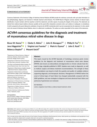
![1 | INTRODUCTION
The panel adopted the following scheme, adapted from the American
Heart Association,1
to rate the strength of the recommendations in these
guidelines. The recommendation class designation appears with each rec-
ommendation, along with a second term that independently rates the
quality of the evidence upon which the recommendation was based.
2 | CLASSIFICATION OF
RECOMMENDATIONS
Class I recommendations are strong, reflecting the panel's conviction
that the recommended action appears to have definite benefit for
most patients, outweighing the risk to most patients.
Class I recommendations can be summarized as “benefit >>> risk”.
Class IIa recommendations are moderately strong, reflecting the
panel's belief that the recommended action should benefit most
patients, probably outweighing the risk to most patients.
Class IIa recommendations can be summarized as “benefit >> risk”.
Class IIb recommendations are weak, reflecting the belief that the
recommended action possibly benefits some patients and may out-
weigh the risk of taking the proposed action in most patients.
Class IIb recommendations can be summarized as “benefit > risk”.
Class III is used to describe recommendations in which the panel
believes that the potential risk and benefit of the proposed action are
essentially equal, such that these actions should probably not be pur-
sued under most circumstances.
Class III recommendations can be summarized as “benefit = risk”.
Class IV recommendations indicate the panel's belief that the pro-
posed action is more likely to cause harm than benefit to most
patients, such that Class IV designates actions that the panel believes
are contraindicated under most circumstances.
Class IV recommendations can be summarized as “risk >> benefit”.
The recommendation classification and level of evidence (LOE)
upon which that recommendation was based were independently
determined by the panel (ie, any class of recommendation may be
paired with any LOE).
The panel acknowledges that future evidence may change the
strength of any of these recommendations, as well as the quality of the
evidence on which they are based. Many important clinical questions
addressed in the guidelines have not yet been adequately addressed by
clinical trials. At times, the panel has made strong recommendations
based on more than the available evidence—thus weak or even absent
evidence does not necessarily accompany a weak recommendation.
Although randomized clinical trial evidence may be unavailable, a clear
clinical consensus that a particular test or treatment is useful may exist.
3 | LEVELS OF EVIDENCE
The methods of assessing the quality of the scientific evidence that
supports clinical decision making are evolving. The panel chose to
use a hybrid of the American Heart Association and Veterinary
Emergency Critical Care RECOVER evidence grading criteria, as out-
lined below.1,2
3.1 | Strong
High-quality evidence is from ≥1 randomized controlled trial, or a
moderate-quality randomized controlled trial, corroborated by a high-
quality observational study or other moderate-quality trials. These
prospective clinical studies were performed in dogs and either ran-
domly allocated subjects to an intervention or control group or used
concurrent controls (ie, controls recruited at the same time as the
experimental subjects) without randomization. Strong evidence also
could have been obtained from prospectively enrolled, controlled,
observational clinical studies in dogs with spontaneously occurring
mitral valve disease. These studies asked clinically relevant questions,
were considered to be adequately powered, and did not experience
excessive loss of subjects to follow up in any group, generating a clear
and statistically valid result.
3.2 | Moderate
Moderate-quality evidence is from ≥1 well-designed, well-executed
nonrandomized studies, observational studies, or registry studies, or
meta-analyses of such studies. Evidence rated as “moderate” by the
panel was generated from controlled, retrospective studies in dogs
(ie, studies in which dogs with mitral valve disease [or appropriate
controls] were selected from a previous period in time). Moderate
evidence also could have been generated from blinded and con-
trolled laboratory studies that were performed in experimental dogs.
3.3 | Weak
Weak-quality evidence is from randomized or nonrandomized obser-
vational or registry studies with limitations of design or execution,
but performed in dogs with clinical myxomatous mitral valve disease
(MMVD), or physiological, mechanistic, or experimental studies per-
formed in research dogs. Evidence rated as “weak” by the panel was
generated from uncontrolled clinical case reports or case series in
dogs with mitral valve disease, as well as by experimental or clinical
studies that were not performed in dogs with MMVD. These could
include experimental models of mitral valve disease in other species,
as well as high-quality studies in humans (such as meta-analyses, ran-
domized controlled trials, and clinical studies with concurrent con-
trols, including observational studies) with spontaneous mitral valve
disease.
3.4 | Expert opinion
Expert opinion based on clinical experience, common sense, or physi-
ologic or mechanistic studies performed in species other than dogs is
considered the weakest LOE.
2 KEENE ET AL.](data:image/gif;base64,R0lGODlhAQABAIAAAAAAAP///yH5BAEAAAAALAAAAAABAAEAAAIBRAA7)