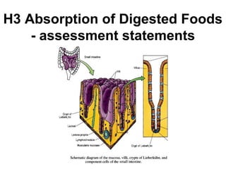
2. H.3.1 Draw and label a diagram showing a transverse section of
the ileum as seen under a light microscope.
(Include mucosa and layers of longitudinal and circular muscle)
H.3.2 Explain the structural features of an epithelial cell of a villus
as seen in electron micrographs, including microvilli, mitochondria,
pinocytotic vesicles and tight junctions.
H.3.3 Explain the mechanisms used by the ileum to absorb and
transport food, including facilitated diffusion, active transport and
endocytosis.
H.3.4 List the materials that are not absorbed and are egested.
(Limit this to cellulose, lignin, bile pigments, bacteria & intestinal
cells)