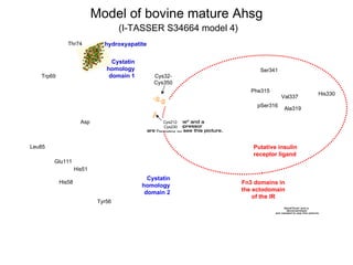
Fig. 14. A cartoon representation of the two structural faces of secreted Ahsg. There is no X-ray crystallographic or NMR-derived structure on which to base the 3-dimensional folding of Ahsg from any species. However, bioinformatics tools can be used to simulate a model which suggests recognition of hydroxyapatite by the C-terminal cystatin homology domains (residues 18-248; dotted red ovals), and the Fn3 homology domains of the insulin receptor by Ahsg residues 249-367 (dotted blue oval). The 350-residue single chain of human Ahsg (residues 18-367) was submitted to the I-TASSER website (Zhang, 2008), which uses amino acid homology of submitted protein sequences with protein sequences in the PDB database, and returns five models. I stipulated that I-TASSER take into consideration the known 5 disulfide pairs in Ahsg using the “Assign contact/distance restraints” feature, and chose Model 5 from Run S34302. PyMol for Mac (http://www.pymol.org/) to visualize the resulting pdb-formatted file generated by I-TASSER. Some of the –S-S- bonds are depicted in orange. Regions of strong α-helicity are colored blue; β-sheet structures yellow, and the unstructured backbone colored green.