SlideShare a Scribd company logo
1 of 4
Download to read offline
International Journal of Scientific and Research Publications, Volume 3, Issue 8, August 2013 1
ISSN 2250-3153
www.ijsrp.org
GIANT ANTERIOR NECK LIPOMA WITH
MEDIASTINAL EXTENSION: A RARE CASE
REPORT
Dr. Smrity Rupa Borah Dutta *, Dr. Sachender Pal Singh **, Dr. Aakanksha Rathor **
*
Assistant Professor, Department OF Otorhinolaryngology, SMCH, Silchar
**
PGT Otorhinolaryngology, Department OF Otorhinolaryngology, SMCH, Silchar
Abstract- Lipomas are benign mesenchymal tumour. Thirteen
percent of lipomas are seen in head and neck region1
. Anterior
neck lipoma is a rare one & anterior neck lipoma with
mediastinal extension is extremely rare. We are reporting a case
of 48 yr. old female who presented with a huge swelling in the
anterior neck region simulating a goitre. It was one of the largest
of its own kind. FNAC & sonography helps in making early
diagnosis of lipoma apart from clinical examination. Enucleation
of lipoma from neck as well as mediastinum was done &
followed up for 1 year without any complication.
Index Terms- Anterior neck Lipoma, giant lipoma,
mediastinum, mesenchymal tumour
I. INTRODUCTION
ipomas are the most common benign mesenchymal
neoplasm of adulthood & are usually subcutaneous but may
develop in other places, e.g. Intermuscular, Subfascial, Parosteal,
Subserous, Submucous, Intra-articular, Subsynovial,
Subendocardium, Subepicardiac, Myocardium, Subdural or
Extradural. They are actually a cluster of fat cells which become
overactive. This is the commonest tumor of subcutaneous tissue.
They are skin colored lesions & are firm & somewhat lobulated
on palpation. Common sites of lipoma are back, arms, shoulder,
anterior chest wall, breasts, thighs, abdominal wall, legs,
forehead & face2
. In head & neck region, where only 13% of
lipomas are seen, posterior cervical space is the commonest site3
.
Anterior neck lipoma is a rare location for lipoma4
and anterior
neck lipoma with mediastinal extension is very rare. Fine needle
aspiration cytology (FNAC) & sonography helps in making early
diagnosis which can be supported with computed tomography
(CT) & confirmed with histopathology report. Surgical
intervention is challenging because of proximity to the great
vessels & vagus nerve and is reserved for patients coming for
cosmesis (most common indication), pressure effects & to rule
out malignancy. We report a case of 48 years old female patient
who presented with a huge swelling in the anterior neck region
extending into the anterior mediastinum mimicking goitre.
II. CASE REPORT
48 years old, female presented in Otorhinolaryngology OPD in
Silchar Medical College & Hospital, Silchar with a huge swelling
in the anterior aspect of neck since 20 years, which gradually
enlarged in size & complained of occasional history of dyspnoea.
Pt. was not having dysphagia or any complain related to her
voice.
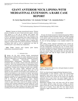
After taking consent, physical examination was carried out.
Though swelling appeared like goiter but found to be smooth,
soft and compressible. Overlying skin was of normal colour,
stretched, not adhered to tumor & dilated veins were present over
the tumor. Swelling was mobile, nontender. The surface of the
mass was smooth. Edges were definite above & slips under the
palpating finger (slip sign) & fingers couldn’t get below the
lower margin.
Figure 1: Preoperative photo of pt. with giant lipoma in anterior
aspect & left side of neck.
FNAC was done & report suggested benign lipomatous lesion.
Ultrasonography of neck showed a large well circumscribed
hypoechoic mass lesion noted in front & left side of neck.
Thyroid was found to be normal. & left carotid was displaced
posteriorly due to mass lesion. On colour Doppler study, no
increase in vascularity was seen. All features were suggestive of
lipomatous lesion.
Radiological imaging: X-ray soft tissue neck (AP & Lateral
view) reports showed a large soft tissue swelling in anterior
aspect & left side of neck with mild anterior compression over
lower part of trachea. X-ray chest PA view report showed
widening of upper mediastinum & trachea was noted to be in
midline.
L
International Journal of Scientific and Research Publications, Volume 3, Issue 8, August 2013 2
ISSN 2250-3153
www.ijsrp.org
Figure 2: X-ray soft tissue neck (AP & Lateral view) : large soft
tissue swelling in anterior aspect & left side of neck with mild
anterior compression over lower part of trachea.
Figure 3: CECT: showing extension of lipoma into the
mediastinum
On contrast enhanced computed tomography (CECT), a large
homogeneous well defined fat density lesion of size 16Ă—14.8Ă—13
cm was noted involving the neck spaces on left side extending
superiorly from the level of inferior aspect of parotid & inferiorly
to the level of left hilum of mediastinum, anterolaterally the
lesion was bounded by skin, posteriorly by pre & paravertebral
muscles. Medially the lesion was crossing the midline, displacing
the visceral space towards right side and in the mediastinum
abutting the great vessels on left side. Left sternocleidomastoid
muscle was compressed & thinned out. The lesion completely
encased the internal jugular vein & encased part of IJV was
showing dilatation. No intralesional soft tissue/
calcification/postcontrast enhancement was noted.
Thyroid profile of pt. was found to be normal.
Operative details : pt. was positioned in supine position under
general anesthesia. After the surgical field was scrubbed sterile
drapes were placed. A single transverse incision was made
following the relaxed skin tension lines & was carried down
through the skin & subcutaneous tissue to the level of the lipoma.
Tumor mass was smooth, soft, yellow, mobile, shining and
encapsulated. Encased part of IJV dissected out properly from
tumor mass & preserved. Enucleation of the tumor mass from
neck as well as mediastinal extension was done, followed by
excision of the excess skin. Total weight of the tumor mass was
1200 gm. Wound was repaired in layers after proper hemostasis
& a vacuum drain was put inside for 48 hrs.
Histopathology report suggested it to be a lipoma.
Figure 4: Intraoperative photo of pt. just after the enucleation of
lipoma from neck & mediastinum
Figure 5: Lipoma after the enucleation
Figure 6: Lipoma after the enucleation
Pt. had a very good recovery without any complication & was
followed for 1 year without recurrence.
Figure 7: Postoperative photo of pt. after 1 year of follow up.
Figure 8: Postoperative photo of pt. after 1 year of follow up.
International Journal of Scientific and Research Publications, Volume 3, Issue 8, August 2013 3
ISSN 2250-3153
www.ijsrp.org
III. DISCUSSION
Now it is the time to articulate the research work with ideas
gathered in Prevalence rate of lipoma is variable, 2.1:1000 to
1:100.5,6
Lipoma is seen in all age group though mostly seen in
fifth and sixth decade7
. It constitutes five percent of all benign
tumors of body and can be found anywhere in the body8
. Lipoma
in head and neck region is not commonly encountered (13%).
The first case of lipoma in the neck was reported over 100 year’s
ago9
. Amongst the head and neck lipomas, commonest location
is posterior neck3
. Anterior neck is a rare location for head and
neck lipoma4
. Lipoma of anterior neck with mediastinal
extension is very rare.
Lipomas are slow growing, painless, mobile, non-fluctuant,
soft masses & are generally well encapsulated. Lipomas can be
singular or multiple & are typically asymptomatic unless they
compress neurovascular structures. Beside frequent aesthetic
consequences, lipomas can also exert pressure on surrounding
tissues and structures. Patient with neck lipoma extending to
mediastinum may present with complaint of dyspnoea as in our
case.
Giant lipomas are defined by Sanchez et al as lesions with
size of at least 10 cm in one dimension or weighing a minimum
of 1,000 gm10
. A large neck mass (>10 cm) with a rapid growth
rate should raise concerns about a possible malignancy10
. A long
standing lipoma may undergo myxomatous degeneration,
saponification, calcification, infection, ulceration due to repeated
trauma & malignant change. Rarely malignant transformation of
lipoma into liposarcoma has been described13, 14
. Differentiation
of lipoma from liposarcoma may be difficult.
Atypical lipomatous tumors are considered to be well-
differentiated liposarcomashttp://emedicine.medscape.com
/article/987446-overview. When a fatty tumor is
encountered in an intramuscular or retroperitoneal location
liposarcomas should be considered in differential diagnosis,
which has predilection for local recurrence but they don’t
metastasize generally. Although the diagnosis is mostly clinical,
imaging tools are useful to confirm the adipose nature of the
lesion and to define its anatomic border, & exclude possible
communication with the spinal canal.
Histologically lipomas are composed of mature adipose tissue,
and several subtypes occur when other mesenchymal elements
are present11
, for example fibrous tissue, nervous tissue or
vascular tissue. According to WHO classification of soft
tumours these can be classified into nine groups, including
lipoma, lipomatosis, lipoblastoma, angiolipoma, myolipoma of
soft tissues, chondroid lipoma, spindle cell lipoma, and finally
hibernoma and pleomorphic lipoma12
. Most common subtype is
conventional lipoma which is well encapsulated mass of mature
adipocytes & varies considerably in size. All subtypes are
painless except angiolipoma. Hibernomas are benign, uncommon
tumors presumably arising from brown fat that may occur in the
back, hips, or neck in adults and infants & has a slightly greater
tendency to bleed during excision and to recur if intralesional
excision is performed.
The characteristic sonographic appearance of head and
neck lipomas is that of an elliptical mass parallel to the skin
surface that is mostly hyperechoic relative to adjacent muscle
and that contains linear echogenic lines at right angles to the
ultrasound beam15, 16
.
Computed tomography is modality of choice to confirm
lipoma. Lipomas appear as homogenous low density areas with a
CT value of -50 to -150 HU with no contrast enhancement17
. A
thin soft tissue capsule may be seen surrounding a subcutaneous
lipoma. Within the lesion there should be homogeneous fat
density with few, if any internal septa. On CT scans capsule of
lipoma is barely visible or adjacent mass effect may be the only
clue to its presence. Larger lesions may contain blood vessels. A
significant soft tissue element or heterogeneity of attenuation
within a fatty lesion raises the possibility of liposarcoma.
In MRI, Lipomas have well defined margins with a uniform
signal intensity of fat on all sequences (best confirmed using fat-
suppressed sequences). Some lipomas may also have internal
septa, an appearance mimicking a well differentiated liposarcoma
(termed atypical lipoma). The use of contrast enhanced fat
suppressed T1-weighted images can be helpful in separating
between enhancing nodular tumour & non-enhancing linear
septas. Margin of lipoma is clearly defined as “black rim”,
distinguishing them from surrounding fat18
. Calcification is rare
& forms centrally within an area of ischaemic necrosis but more
commonly it’s a feature of a liposarcoma.
Surgical excision of lipoma is the definitive treatment.
Surgery is reserved for patients coming for cosmesis (most
common indication) and pressure effects & to rule out
malignancy. Smaller lipomas can be excised easily with low
recurrence rate because they usually grow expansively between
different fascial planes without infiltrating the neighbouring
structures. Surgical intervention of giant lipoma of anterior neck
with mediastinal extension is challenging because of proximity to
the great vessels, vagus & spinal accessory nerves, lungs & heart.
Preoperative consent regarding possible complications such as
injury to neurovascular structures etc. must be taken. Lipomas
may be lobulated, and it is essential that all lobules be removed.
Complete surgical excision with the capsule is advocated to
prevent local recurrence. Other modalities of treatment have been
reported, like liposuction19, 20,21,22,23
& steroid injections24
.
Liposuction is sometimes preferred as there is less scarring22,23
following the procedure but there is higher chance of recurrence
compared to excision if residual tumour or capsule, remains after
the procedure. For smaller lipomas steroid injections may also be
used, but several injections are required and the overlying skin
may be depigmented. Surgery for giant lipoma in anterior neck
with mediastinal extension should be done in a meticulous way.
REFERENCES
1. Barnes L. Tumors and tumorlike lesions of the head and neck: In:
Barnes L, ed. Surgical Pathology of the Head and Neck. New York,
NY: Dekker; 1985:747–758
2. Rapidis AD. Lipoma of the oral cavity. Int J Oral Surg. 1982; 11:30-5.
3. Barnes L. tumors & tumor like lesions of the head & neck: In: Barnes
L, ed. Surgical pathology of the head & neck. New York, NY:Dekker;
1985: 747-758.
4. Medina CR, Schneider S, Mitra A, Spears J and Mitra A. Giant
submental lipoma: Case report and review of the literature. Can J Plast
Surg 2007; 15(4):219-222.
5. Nickloes TA, Sutphin DD, Radebold K. 2010. Lipomas. Available
from http://emedicine.medscape.com/article/19 1233-overview.
International Journal of Scientific and Research Publications, Volume 3, Issue 8, August 2013 4
ISSN 2250-3153
www.ijsrp.org
6. Rydholm A, Berg NO. Size, site and clinical incidence of lipoma.
Factors in the differential diagnosis of lipoma and sarcoma. Acta
Orthop Scand, 1983. 54(6): p. 929-34.
7. Salam G. Lipoma excision. Am Fam Physician 2002; 65:901-905.
8. Enzinger FM, Weiss SW. Benign lipomatous tumors. In: Enzinger
FM, Weiss SW, eds. Soft Tissue Tumors. 2nd edn. St Louis: Mosby;
1988.p.301-45.
9. Horne, W.J., Lipoma or Cystoma of the Neck. Proc R Soc Med, 1908.
1(Laryngol Sect): p. 38-39.
10. Sanchez MR, Golomb FM, Moy JA, Potozkin JR. Giant lipoma: Case
report and review of theliterature. J Am Acad Dermatol. 1993; 28:266.
11. Edmonds JL, Woodroof JM, Ator GA: Middle-ear lipoma as a cause
ofotomastoiditis. J Laryngol Otol 1997, 111(12):1162-1165.
12. Murphey MD, Carroll JF, Flemming DJ, Pope TL, Gannon FH,
Kransdorf MJ. From the archives of the AFIP: Benign
musculoskeletal lipomatous lesions. Radiographics 2004;
24:1433‑ 66.
13. MENTZEL T. Cutaneous Lipomatous Neoplasms. Semi Diagn Pathol,
2001, 18: 250-7.
14. MENTZEL T. Biological continuum of benign, atypical, and
malignant mesenchymal neoplasms – does it exist? J Pathol, 2000,
190: 523-5.
15. Ahuja AT, King AD, Kew J, King W, Metreweli C. Head and neck
lipomas: sonographic appearance. AJNR Am J Neuroradiol 1998;
19:505–508.
16. , Gritzman N, Schratter M, Traxler M. Sonography and computed
tomography in deep cervical lipomas and lipomatosis of the neck. J
Ultrasound Med 1988; 7:451–456.
17. Barisa AD, Pawar Nh, Bakhshi CD, Yogesh S Puri, Aftab Shaikh,
Narendra N Nikam, Giant Axillary Lipoma. Bombay Hosp J 2009;
51:91-3.
18. Chikui T, Yonetsu K, Yoshiura K, Miwa K, Kanda S, Ozeki S, et al.
Imaging findings of lipomas in the orofacial region with CT, US, and
MRI. Oral Surg Oral Med Oral Pathol Oral Radiol Endod 1997;84:88-
95.
19. Wilhelmi BJ, Blackwell SJ, Mancoll JS, Phillips LG. Another
indication for liposuction: Small facial lipomas. Plast Reconstr Surg
1999;103:1864-7.
20. Sharma PK, Janninger CK, Schwartz RA, Rauscher GE, Lambert WC.
The treatment of atypical lipoma with liposuction. J Dermatol Surg
Oncol 1991; 17:332-4.
21. Field LM. Lipo-suction surgery: A review. J Dermatol Surg Oncol
1984; 10:530-8.
22. Rubenstein R, Roenigk HH, Garden JM, Goldberg NS, Pinski JB.
Liposuction for lipomas. J Dermatol Surg Oncol 1985; 11:1070-4.
23. Calhoun KH, Bradfield JJ, Thompson C. Liposuction-assisted
excision of cervicofacial lipomas. Otolaryngol Head Neck Surg 1995;
113:401-3.
24. Koh HK, Bhawan J. Tumors of the skin. In: Moschella SL, Hurley HJ,
eds. Dermatology, 3rd edn. Philadelphia: Saunders, 1992:1721-808.
AUTHORS
First Author – Dr. Smrity Rupa Borah Dutta, Assistant
Professor, SMCH, Silchar, Assam, email address:
smritylana@gmail.com
Second Author – Dr. Sachender Pal Singh, PGT
Otorhinolaryngology, SMCH, Silchar, Assam email address:
sachender123@gmail.com
Third Author – Dr. Aakanksha Rathor, PGT
Otorhinolaryngology, SMCH, Silchar, Assam email address:
aakanksha2128@gmail.com
Correspondence Author – Dr. Sachender Pal Singh, email
address: sachender123@gmail.com, alternate email address:
aakanksha2128@gmail.com , contact number: 08011205680

More Related Content

What's hot

Presentation1.pptx, radiological imaging of the nasopharyngeal diseases.
Presentation1.pptx, radiological imaging of the nasopharyngeal diseases.Presentation1.pptx, radiological imaging of the nasopharyngeal diseases.
Presentation1.pptx, radiological imaging of the nasopharyngeal diseases.Abdellah Nazeer
 
Neuroradiology primary spinal cord tumours
Neuroradiology   primary spinal cord tumoursNeuroradiology   primary spinal cord tumours
Neuroradiology primary spinal cord tumoursRoopchand Ps
 
Spinal axis tumors
Spinal axis tumorsSpinal axis tumors
Spinal axis tumorsAhmed Debes
 
Tumor of spine and spinal cord
Tumor of spine and spinal cordTumor of spine and spinal cord
Tumor of spine and spinal cordHong Sothea
 
Intramedullary tumors
Intramedullary tumorsIntramedullary tumors
Intramedullary tumorsRitesh Mahajan
 
Spinal neoplasms
Spinal neoplasmsSpinal neoplasms
Spinal neoplasmsfahad shafi
 
EXTRANODAL EXTENSION
EXTRANODAL EXTENSIONEXTRANODAL EXTENSION
EXTRANODAL EXTENSIONDrAyush Garg
 
Presentation1.pptx, radiological imaging of adult neck masses.
Presentation1.pptx, radiological imaging of adult neck masses.Presentation1.pptx, radiological imaging of adult neck masses.
Presentation1.pptx, radiological imaging of adult neck masses.Abdellah Nazeer
 
Chest wall tumor
Chest wall tumorChest wall tumor
Chest wall tumordegnarog
 
Spinal tumors lecture
Spinal tumors lectureSpinal tumors lecture
Spinal tumors lecturetest
 
Metastatic neck disease
Metastatic neck diseaseMetastatic neck disease
Metastatic neck diseaseMamoon Ameen
 
Petroclival Meningioma
Petroclival MeningiomaPetroclival Meningioma
Petroclival MeningiomaFarrukh Javeed
 
Presentation1.pptx, radiological imaging of spinal cord tumour.
Presentation1.pptx, radiological imaging of spinal cord tumour.Presentation1.pptx, radiological imaging of spinal cord tumour.
Presentation1.pptx, radiological imaging of spinal cord tumour.Abdellah Nazeer
 
Intramedullary Spinal Cord Tumors
Intramedullary  Spinal  Cord  TumorsIntramedullary  Spinal  Cord  Tumors
Intramedullary Spinal Cord TumorsLeslieHutchinsMD
 
Parasagittal Meningioma
Parasagittal MeningiomaParasagittal Meningioma
Parasagittal MeningiomaFarrukh Javeed
 
Parapharyngeal space tumours
Parapharyngeal space tumoursParapharyngeal space tumours
Parapharyngeal space tumoursDr./ Ihab Samy
 

What's hot (20)

Presentation1.pptx, radiological imaging of the nasopharyngeal diseases.
Presentation1.pptx, radiological imaging of the nasopharyngeal diseases.Presentation1.pptx, radiological imaging of the nasopharyngeal diseases.
Presentation1.pptx, radiological imaging of the nasopharyngeal diseases.
 
Neuroradiology primary spinal cord tumours
Neuroradiology   primary spinal cord tumoursNeuroradiology   primary spinal cord tumours
Neuroradiology primary spinal cord tumours
 
Spinal axis tumors
Spinal axis tumorsSpinal axis tumors
Spinal axis tumors
 
Tumor of spine and spinal cord
Tumor of spine and spinal cordTumor of spine and spinal cord
Tumor of spine and spinal cord
 
International Journal of Rhinology & Otolaryngology
International Journal of Rhinology & OtolaryngologyInternational Journal of Rhinology & Otolaryngology
International Journal of Rhinology & Otolaryngology
 
Intramedullary tumors
Intramedullary tumorsIntramedullary tumors
Intramedullary tumors
 
Spinal tumors
Spinal tumorsSpinal tumors
Spinal tumors
 
Spinal neoplasms
Spinal neoplasmsSpinal neoplasms
Spinal neoplasms
 
Ependymoma
EpendymomaEpendymoma
Ependymoma
 
EXTRANODAL EXTENSION
EXTRANODAL EXTENSIONEXTRANODAL EXTENSION
EXTRANODAL EXTENSION
 
Presentation1.pptx, radiological imaging of adult neck masses.
Presentation1.pptx, radiological imaging of adult neck masses.Presentation1.pptx, radiological imaging of adult neck masses.
Presentation1.pptx, radiological imaging of adult neck masses.
 
Chest wall tumor
Chest wall tumorChest wall tumor
Chest wall tumor
 
Spinal tumors lecture
Spinal tumors lectureSpinal tumors lecture
Spinal tumors lecture
 
Metastatic neck disease
Metastatic neck diseaseMetastatic neck disease
Metastatic neck disease
 
Petroclival Meningioma
Petroclival MeningiomaPetroclival Meningioma
Petroclival Meningioma
 
Dr.mumtaz ali
Dr.mumtaz aliDr.mumtaz ali
Dr.mumtaz ali
 
Presentation1.pptx, radiological imaging of spinal cord tumour.
Presentation1.pptx, radiological imaging of spinal cord tumour.Presentation1.pptx, radiological imaging of spinal cord tumour.
Presentation1.pptx, radiological imaging of spinal cord tumour.
 
Intramedullary Spinal Cord Tumors
Intramedullary  Spinal  Cord  TumorsIntramedullary  Spinal  Cord  Tumors
Intramedullary Spinal Cord Tumors
 
Parasagittal Meningioma
Parasagittal MeningiomaParasagittal Meningioma
Parasagittal Meningioma
 
Parapharyngeal space tumours
Parapharyngeal space tumoursParapharyngeal space tumours
Parapharyngeal space tumours
 

Similar to GIANT ANTERIOR NECK LIPOMA WITH MEDIASTINAL EXTENSION

Liposarcoma of spermatic cord - A rare entity?
Liposarcoma of spermatic cord - A rare entity?Liposarcoma of spermatic cord - A rare entity?
Liposarcoma of spermatic cord - A rare entity?Apollo Hospitals
 
Approach To Management Of Neck Mass
Approach To  Management Of Neck MassApproach To  Management Of Neck Mass
Approach To Management Of Neck MassPrasanna Datta
 
Liposarcoma of Spermatic Cord - A Rare Entity
Liposarcoma of Spermatic Cord - A Rare EntityLiposarcoma of Spermatic Cord - A Rare Entity
Liposarcoma of Spermatic Cord - A Rare EntityApollo Hospitals
 
Liposarcoma of spermatic cord - A rare entity
Liposarcoma of spermatic cord - A rare entityLiposarcoma of spermatic cord - A rare entity
Liposarcoma of spermatic cord - A rare entityApollo Hospitals
 
Liposarcoma of Spermatic Cord - A Rare Entity
Liposarcoma of Spermatic Cord - A Rare EntityLiposarcoma of Spermatic Cord - A Rare Entity
Liposarcoma of Spermatic Cord - A Rare EntityApollo Hospitals
 
Third ventricular-masses
Third ventricular-massesThird ventricular-masses
Third ventricular-massesNabaz Mohammed
 
Cervical Cystic Hygroma-a Case Report
Cervical Cystic Hygroma-a Case ReportCervical Cystic Hygroma-a Case Report
Cervical Cystic Hygroma-a Case ReportCrimsonpublishersMedical
 
Neck Imaging.pptx
Neck Imaging.pptxNeck Imaging.pptx
Neck Imaging.pptxJwan AlSofi
 
Temporal bone neoplasms
Temporal bone neoplasmsTemporal bone neoplasms
Temporal bone neoplasmsabhijeet89singh
 
A Thyroglossal Duct Cyst with Discharging Sinus Presenting As an Infected Thy...
A Thyroglossal Duct Cyst with Discharging Sinus Presenting As an Infected Thy...A Thyroglossal Duct Cyst with Discharging Sinus Presenting As an Infected Thy...
A Thyroglossal Duct Cyst with Discharging Sinus Presenting As an Infected Thy...iosrjce
 
A rare site of Cystic hygroma
A rare site of Cystic hygromaA rare site of Cystic hygroma
A rare site of Cystic hygromaiosrjce
 
Parotid Gland Oncocytoma: A Case Report
Parotid Gland Oncocytoma: A Case ReportParotid Gland Oncocytoma: A Case Report
Parotid Gland Oncocytoma: A Case Reportiosrjce
 
The advantage of mri in detection of coincidental adenocarcinoma of palate an...
The advantage of mri in detection of coincidental adenocarcinoma of palate an...The advantage of mri in detection of coincidental adenocarcinoma of palate an...
The advantage of mri in detection of coincidental adenocarcinoma of palate an...Clinical Surgery Research Communications
 
An approach to the neck mass
An approach to the neck massAn approach to the neck mass
An approach to the neck massHardi Hussein
 
Cervical Sympathetic chain ganglioneuroma : case report and review of literature
Cervical Sympathetic chain ganglioneuroma : case report and review of literatureCervical Sympathetic chain ganglioneuroma : case report and review of literature
Cervical Sympathetic chain ganglioneuroma : case report and review of literatureiosrphr_editor
 
Primary Endometrial Stromal Sarcoma arising from Cervix
Primary Endometrial Stromal Sarcoma arising from CervixPrimary Endometrial Stromal Sarcoma arising from Cervix
Primary Endometrial Stromal Sarcoma arising from Cervixiosrjce
 

Similar to GIANT ANTERIOR NECK LIPOMA WITH MEDIASTINAL EXTENSION (20)

Primary Mediastinal Liposarcoma
Primary Mediastinal LiposarcomaPrimary Mediastinal Liposarcoma
Primary Mediastinal Liposarcoma
 
Liposarcoma of spermatic cord - A rare entity?
Liposarcoma of spermatic cord - A rare entity?Liposarcoma of spermatic cord - A rare entity?
Liposarcoma of spermatic cord - A rare entity?
 
Approach To Management Of Neck Mass
Approach To  Management Of Neck MassApproach To  Management Of Neck Mass
Approach To Management Of Neck Mass
 
Liposarcoma of Spermatic Cord - A Rare Entity
Liposarcoma of Spermatic Cord - A Rare EntityLiposarcoma of Spermatic Cord - A Rare Entity
Liposarcoma of Spermatic Cord - A Rare Entity
 
Liposarcoma of spermatic cord - A rare entity
Liposarcoma of spermatic cord - A rare entityLiposarcoma of spermatic cord - A rare entity
Liposarcoma of spermatic cord - A rare entity
 
Liposarcoma of Spermatic Cord - A Rare Entity
Liposarcoma of Spermatic Cord - A Rare EntityLiposarcoma of Spermatic Cord - A Rare Entity
Liposarcoma of Spermatic Cord - A Rare Entity
 
Third ventricular-masses
Third ventricular-massesThird ventricular-masses
Third ventricular-masses
 
Third ventricular-masses
Third ventricular-massesThird ventricular-masses
Third ventricular-masses
 
Cervical Cystic Hygroma-a Case Report
Cervical Cystic Hygroma-a Case ReportCervical Cystic Hygroma-a Case Report
Cervical Cystic Hygroma-a Case Report
 
Neck Imaging.pptx
Neck Imaging.pptxNeck Imaging.pptx
Neck Imaging.pptx
 
Temporal bone neoplasms
Temporal bone neoplasmsTemporal bone neoplasms
Temporal bone neoplasms
 
A Thyroglossal Duct Cyst with Discharging Sinus Presenting As an Infected Thy...
A Thyroglossal Duct Cyst with Discharging Sinus Presenting As an Infected Thy...A Thyroglossal Duct Cyst with Discharging Sinus Presenting As an Infected Thy...
A Thyroglossal Duct Cyst with Discharging Sinus Presenting As an Infected Thy...
 
A rare site of Cystic hygroma
A rare site of Cystic hygromaA rare site of Cystic hygroma
A rare site of Cystic hygroma
 
A Rare Case of Spindle Cell Neoplasm of Rectum
A Rare Case of Spindle Cell Neoplasm of RectumA Rare Case of Spindle Cell Neoplasm of Rectum
A Rare Case of Spindle Cell Neoplasm of Rectum
 
Parotid Gland Oncocytoma: A Case Report
Parotid Gland Oncocytoma: A Case ReportParotid Gland Oncocytoma: A Case Report
Parotid Gland Oncocytoma: A Case Report
 
The advantage of mri in detection of coincidental adenocarcinoma of palate an...
The advantage of mri in detection of coincidental adenocarcinoma of palate an...The advantage of mri in detection of coincidental adenocarcinoma of palate an...
The advantage of mri in detection of coincidental adenocarcinoma of palate an...
 
An approach to the neck mass
An approach to the neck massAn approach to the neck mass
An approach to the neck mass
 
Cervical Sympathetic chain ganglioneuroma : case report and review of literature
Cervical Sympathetic chain ganglioneuroma : case report and review of literatureCervical Sympathetic chain ganglioneuroma : case report and review of literature
Cervical Sympathetic chain ganglioneuroma : case report and review of literature
 
Primary Endometrial Stromal Sarcoma arising from Cervix
Primary Endometrial Stromal Sarcoma arising from CervixPrimary Endometrial Stromal Sarcoma arising from Cervix
Primary Endometrial Stromal Sarcoma arising from Cervix
 
Benign neck disease
Benign neck diseaseBenign neck disease
Benign neck disease
 

More from Aakanksha Rathor

Voice Therapy: Management Of Benign Voice Disorders
Voice Therapy: Management Of Benign Voice DisordersVoice Therapy: Management Of Benign Voice Disorders
Voice Therapy: Management Of Benign Voice DisordersAakanksha Rathor
 
SILICONE IMPLANT IN AUGMENTATON OF SADDLE NOSE
SILICONE IMPLANT IN AUGMENTATON OF SADDLE NOSESILICONE IMPLANT IN AUGMENTATON OF SADDLE NOSE
SILICONE IMPLANT IN AUGMENTATON OF SADDLE NOSEAakanksha Rathor
 
Benign Sinonasal Paraganglioma
Benign Sinonasal ParagangliomaBenign Sinonasal Paraganglioma
Benign Sinonasal ParagangliomaAakanksha Rathor
 
Unusual Presentation of Tuberculosis in Head and Neck Region
Unusual Presentation of Tuberculosis in Head and Neck RegionUnusual Presentation of Tuberculosis in Head and Neck Region
Unusual Presentation of Tuberculosis in Head and Neck RegionAakanksha Rathor
 
Analysis and Management of Tripod Fractures
Analysis and Management of Tripod FracturesAnalysis and Management of Tripod Fractures
Analysis and Management of Tripod FracturesAakanksha Rathor
 
MUSCLE TENSION DYSPHONIA CASE
MUSCLE TENSION DYSPHONIA CASEMUSCLE TENSION DYSPHONIA CASE
MUSCLE TENSION DYSPHONIA CASEAakanksha Rathor
 
FIBROUS DYSPLASIA OF MAXILLA
FIBROUS DYSPLASIA OF MAXILLAFIBROUS DYSPLASIA OF MAXILLA
FIBROUS DYSPLASIA OF MAXILLAAakanksha Rathor
 

More from Aakanksha Rathor (7)

Voice Therapy: Management Of Benign Voice Disorders
Voice Therapy: Management Of Benign Voice DisordersVoice Therapy: Management Of Benign Voice Disorders
Voice Therapy: Management Of Benign Voice Disorders
 
SILICONE IMPLANT IN AUGMENTATON OF SADDLE NOSE
SILICONE IMPLANT IN AUGMENTATON OF SADDLE NOSESILICONE IMPLANT IN AUGMENTATON OF SADDLE NOSE
SILICONE IMPLANT IN AUGMENTATON OF SADDLE NOSE
 
Benign Sinonasal Paraganglioma
Benign Sinonasal ParagangliomaBenign Sinonasal Paraganglioma
Benign Sinonasal Paraganglioma
 
Unusual Presentation of Tuberculosis in Head and Neck Region
Unusual Presentation of Tuberculosis in Head and Neck RegionUnusual Presentation of Tuberculosis in Head and Neck Region
Unusual Presentation of Tuberculosis in Head and Neck Region
 
Analysis and Management of Tripod Fractures
Analysis and Management of Tripod FracturesAnalysis and Management of Tripod Fractures
Analysis and Management of Tripod Fractures
 
MUSCLE TENSION DYSPHONIA CASE
MUSCLE TENSION DYSPHONIA CASEMUSCLE TENSION DYSPHONIA CASE
MUSCLE TENSION DYSPHONIA CASE
 
FIBROUS DYSPLASIA OF MAXILLA
FIBROUS DYSPLASIA OF MAXILLAFIBROUS DYSPLASIA OF MAXILLA
FIBROUS DYSPLASIA OF MAXILLA
 

Recently uploaded

Low Rate Call Girls Pune Esha 9907093804 Short 1500 Night 6000 Best call girl...
Low Rate Call Girls Pune Esha 9907093804 Short 1500 Night 6000 Best call girl...Low Rate Call Girls Pune Esha 9907093804 Short 1500 Night 6000 Best call girl...
Low Rate Call Girls Pune Esha 9907093804 Short 1500 Night 6000 Best call girl...Miss joya
 
Aspirin presentation slides by Dr. Rewas Ali
Aspirin presentation slides by Dr. Rewas AliAspirin presentation slides by Dr. Rewas Ali
Aspirin presentation slides by Dr. Rewas AliRewAs ALI
 
VIP Call Girls Lucknow Nandini 7001305949 Independent Escort Service Lucknow
VIP Call Girls Lucknow Nandini 7001305949 Independent Escort Service LucknowVIP Call Girls Lucknow Nandini 7001305949 Independent Escort Service Lucknow
VIP Call Girls Lucknow Nandini 7001305949 Independent Escort Service Lucknownarwatsonia7
 
See the 2,456 pharmacies on the National E-Pharmacy Platform
See the 2,456 pharmacies on the National E-Pharmacy PlatformSee the 2,456 pharmacies on the National E-Pharmacy Platform
See the 2,456 pharmacies on the National E-Pharmacy PlatformKweku Zurek
 
call girls in Connaught Place DELHI 🔝 >༒9540349809 🔝 genuine Escort Service ...
call girls in Connaught Place  DELHI 🔝 >༒9540349809 🔝 genuine Escort Service ...call girls in Connaught Place  DELHI 🔝 >༒9540349809 🔝 genuine Escort Service ...
call girls in Connaught Place DELHI 🔝 >༒9540349809 🔝 genuine Escort Service ...saminamagar
 
Low Rate Call Girls Mumbai Suman 9910780858 Independent Escort Service Mumbai
Low Rate Call Girls Mumbai Suman 9910780858 Independent Escort Service MumbaiLow Rate Call Girls Mumbai Suman 9910780858 Independent Escort Service Mumbai
Low Rate Call Girls Mumbai Suman 9910780858 Independent Escort Service Mumbaisonalikaur4
 
Call Girls Jayanagar Just Call 7001305949 Top Class Call Girl Service Available
Call Girls Jayanagar Just Call 7001305949 Top Class Call Girl Service AvailableCall Girls Jayanagar Just Call 7001305949 Top Class Call Girl Service Available
Call Girls Jayanagar Just Call 7001305949 Top Class Call Girl Service Availablenarwatsonia7
 
Call Girls Whitefield Just Call 7001305949 Top Class Call Girl Service Available
Call Girls Whitefield Just Call 7001305949 Top Class Call Girl Service AvailableCall Girls Whitefield Just Call 7001305949 Top Class Call Girl Service Available
Call Girls Whitefield Just Call 7001305949 Top Class Call Girl Service Availablenarwatsonia7
 
VIP Call Girls Mumbai Arpita 9910780858 Independent Escort Service Mumbai
VIP Call Girls Mumbai Arpita 9910780858 Independent Escort Service MumbaiVIP Call Girls Mumbai Arpita 9910780858 Independent Escort Service Mumbai
VIP Call Girls Mumbai Arpita 9910780858 Independent Escort Service Mumbaisonalikaur4
 
Call Girls Thane Just Call 9910780858 Get High Class Call Girls Service
Call Girls Thane Just Call 9910780858 Get High Class Call Girls ServiceCall Girls Thane Just Call 9910780858 Get High Class Call Girls Service
Call Girls Thane Just Call 9910780858 Get High Class Call Girls Servicesonalikaur4
 
College Call Girls Vyasarpadi Whatsapp 7001305949 Independent Escort Service
College Call Girls Vyasarpadi Whatsapp 7001305949 Independent Escort ServiceCollege Call Girls Vyasarpadi Whatsapp 7001305949 Independent Escort Service
College Call Girls Vyasarpadi Whatsapp 7001305949 Independent Escort ServiceNehru place Escorts
 
Asthma Review - GINA guidelines summary 2024
Asthma Review - GINA guidelines summary 2024Asthma Review - GINA guidelines summary 2024
Asthma Review - GINA guidelines summary 2024Gabriel Guevara MD
 
Call Girls Electronic City Just Call 7001305949 Top Class Call Girl Service A...
Call Girls Electronic City Just Call 7001305949 Top Class Call Girl Service A...Call Girls Electronic City Just Call 7001305949 Top Class Call Girl Service A...
Call Girls Electronic City Just Call 7001305949 Top Class Call Girl Service A...narwatsonia7
 
Mumbai Call Girls Service 9910780858 Real Russian Girls Looking Models
Mumbai Call Girls Service 9910780858 Real Russian Girls Looking ModelsMumbai Call Girls Service 9910780858 Real Russian Girls Looking Models
Mumbai Call Girls Service 9910780858 Real Russian Girls Looking Modelssonalikaur4
 
Call Girl Service Bidadi - For 7001305949 Cheap & Best with original Photos
Call Girl Service Bidadi - For 7001305949 Cheap & Best with original PhotosCall Girl Service Bidadi - For 7001305949 Cheap & Best with original Photos
Call Girl Service Bidadi - For 7001305949 Cheap & Best with original Photosnarwatsonia7
 
Russian Call Girls Chickpet - 7001305949 Booking and charges genuine rate for...
Russian Call Girls Chickpet - 7001305949 Booking and charges genuine rate for...Russian Call Girls Chickpet - 7001305949 Booking and charges genuine rate for...
Russian Call Girls Chickpet - 7001305949 Booking and charges genuine rate for...narwatsonia7
 
Book Call Girls in Yelahanka - For 7001305949 Cheap & Best with original Photos
Book Call Girls in Yelahanka - For 7001305949 Cheap & Best with original PhotosBook Call Girls in Yelahanka - For 7001305949 Cheap & Best with original Photos
Book Call Girls in Yelahanka - For 7001305949 Cheap & Best with original Photosnarwatsonia7
 
Housewife Call Girls Hoskote | 7001305949 At Low Cost Cash Payment Booking
Housewife Call Girls Hoskote | 7001305949 At Low Cost Cash Payment BookingHousewife Call Girls Hoskote | 7001305949 At Low Cost Cash Payment Booking
Housewife Call Girls Hoskote | 7001305949 At Low Cost Cash Payment Bookingnarwatsonia7
 

Recently uploaded (20)

Low Rate Call Girls Pune Esha 9907093804 Short 1500 Night 6000 Best call girl...
Low Rate Call Girls Pune Esha 9907093804 Short 1500 Night 6000 Best call girl...Low Rate Call Girls Pune Esha 9907093804 Short 1500 Night 6000 Best call girl...
Low Rate Call Girls Pune Esha 9907093804 Short 1500 Night 6000 Best call girl...
 
Aspirin presentation slides by Dr. Rewas Ali
Aspirin presentation slides by Dr. Rewas AliAspirin presentation slides by Dr. Rewas Ali
Aspirin presentation slides by Dr. Rewas Ali
 
VIP Call Girls Lucknow Nandini 7001305949 Independent Escort Service Lucknow
VIP Call Girls Lucknow Nandini 7001305949 Independent Escort Service LucknowVIP Call Girls Lucknow Nandini 7001305949 Independent Escort Service Lucknow
VIP Call Girls Lucknow Nandini 7001305949 Independent Escort Service Lucknow
 
See the 2,456 pharmacies on the National E-Pharmacy Platform
See the 2,456 pharmacies on the National E-Pharmacy PlatformSee the 2,456 pharmacies on the National E-Pharmacy Platform
See the 2,456 pharmacies on the National E-Pharmacy Platform
 
call girls in Connaught Place DELHI 🔝 >༒9540349809 🔝 genuine Escort Service ...
call girls in Connaught Place  DELHI 🔝 >༒9540349809 🔝 genuine Escort Service ...call girls in Connaught Place  DELHI 🔝 >༒9540349809 🔝 genuine Escort Service ...
call girls in Connaught Place DELHI 🔝 >༒9540349809 🔝 genuine Escort Service ...
 
sauth delhi call girls in Bhajanpura 🔝 9953056974 🔝 escort Service
sauth delhi call girls in Bhajanpura 🔝 9953056974 🔝 escort Servicesauth delhi call girls in Bhajanpura 🔝 9953056974 🔝 escort Service
sauth delhi call girls in Bhajanpura 🔝 9953056974 🔝 escort Service
 
Low Rate Call Girls Mumbai Suman 9910780858 Independent Escort Service Mumbai
Low Rate Call Girls Mumbai Suman 9910780858 Independent Escort Service MumbaiLow Rate Call Girls Mumbai Suman 9910780858 Independent Escort Service Mumbai
Low Rate Call Girls Mumbai Suman 9910780858 Independent Escort Service Mumbai
 
Call Girls Jayanagar Just Call 7001305949 Top Class Call Girl Service Available
Call Girls Jayanagar Just Call 7001305949 Top Class Call Girl Service AvailableCall Girls Jayanagar Just Call 7001305949 Top Class Call Girl Service Available
Call Girls Jayanagar Just Call 7001305949 Top Class Call Girl Service Available
 
Call Girls Whitefield Just Call 7001305949 Top Class Call Girl Service Available
Call Girls Whitefield Just Call 7001305949 Top Class Call Girl Service AvailableCall Girls Whitefield Just Call 7001305949 Top Class Call Girl Service Available
Call Girls Whitefield Just Call 7001305949 Top Class Call Girl Service Available
 
VIP Call Girls Mumbai Arpita 9910780858 Independent Escort Service Mumbai
VIP Call Girls Mumbai Arpita 9910780858 Independent Escort Service MumbaiVIP Call Girls Mumbai Arpita 9910780858 Independent Escort Service Mumbai
VIP Call Girls Mumbai Arpita 9910780858 Independent Escort Service Mumbai
 
Call Girls Thane Just Call 9910780858 Get High Class Call Girls Service
Call Girls Thane Just Call 9910780858 Get High Class Call Girls ServiceCall Girls Thane Just Call 9910780858 Get High Class Call Girls Service
Call Girls Thane Just Call 9910780858 Get High Class Call Girls Service
 
College Call Girls Vyasarpadi Whatsapp 7001305949 Independent Escort Service
College Call Girls Vyasarpadi Whatsapp 7001305949 Independent Escort ServiceCollege Call Girls Vyasarpadi Whatsapp 7001305949 Independent Escort Service
College Call Girls Vyasarpadi Whatsapp 7001305949 Independent Escort Service
 
Asthma Review - GINA guidelines summary 2024
Asthma Review - GINA guidelines summary 2024Asthma Review - GINA guidelines summary 2024
Asthma Review - GINA guidelines summary 2024
 
Call Girls Electronic City Just Call 7001305949 Top Class Call Girl Service A...
Call Girls Electronic City Just Call 7001305949 Top Class Call Girl Service A...Call Girls Electronic City Just Call 7001305949 Top Class Call Girl Service A...
Call Girls Electronic City Just Call 7001305949 Top Class Call Girl Service A...
 
Mumbai Call Girls Service 9910780858 Real Russian Girls Looking Models
Mumbai Call Girls Service 9910780858 Real Russian Girls Looking ModelsMumbai Call Girls Service 9910780858 Real Russian Girls Looking Models
Mumbai Call Girls Service 9910780858 Real Russian Girls Looking Models
 
Escort Service Call Girls In Sarita Vihar,, 99530°56974 Delhi NCR
Escort Service Call Girls In Sarita Vihar,, 99530°56974 Delhi NCREscort Service Call Girls In Sarita Vihar,, 99530°56974 Delhi NCR
Escort Service Call Girls In Sarita Vihar,, 99530°56974 Delhi NCR
 
Call Girl Service Bidadi - For 7001305949 Cheap & Best with original Photos
Call Girl Service Bidadi - For 7001305949 Cheap & Best with original PhotosCall Girl Service Bidadi - For 7001305949 Cheap & Best with original Photos
Call Girl Service Bidadi - For 7001305949 Cheap & Best with original Photos
 
Russian Call Girls Chickpet - 7001305949 Booking and charges genuine rate for...
Russian Call Girls Chickpet - 7001305949 Booking and charges genuine rate for...Russian Call Girls Chickpet - 7001305949 Booking and charges genuine rate for...
Russian Call Girls Chickpet - 7001305949 Booking and charges genuine rate for...
 
Book Call Girls in Yelahanka - For 7001305949 Cheap & Best with original Photos
Book Call Girls in Yelahanka - For 7001305949 Cheap & Best with original PhotosBook Call Girls in Yelahanka - For 7001305949 Cheap & Best with original Photos
Book Call Girls in Yelahanka - For 7001305949 Cheap & Best with original Photos
 
Housewife Call Girls Hoskote | 7001305949 At Low Cost Cash Payment Booking
Housewife Call Girls Hoskote | 7001305949 At Low Cost Cash Payment BookingHousewife Call Girls Hoskote | 7001305949 At Low Cost Cash Payment Booking
Housewife Call Girls Hoskote | 7001305949 At Low Cost Cash Payment Booking
 

GIANT ANTERIOR NECK LIPOMA WITH MEDIASTINAL EXTENSION

  • 1. International Journal of Scientific and Research Publications, Volume 3, Issue 8, August 2013 1 ISSN 2250-3153 www.ijsrp.org GIANT ANTERIOR NECK LIPOMA WITH MEDIASTINAL EXTENSION: A RARE CASE REPORT Dr. Smrity Rupa Borah Dutta *, Dr. Sachender Pal Singh **, Dr. Aakanksha Rathor ** * Assistant Professor, Department OF Otorhinolaryngology, SMCH, Silchar ** PGT Otorhinolaryngology, Department OF Otorhinolaryngology, SMCH, Silchar Abstract- Lipomas are benign mesenchymal tumour. Thirteen percent of lipomas are seen in head and neck region1 . Anterior neck lipoma is a rare one & anterior neck lipoma with mediastinal extension is extremely rare. We are reporting a case of 48 yr. old female who presented with a huge swelling in the anterior neck region simulating a goitre. It was one of the largest of its own kind. FNAC & sonography helps in making early diagnosis of lipoma apart from clinical examination. Enucleation of lipoma from neck as well as mediastinum was done & followed up for 1 year without any complication. Index Terms- Anterior neck Lipoma, giant lipoma, mediastinum, mesenchymal tumour I. INTRODUCTION ipomas are the most common benign mesenchymal neoplasm of adulthood & are usually subcutaneous but may develop in other places, e.g. Intermuscular, Subfascial, Parosteal, Subserous, Submucous, Intra-articular, Subsynovial, Subendocardium, Subepicardiac, Myocardium, Subdural or Extradural. They are actually a cluster of fat cells which become overactive. This is the commonest tumor of subcutaneous tissue. They are skin colored lesions & are firm & somewhat lobulated on palpation. Common sites of lipoma are back, arms, shoulder, anterior chest wall, breasts, thighs, abdominal wall, legs, forehead & face2 . In head & neck region, where only 13% of lipomas are seen, posterior cervical space is the commonest site3 . Anterior neck lipoma is a rare location for lipoma4 and anterior neck lipoma with mediastinal extension is very rare. Fine needle aspiration cytology (FNAC) & sonography helps in making early diagnosis which can be supported with computed tomography (CT) & confirmed with histopathology report. Surgical intervention is challenging because of proximity to the great vessels & vagus nerve and is reserved for patients coming for cosmesis (most common indication), pressure effects & to rule out malignancy. We report a case of 48 years old female patient who presented with a huge swelling in the anterior neck region extending into the anterior mediastinum mimicking goitre. II. CASE REPORT 48 years old, female presented in Otorhinolaryngology OPD in Silchar Medical College & Hospital, Silchar with a huge swelling in the anterior aspect of neck since 20 years, which gradually enlarged in size & complained of occasional history of dyspnoea. Pt. was not having dysphagia or any complain related to her voice. After taking consent, physical examination was carried out. Though swelling appeared like goiter but found to be smooth, soft and compressible. Overlying skin was of normal colour, stretched, not adhered to tumor & dilated veins were present over the tumor. Swelling was mobile, nontender. The surface of the mass was smooth. Edges were definite above & slips under the palpating finger (slip sign) & fingers couldn’t get below the lower margin. Figure 1: Preoperative photo of pt. with giant lipoma in anterior aspect & left side of neck. FNAC was done & report suggested benign lipomatous lesion. Ultrasonography of neck showed a large well circumscribed hypoechoic mass lesion noted in front & left side of neck. Thyroid was found to be normal. & left carotid was displaced posteriorly due to mass lesion. On colour Doppler study, no increase in vascularity was seen. All features were suggestive of lipomatous lesion. Radiological imaging: X-ray soft tissue neck (AP & Lateral view) reports showed a large soft tissue swelling in anterior aspect & left side of neck with mild anterior compression over lower part of trachea. X-ray chest PA view report showed widening of upper mediastinum & trachea was noted to be in midline. L
  • 2. International Journal of Scientific and Research Publications, Volume 3, Issue 8, August 2013 2 ISSN 2250-3153 www.ijsrp.org Figure 2: X-ray soft tissue neck (AP & Lateral view) : large soft tissue swelling in anterior aspect & left side of neck with mild anterior compression over lower part of trachea. Figure 3: CECT: showing extension of lipoma into the mediastinum On contrast enhanced computed tomography (CECT), a large homogeneous well defined fat density lesion of size 16Ă—14.8Ă—13 cm was noted involving the neck spaces on left side extending superiorly from the level of inferior aspect of parotid & inferiorly to the level of left hilum of mediastinum, anterolaterally the lesion was bounded by skin, posteriorly by pre & paravertebral muscles. Medially the lesion was crossing the midline, displacing the visceral space towards right side and in the mediastinum abutting the great vessels on left side. Left sternocleidomastoid muscle was compressed & thinned out. The lesion completely encased the internal jugular vein & encased part of IJV was showing dilatation. No intralesional soft tissue/ calcification/postcontrast enhancement was noted. Thyroid profile of pt. was found to be normal. Operative details : pt. was positioned in supine position under general anesthesia. After the surgical field was scrubbed sterile drapes were placed. A single transverse incision was made following the relaxed skin tension lines & was carried down through the skin & subcutaneous tissue to the level of the lipoma. Tumor mass was smooth, soft, yellow, mobile, shining and encapsulated. Encased part of IJV dissected out properly from tumor mass & preserved. Enucleation of the tumor mass from neck as well as mediastinal extension was done, followed by excision of the excess skin. Total weight of the tumor mass was 1200 gm. Wound was repaired in layers after proper hemostasis & a vacuum drain was put inside for 48 hrs. Histopathology report suggested it to be a lipoma. Figure 4: Intraoperative photo of pt. just after the enucleation of lipoma from neck & mediastinum Figure 5: Lipoma after the enucleation Figure 6: Lipoma after the enucleation Pt. had a very good recovery without any complication & was followed for 1 year without recurrence. Figure 7: Postoperative photo of pt. after 1 year of follow up. Figure 8: Postoperative photo of pt. after 1 year of follow up.
  • 3. International Journal of Scientific and Research Publications, Volume 3, Issue 8, August 2013 3 ISSN 2250-3153 www.ijsrp.org III. DISCUSSION Now it is the time to articulate the research work with ideas gathered in Prevalence rate of lipoma is variable, 2.1:1000 to 1:100.5,6 Lipoma is seen in all age group though mostly seen in fifth and sixth decade7 . It constitutes five percent of all benign tumors of body and can be found anywhere in the body8 . Lipoma in head and neck region is not commonly encountered (13%). The first case of lipoma in the neck was reported over 100 year’s ago9 . Amongst the head and neck lipomas, commonest location is posterior neck3 . Anterior neck is a rare location for head and neck lipoma4 . Lipoma of anterior neck with mediastinal extension is very rare. Lipomas are slow growing, painless, mobile, non-fluctuant, soft masses & are generally well encapsulated. Lipomas can be singular or multiple & are typically asymptomatic unless they compress neurovascular structures. Beside frequent aesthetic consequences, lipomas can also exert pressure on surrounding tissues and structures. Patient with neck lipoma extending to mediastinum may present with complaint of dyspnoea as in our case. Giant lipomas are defined by Sanchez et al as lesions with size of at least 10 cm in one dimension or weighing a minimum of 1,000 gm10 . A large neck mass (>10 cm) with a rapid growth rate should raise concerns about a possible malignancy10 . A long standing lipoma may undergo myxomatous degeneration, saponification, calcification, infection, ulceration due to repeated trauma & malignant change. Rarely malignant transformation of lipoma into liposarcoma has been described13, 14 . Differentiation of lipoma from liposarcoma may be difficult. Atypical lipomatous tumors are considered to be well- differentiated liposarcomashttp://emedicine.medscape.com /article/987446-overview. When a fatty tumor is encountered in an intramuscular or retroperitoneal location liposarcomas should be considered in differential diagnosis, which has predilection for local recurrence but they don’t metastasize generally. Although the diagnosis is mostly clinical, imaging tools are useful to confirm the adipose nature of the lesion and to define its anatomic border, & exclude possible communication with the spinal canal. Histologically lipomas are composed of mature adipose tissue, and several subtypes occur when other mesenchymal elements are present11 , for example fibrous tissue, nervous tissue or vascular tissue. According to WHO classification of soft tumours these can be classified into nine groups, including lipoma, lipomatosis, lipoblastoma, angiolipoma, myolipoma of soft tissues, chondroid lipoma, spindle cell lipoma, and finally hibernoma and pleomorphic lipoma12 . Most common subtype is conventional lipoma which is well encapsulated mass of mature adipocytes & varies considerably in size. All subtypes are painless except angiolipoma. Hibernomas are benign, uncommon tumors presumably arising from brown fat that may occur in the back, hips, or neck in adults and infants & has a slightly greater tendency to bleed during excision and to recur if intralesional excision is performed. The characteristic sonographic appearance of head and neck lipomas is that of an elliptical mass parallel to the skin surface that is mostly hyperechoic relative to adjacent muscle and that contains linear echogenic lines at right angles to the ultrasound beam15, 16 . Computed tomography is modality of choice to confirm lipoma. Lipomas appear as homogenous low density areas with a CT value of -50 to -150 HU with no contrast enhancement17 . A thin soft tissue capsule may be seen surrounding a subcutaneous lipoma. Within the lesion there should be homogeneous fat density with few, if any internal septa. On CT scans capsule of lipoma is barely visible or adjacent mass effect may be the only clue to its presence. Larger lesions may contain blood vessels. A significant soft tissue element or heterogeneity of attenuation within a fatty lesion raises the possibility of liposarcoma. In MRI, Lipomas have well defined margins with a uniform signal intensity of fat on all sequences (best confirmed using fat- suppressed sequences). Some lipomas may also have internal septa, an appearance mimicking a well differentiated liposarcoma (termed atypical lipoma). The use of contrast enhanced fat suppressed T1-weighted images can be helpful in separating between enhancing nodular tumour & non-enhancing linear septas. Margin of lipoma is clearly defined as “black rim”, distinguishing them from surrounding fat18 . Calcification is rare & forms centrally within an area of ischaemic necrosis but more commonly it’s a feature of a liposarcoma. Surgical excision of lipoma is the definitive treatment. Surgery is reserved for patients coming for cosmesis (most common indication) and pressure effects & to rule out malignancy. Smaller lipomas can be excised easily with low recurrence rate because they usually grow expansively between different fascial planes without infiltrating the neighbouring structures. Surgical intervention of giant lipoma of anterior neck with mediastinal extension is challenging because of proximity to the great vessels, vagus & spinal accessory nerves, lungs & heart. Preoperative consent regarding possible complications such as injury to neurovascular structures etc. must be taken. Lipomas may be lobulated, and it is essential that all lobules be removed. Complete surgical excision with the capsule is advocated to prevent local recurrence. Other modalities of treatment have been reported, like liposuction19, 20,21,22,23 & steroid injections24 . Liposuction is sometimes preferred as there is less scarring22,23 following the procedure but there is higher chance of recurrence compared to excision if residual tumour or capsule, remains after the procedure. For smaller lipomas steroid injections may also be used, but several injections are required and the overlying skin may be depigmented. Surgery for giant lipoma in anterior neck with mediastinal extension should be done in a meticulous way. REFERENCES 1. Barnes L. Tumors and tumorlike lesions of the head and neck: In: Barnes L, ed. Surgical Pathology of the Head and Neck. New York, NY: Dekker; 1985:747–758 2. Rapidis AD. Lipoma of the oral cavity. Int J Oral Surg. 1982; 11:30-5. 3. Barnes L. tumors & tumor like lesions of the head & neck: In: Barnes L, ed. Surgical pathology of the head & neck. New York, NY:Dekker; 1985: 747-758. 4. Medina CR, Schneider S, Mitra A, Spears J and Mitra A. Giant submental lipoma: Case report and review of the literature. Can J Plast Surg 2007; 15(4):219-222. 5. Nickloes TA, Sutphin DD, Radebold K. 2010. Lipomas. Available from http://emedicine.medscape.com/article/19 1233-overview.
  • 4. International Journal of Scientific and Research Publications, Volume 3, Issue 8, August 2013 4 ISSN 2250-3153 www.ijsrp.org 6. Rydholm A, Berg NO. Size, site and clinical incidence of lipoma. Factors in the differential diagnosis of lipoma and sarcoma. Acta Orthop Scand, 1983. 54(6): p. 929-34. 7. Salam G. Lipoma excision. Am Fam Physician 2002; 65:901-905. 8. Enzinger FM, Weiss SW. Benign lipomatous tumors. In: Enzinger FM, Weiss SW, eds. Soft Tissue Tumors. 2nd edn. St Louis: Mosby; 1988.p.301-45. 9. Horne, W.J., Lipoma or Cystoma of the Neck. Proc R Soc Med, 1908. 1(Laryngol Sect): p. 38-39. 10. Sanchez MR, Golomb FM, Moy JA, Potozkin JR. Giant lipoma: Case report and review of theliterature. J Am Acad Dermatol. 1993; 28:266. 11. Edmonds JL, Woodroof JM, Ator GA: Middle-ear lipoma as a cause ofotomastoiditis. J Laryngol Otol 1997, 111(12):1162-1165. 12. Murphey MD, Carroll JF, Flemming DJ, Pope TL, Gannon FH, Kransdorf MJ. From the archives of the AFIP: Benign musculoskeletal lipomatous lesions. Radiographics 2004; 24:1433‑ 66. 13. MENTZEL T. Cutaneous Lipomatous Neoplasms. Semi Diagn Pathol, 2001, 18: 250-7. 14. MENTZEL T. Biological continuum of benign, atypical, and malignant mesenchymal neoplasms – does it exist? J Pathol, 2000, 190: 523-5. 15. Ahuja AT, King AD, Kew J, King W, Metreweli C. Head and neck lipomas: sonographic appearance. AJNR Am J Neuroradiol 1998; 19:505–508. 16. , Gritzman N, Schratter M, Traxler M. Sonography and computed tomography in deep cervical lipomas and lipomatosis of the neck. J Ultrasound Med 1988; 7:451–456. 17. Barisa AD, Pawar Nh, Bakhshi CD, Yogesh S Puri, Aftab Shaikh, Narendra N Nikam, Giant Axillary Lipoma. Bombay Hosp J 2009; 51:91-3. 18. Chikui T, Yonetsu K, Yoshiura K, Miwa K, Kanda S, Ozeki S, et al. Imaging findings of lipomas in the orofacial region with CT, US, and MRI. Oral Surg Oral Med Oral Pathol Oral Radiol Endod 1997;84:88- 95. 19. Wilhelmi BJ, Blackwell SJ, Mancoll JS, Phillips LG. Another indication for liposuction: Small facial lipomas. Plast Reconstr Surg 1999;103:1864-7. 20. Sharma PK, Janninger CK, Schwartz RA, Rauscher GE, Lambert WC. The treatment of atypical lipoma with liposuction. J Dermatol Surg Oncol 1991; 17:332-4. 21. Field LM. Lipo-suction surgery: A review. J Dermatol Surg Oncol 1984; 10:530-8. 22. Rubenstein R, Roenigk HH, Garden JM, Goldberg NS, Pinski JB. Liposuction for lipomas. J Dermatol Surg Oncol 1985; 11:1070-4. 23. Calhoun KH, Bradfield JJ, Thompson C. Liposuction-assisted excision of cervicofacial lipomas. Otolaryngol Head Neck Surg 1995; 113:401-3. 24. Koh HK, Bhawan J. Tumors of the skin. In: Moschella SL, Hurley HJ, eds. Dermatology, 3rd edn. Philadelphia: Saunders, 1992:1721-808. AUTHORS First Author – Dr. Smrity Rupa Borah Dutta, Assistant Professor, SMCH, Silchar, Assam, email address: smritylana@gmail.com Second Author – Dr. Sachender Pal Singh, PGT Otorhinolaryngology, SMCH, Silchar, Assam email address: sachender123@gmail.com Third Author – Dr. Aakanksha Rathor, PGT Otorhinolaryngology, SMCH, Silchar, Assam email address: aakanksha2128@gmail.com Correspondence Author – Dr. Sachender Pal Singh, email address: sachender123@gmail.com, alternate email address: aakanksha2128@gmail.com , contact number: 08011205680