
Recommended
Recommended
More Related Content
Similar to HEG.pdf
Similar to HEG.pdf (20)
Recently uploaded
Recently uploaded (20)
HEG.pdf
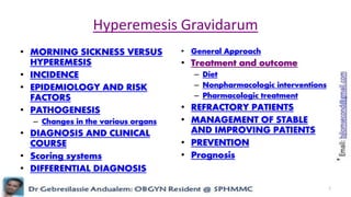
- 1. Hyperemesis Gravidarum • MORNING SICKNESS VERSUS HYPEREMESIS • INCIDENCE • EPIDEMIOLOGY AND RISK FACTORS • PATHOGENESIS – Changes in the various organs • DIAGNOSIS AND CLINICAL COURSE • Scoring systems • DIFFERENTIAL DIAGNOSIS • General Approach • Treatment and outcome – Diet – Nonpharmacologic interventions – Pharmacologic treatment • REFRACTORY PATIENTS • MANAGEMENT OF STABLE AND IMPROVING PATIENTS • PREVENTION • Prognosis 1
- 2. Hyperemesis gravidarum • intractable vomiting + disturbed nutrition • Early signs : weight loss (up to 5% of body weight) and ketonuria • Late signs: – Because vomiting causes potassium loss, electrocardiographic evidence of potassium depletion, such as inverted T waves and prolonged QT and PR intervals, is usually a later finding. – Jaundice also is a later finding and is probably due to fatty infiltration of the liver; occasionally, acute hepatic necrosis occurs. – Metabolic acidosis is rare. Hypokalemic nephropathy with isosthenuria may occur late. Hypoproteinemia also may result, caused by poor diet as well as by albuminuria. • Patients who have hyperemesis gravidarum are best treated (if the disease is early in its course) with parenteral fluids and electrolytes, sedation, rest, vitamins, and antiemetics if necessary • In some cases, isolation of the patient is necessary. • Very slow reinstitution of oral feeding is permitted after dehydration and electrolyte disturbances are corrected. • Therapeutic abortion may be necessary in rare instances; however, the disease usually improves spontaneously as pregnancy progresses. • Hyperemesis gravidarum is characterized by nausea and vomiting unresponsive to simple therapy. It usually occurs early in the first trimester and resolves by about 16 weeks. In some cases, there can be a transient hepatic dysfunction 2
- 3. • 125. Nausea and vomiting are common in pregnancy. Hyperemesis gravidarum, however, is a much more serious and potentially fatal problem. Findings that should alert the physician to the diagnosis of hyperemesis gravidarum early in its course include – a. Electrocardiographic evidence of hypokalemia – b. Metabolic acidosis – c. Jaundice – d. Ketonuria • 125. The answer is d. (Reece, 2/e, pp 1112–1113.) Hyperemesis gravidarum is intractable vomiting of pregnancy and is associated with disturbed nutrition. Early signs of the disorder include weight loss (up to 5% of body weight) and ketonuria. Because vomiting causes potassium loss, electrocardiographic evidence of potassium depletion, such as inverted T waves and prolonged QT and PR intervals, is usually a later finding. Jaundice also is a later finding and is probably due to fatty infiltration of the liver; occasionally, acute hepatic necrosis occurs. Metabolic acidosis is rare. Hypokalemic nephropathy with isosthenuria may occur late. Hypoproteinemia also may result, caused by poor diet as well as by albuminuria. Patients who have hyperemesis gravidarum are best treated (if the disease is early in its course) with parenteral fluids and electrolytes, sedation, rest, vitamins, and antiemetics if necessary. In some cases, isolation of the patient is necessary. Very slow reinstitution of oral feeding is permitted after dehydration and electrolyte disturbances are corrected. Therapeutic abortion may be necessary in rare instances; however, the disease usually improves spontaneously as pregnancy progresses. 3
- 4. • Hyperemesis gravidarum is defined – persistent, unexplained intractable nausea, retching, or vomiting sufficiently severe to produce • weight loss more than 5% of prepregnancy weight – > 3 kg ??? • Dehydration • alkalosis from loss of hydrochloric acid – Hypochloraemic alkalosis • Hypokalemia • ketonuria - Acidosis develops from partial starvation – usually ≥ 2+ unrelated to other causes • In most women, mild to moderate nausea and vomiting are especially common until approximately 16 weeks 4
- 5. MORNING SICKNESS VERSUS HYPEREMESIS • Morning sickness – Nausea and vomiting often develop by five to six weeks of pregnancy – The symptoms are worst around nine weeks, and typically improve by 16 to 18 weeks of pregnancy. However, symptoms continue until the third trimester in 15 to 20 percent of women and until delivery in 5 percent of women – Although mild pregnancy-related nausea and vomiting is often called "morning sickness," you may feel sick at any time of day and many women (80 percent) feel sick throughout the day. – Interestingly, women with mild nausea and vomiting during pregnancy experience fewer miscarriages and stillbirths than women without these symptoms. • Hyperemesis gravidarum – is the term used to describe more severe nausea and vomiting during pregnancy – may lose more than 5 percent of their pre-pregnancy body weigh 5
- 6. INCIDENCE • The incidence in the United States is 1 in 1000 pregnancies 6
- 7. EPIDEMIOLOGY AND RISK FACTORS • HEG – It affects 0.3–2% of pregnant women – is the second most common cause of antenatal hospitalization in the United States • Upto 50 – 80% of pregnant women experience mild form of nausea & vomiting during 1st TM but severe hyperemesis requiring hospitalization occurs in 0.3 – 2% • more common among younger mothers and those with a history of motion sickness, migraines, and nausea and vomiting associated with oral contraceptives • It is more commonly seen in women carrying multiple gestations, and patients with siblings or a mother with HEG are more likely to be affected • Factors that increase the risk for admission include • Hyperthyroidism • previous molar pregnancy • Diabetes • gastrointestinal illnesses, and asthma • for unknown reasons—perhaps estrogen-related—a female fetus increases the risk by 1.5-fold 7
- 8. • Risk factors – Genetic: Twin and sibling – Epidemiologic: younger age, low prepregnancy body mass, female fetus, history of motion sickness or migraines, and Helicobacter pylori infection. • Protective factors – Smoking – Obesity 8
- 9. PATHOGENESIS • pathogenesis is largely unknown – The etiology is not understood – etiopathogenesis is likely multifactorial and certainly is enigmatic • The recurrence rate is approximately 20% in prospective studies – but this value probably is an underestimation because the sickest women often limit family size • THEORIES – (1) Hormonal – (2) Psychogenic – (3) Dietetic deficiency – (4) Allergic or immunological basis – (5) Decreased gastric motility 9
- 10. (1) Hormonal • (a) Excess of chorionic gonadotropin or higher biological activity of hCG is associated – This is proved by the frequency of vomiting at the peak level of hCG and also the increased association with hydatidiform mole or multiple pregnancy when the hCG titer is very much raised • (b) High serum level of estrogen • (c) Progesterone excess leading to relaxation of the cardiac sphincter and simultaneous retention of gastric fluids due to impaired gastric motility • Other hormones involved are: thyroxin, prolactin, leptin and adrenocortical hormones. 10
- 11. • No single hormonal profile can accurately predict the presence of hyperemesis • Estrogen and Progesterone – Elevated serum concentrations of estrogen and progesterone – Relax smooth muscle and thus slow GI transit time and may alter gastric emptying – The fact that sex hormone levels peak in the 3rd trimester, long after symptoms of hyperemesis have typically resolved, is inconsistent with this theory • HCG – Serum concentrations of hCG peak during the 1st trimester, the time when hyperemesis is typically seen – Serum hCG concentration is higher in women with hyperemesis than in other pregnant women – Nausea & vomiting are worse in women with multiple gestations & hydatidiform moles, conditions associated with high hCG levels – HCG isoforms or hCG receptor mutations explain the differences in Symptoms among women with similar hCG levels. – Causal association between hCG levels and Hyperemesis has not been firmly established 11
- 12. • Hyperemesis appears to be related to high or rapidly rising serum levels of pregnancy-related hormones – Although the exact stimulus is unknown, – putative culprits include • human chorionic gonadotropin (hCG), • estrogens, • progesterone, • leptin, • placental growth hormone, • prolactin, • thyroxine, and • adrenocortical hormones 12
- 13. (2) Psychogenic • Conversion or somatization disorder or response to stress – Feeling of ambivalence about the pregnancy • It probably aggravates the nausea once it begins • But neurogenic element sometimes plays a role, as evidenced by its subsidence after shifting the patient from the home surroundings • If associated psychiatric and social factors contribute to the illness, the woman usually improves remarkably while hospitalized – That said, symptoms may relapse in these women, and some go on to develop posttraumatic stress syndrome (3) Dietetic deficiency • Probably due to low carbohydrate reserve, as it happens after a night without food • Deficiency of vitamin B6, Vit B1 and proteins may be the effects rather than the cause. 13
- 14. (4) Allergic or immunological basis (5) Decreased gastric motility is found to cause nausea • The Lower esophageal sphincter is relaxed in pregnancy, leading to an increase in Gastro-esophageal reflux, which results in heartburn and, in some individuals, nausea • However, if this were the mechanism for hyperemesis, symptoms should worsen as pregnancy advances, rather than improve so these abnormalities are not highly predictive of the disease 14
- 15. • An association of Helicobacter pylori infection has also been proposed, but evidence is not conclusive – Most women with H. pylori do not develop severe nausea & vomiting in pregnancy, but the infection may play a role in pathogenesis of disease in some women. – Some reports showed significant association between H. pylori infection and hyperemesis and improvement in sxs in women with severe disease after rx of the infection. – But distinguishing between active infection and past infection is a question • Helicobacter pylori Infection – H. pylori infection has been linked to • risk for preeclampsia • iron deficiency in pregnancy • Hyperemesis 15
- 16. • Whatever may be the cause of initiation of vomiting, it is probably aggravated by the neurogenic element. • Unless it is not quickly rectified, features of dehydration and carbohydrate starvation supervene and a vicious cycle of vomiting appears – vomiting → carbohydrate starvation → ketoacidosis → vomiting 16
- 17. Changes in the various organs • are the generalized manifestations of starvation and severe malnutrition • Maternal complications of HEG can include – Wernicke’s encephalopathy, acute tubular necrosis, central pontine myelinolysis, Mallory-Weiss tear of the esophagus, pneumomediastinum, and splenic avulsion – Additionally, significant psychological burden of the disease has been reported, with depression, anxiety, and lost work frequently seen among those with persistent or severe HEG. • Fetal – Fortunately, no clear fetal complications have been associated with HEG – One study did show that women with HEG who gain <7 kg during the entire pregnancy have a slightly higher risk of low birth weight and preterm birth. However, there are no congenital anomalies or increased risk of miscarriage or stillbirth noted. 17
- 18. 18
- 19. • Liver – There is centrilobular fatty infiltration without necrosis • Kidneys – Usually normal with occasional findings of fatty change in the cells of first convoluted tubule, which may be related to acidosis. • Heart – A small heart is a constant finding – There may be subendocardial hemorrhage. • Brain – Small hemorrhages in the hypothalamic region giving the manifestation of Wernicke’s encephalopathy. – The lesion may be related to vitamin B1 deficiency. 19
- 20. • Two serious vitamin deficiencies – Wernicke encephalopathy from thiamine deficiency – vitamin K defifificiency - cause maternal coagulopathy and fetal intracranial hemorrhage • Vitamin K: Cofactor for posttranslation carboxylation of many proteins including essential clotting factors • Wernicke's encephalopathy (WE) – is an acute syndrome requiring emergent treatment to prevent death and neurologic morbidity – Korsakoff's syndrome (KS) refers to a chronic neurologic condition that usually occurs as a consequence of WE – classic triad of Wernicke's encephalopathy (WE) includes: • Encephalopathy (confusion • Oculomotor dysfunction • Gait ataxia – abnormal electroencephalogram (EEG) may be seen, and usually there are MR imaging findings 20
- 21. • Wernicke’s encephalopathy (Harrison 19th – thiamine deficiency – manifestations • horizontal nystagmus • ophthalmoplegia (due to weakness of one or more extraocular muscles) • cerebellar ataxia, and mental impairment – When there is an additional loss of memory and a confabulatory psychosis, the syndrome is known as Wernicke-Korsakoff syndrome – Rx • In acute thiamine deficiency with either cardiovascular or neurologic signs, 200 mg of thiamine IV TID daily should be given until there is no further improvement in acute symptoms; • oral thiamine (10 mg/d) should subsequently be given until recovery 21
- 22. METABOLIC, BIOCHEMICAL AND CIRCULATORY CHANGES • are due to the combined effect of dehydration and starvation consequent upon vomiting. • Metabolic: – Inadequate intake of food results in glycogen depletion – For the energy supply, the fat reserve is broken down – Due to low carbohydrate, there is incomplete oxidation of fat and accumulation of ketone bodies in the blood – The acetone is ultimately excreted through the kidneys and in the breath – There is also increase in endogenous tissue protein metabolism resulting in excessive excretion of non-protein nitrogen in the urine – Water and electrolyte metabolizm are seriously affected leading to biochemical and circulatory changes. 22
- 23. • Biochemical – Loss of water and salts in the vomitus results in fall in plasma sodium, potassium and chlorides. – The urinary chloride may be well below the normal 5 g/liter or may even be absent • Hypochloraemic alkalosis – Hepatic dysfunction results in acidosis and ketosis with rise in blood urea and uric acid; hypoglycemia; hypoproteinemia; hypovitaminosis and rarely hyperbilirubinemia. • Circulatory – There is hemoconcentration leading to rise in hemoglobin percentage, RBC count and hematocrit values. – There is slight increase in the white cell count with increase in eosinophils – There is concomitant reduction of extracellular fluid 23
- 24. DIAGNOSIS AND CLINICAL COURSE • Symptoms typically start between 3 and 5 weeks of pregnancy and 80% resolve by 20 weeks Mean onset of symptoms is at 5 – 6 wks of gestation, peaking at about 9 weeks, usually abating by 16 – 20 wks of gestation. Symptoms may continue until 3rd trimester in 15 – 20% of gravida and delivery in 5 % In contrast to mild disease, women with hyperemesis gravidarum have • Orthostatic hypotension • Physical signs of dehydration • Laboratory abnormalities and • Often require hospitalization for stabilization Other causes should be considered because it is a diagnosis of exclusion 24
- 25. • Symptoms & Signs – severe nausea and vomiting that may result in dehydration, weight loss, and frequently social isolation and negative impacts on relationships with family and friends – Excess salivation (ptyalism) may also be seen • Chemical hyperthyroidism – hCG has more thyroid-stimulating activity – So serum TSH concentrations are often lower than those in normal pregnant women (60 versus 9 percent had a low serum TSH – few women have high serum free T4 concentrations and therefore have overt hyperthyroidism. 25
- 26. – Features that distinguish the transient hyperthyroidism of hyperemesis gravidarum from hyperthyroidism of other causes (which in a pregnant woman is most likely due to Graves' disease) are • vomiting, • absence of goiter and ophthalmopathy, and • absence of the common symptoms and signs of hyperthyroidism (tachycardia greater than 100 beats/minute, hyperdefecation, muscle weakness, tremor) • In addition, serum free T4 concentrations are only minimally elevated and serum T3 concentrations are frequently not elevated in women with hyperemesis gravidarum, whereas both are usually unequivocally elevated in pregnant women with true hyperthyroidism from Graves’ disease. 26
- 27. – The thyroid hyperfunction in women with hyperemesis gravidarum usually does not require treatment because it is mild and subsides as hCG production falls (as does the vomiting) – Like the thyroid hyperfunction, the vomiting is also proportional to the elevation in serum hCG and estradiol concentrations, and it is thought to be caused by estradiol – If overt hyperthyroidism persists for more than several weeks or beyond the first trimester, it is probably not hCG-mediated. 27
- 28. • Laboratory Findings – Suppressed thyroid-stimulating hormone/elevated free thyroxine and elevated liver enzymes, bilirubin, amylase, and lipase may all be noted in patients with severe nausea and vomiting; these are transiently abnormal and resolve with improvement of HEG – Elevated levels of transaminases, bilirubin, amylase, lipase, and various electrolytes are seen in 15–40% of patients. – Biochemical evidence of hyperthyroidism due to the effect of human chorionic gonadotropin on the thyroid-stimulating hormone receptor is seen in 60–70% of patients. 28
- 29. Risk Factors Age < 30yrs Primigravid Not taking MVT prior to 6 wks or during the peri-conceptional period Heartburn and acid reflux Multiple gestations Hydatidiform mole Obesity Recurrence (15 – 20 %) 1st degree family History : Daughters of mother with hyperemesis history 29
- 30. Scoring systems • Motherisk-PUQE scoring index • Pregnancy- Unique Quantification of Emesis and Nausea • Rhodes Index • tools for quantifying the severity of nausea and vomiting in pregnancy • used predominantly in research studies • A high score indicates the woman should be evaluated for dehydration and her serum electrolyte levels should be checked • Some clinicians find that these tools are helpful in assessing symptoms in their patients, but they have not been validated for guiding management of nausea and vomiting of pregnancy in the clinical setting. 30
- 31. DIFFERENTIAL DIAGNOSIS • The differential diagnosis includes a wide variety of conditions capable of causing persistent nausea and vomiting and must be considered carefully in each patient • Pancreatitis • Cholecystitis • Hepatitis • Gastritis • Gastroenteritis (IP) • Appendicitis • Pyelonephritis • Diabetic ketoacidosis • Ovarian torsion • Degenerating uterine leiomyomas • Psychiatric disease • Thyroid disease • Preeclampsia • HELLP syndrome • Acute fatty liver of pregnancy 31
- 32. General Approach • History – Assess gestational age – Dietary history, weight loss – History of PUD – Use of any drugs like iron supplement – Symptoms of thyrotoxicosis – Bowel habits and the presence of diarrhea and/or constipation. – Bilious emesis, Abdominal pain – Fever, rigors, shivering, leukocytosis – Hypertension – Urinary symptoms – Abdominal/pelvic/back pain – Psychiatric history • Physical examination – Usually unremarkable – Vital signs – Volume status - Signs of dehydration ( skin turgor, sunken eyeballs, mucus membrane, mental status ) – Nutritional status ( Weight, BMI ) – Thyroid evaluation – Abdominal evaluation – Cardiac evaluation – Neurologic evaluation 32
- 33. • Investigation – CBC – Electrolytes- Serum Potassium, Sodium, Chloride, Calcium, Magnesium – RFT – RBS – Liver enzymes – Viral markers (HbsAg, anti HCV) – Urinalysis - Presence of UTI, ketone, specific gravity, glucose – Obstetric ultrasound - to exclude molar pregnancy and look for multiple gestation – Urine culture- For cases of recurrent admission – Other investigations- Guided by history and P/E findings (Eg. TFTs for symptomatic thyrotoxicosis, who have goiter) 33
- 34. Treatment and outcome – Identify or rule out any co-morbidities – Identify complications – Classify severity • Goals of treatment – Reduce symptoms through changes in diet/environment and by medication – Correct consequences or complications of nausea & vomiting (eg, fluid depletion, hypokalemia, nutritional deficiencies and metabolic alkalosis) – Minimize the fetal effects of maternal nausea and vomiting and their treatment 34
- 35. • Principles in the management are: – avoidance of noxious stimuli – Antiemetics: control nausea and vomiting – Hydration- correct the fluids and electrolytes imbalance • intravenous Ringer lactate or normal saline solutions are given to correct dehydration, ketonemia, electrolyte deficits, and acid-base imbalances • There are no benefits to using 5-percent dextrose along with crystalloids – Correct metabolic disturbances (acidosis or alkalosis) – Prevent the serious complications of severe vomiting • Thiamine, 100 mg - to prevent WE – Maintain pregnancy 35
- 36. • Fetal status is generally not adversely affected by vomiting until persistent maternal weight loss occurs – In this setting, the rate of intrauterine growth retardation (IUGR) increases and fetal death appears to increase – Long-term effects of hyperemesis gravidarum on offspring are unknown • If weight loss persists despite therapy, nutritional supplementation by enteral tube feeding or parenteral feeding is necessary. 36
- 37. • Non Pharmacologic – Dietary modifications – Life style modifications • Supportive – Fluid & Electrolytes replacement • Pharmacologic – Anti-emetics – Anti-acids 37
- 38. Diet • Women with nausea should eat before or as soon as they feel hungry to avoid an empty stomach, which can aggravate nausea. – Small frequent meals every 1 to 2 hrs to avoid a full stomach – Food rich in carbohydrate – Sour cold drinks in between meals – Cold solid foods are tolerated better than hot solid foods because they have less odor and require less preparation & exposure time. – Aroma therapy with lemonade, orange and ginger – Dietary Manipulations • Eliminating coffee and spicy, odorous, high fat, acidic, and very sweet foods, and • substituting snacks/meals that are protein dominant, salty, low fat, and/or dry (eg, peanuts, cereal, pea, bea…) 38
- 39. • Diet – A snack before getting out of bed in the morning can be helpful. – Fluids are better tolerated if cold, clear, and carbonated or sour (eg, ginger ale, lemonade, popsicles), and if taken in small amounts between meals – Liquids can be taken in small amounts with a straw. Small volumes of electrolyte-replacement sports drinks replace both fluids and electrolytes – Aromatic therapies involving lemon (lemonade), mint (tea), or orange have also been described as useful – Fluids should be consumed at least 30 minutes before or after solid food to minimize the effect of a full stomach 39
- 40. Nonpharmacologic interventions • Avoidance of triggers – Avoidance of Environmental triggers such as: stuffy Rooms, odors (eg, perfume, chemicals, food, smoke), heat, noise and visual or physical motion (eg, lights, driving). – Getting enough rest, particularly after eating, frequent naps and shortening working hours – Brushing teeth after a meal and frequently washing out the mouth. – Avoid supplements containing Iron until symptoms resolve. – Taking prenatal vitamins before bed with a snack, may also be helpful • Acupuncture and acupressure - become a popular self-administered intervention • Hypnosis • Psychotherapy 40
- 41. Pharmacologic treatment • Complementary and alternative medications (CAM) – Ginger • has been successful in treating the nausea and vomiting of pregnancy • Pyridoxine (vitamin B6) – Navidoxine 25 mg po TID • Anti – emetics 41
- 42. Outpatient management • Outpatient rehydration if – Weight loss – Ketonuria +2 or less • Signs of dehydration – Infuse first liter over 1 – 2 hours then 1000mls over 4hours, followed by further assessment , including urine ketone testing. – If improved- Antiemetics will be given and discharged • Pyridoxine + Doxylamine combination or • Meclizine + Pyridoxine (Navidoxine) 25mg po BID or • Pyridoxine 10 – 25 mg po BID/TID + Metoclopramide 5 – 10mg po TID or • Promethazine 12.5 – 25mg po QID. – If no improvement- Reclassify for inpatient management 42
- 43. Indications for Admission • Severe vomiting, weight loss (10% or more body weight) , ketonuria, dehydration, hypotension, alkalosis from HCL loss, hypokalemia, or nutritional deficiencies. • Unable to maintain adequate hydration, normal electrolyte levels, and acid-base balance with the initial interventions. • Having persistent vomiting after rehydration at OPD • Presence of co-morbidity • Unsettled diagnosis In women with persistent vomiting after hospitalization, exclude underlying diseases that can cause hyperemesis 43
- 44. Components of Inpatient Management • Fluid therapy – Fluid replacement • Replace fluid deficit • Give maintenance fluid • Give replacement fluid for ongoing loss • Calorie replacement • Electrolyte supplementation • Vitamins supplementation • Parenteral Antiemetics • Acid reducing agents • Dietary advice 44
- 45. Fluid therapy • Replacement of fluid deficit – NS and RL should be used – At least 1 – 2 liters of isotonic saline as rapidly as possible. – Fluid repletion is continued at a rapid rate ( 1 – 2L over the next 2 – 3hrs ) until the clinical signs of hypovolemia improve • (eg, low BP, low urine output, and/or impaired mental status) – Dextrose containing fluid should be avoided until thiamine is supplemented with the initial rehydration fluid – glucose can exacerbate/precipitate WE • How • Maintanance fluid – 3L/day ( 5% dextrose in normal saline) • Oral feeding is withheld for at least 24 hours after the cessation of vomiting • With this regime — dehydration, ketoacidosis, water and electrolyte imbalance are likely to be rectified 45
- 46. Calorie replacement • Average Daily calorie requirement of a pregnant woman is 2500kcal/day • Difficult to supply this whole calorie with dextrose containing fluid, so the pt should be started on po feeding as soon as DHN is corrected & food can be tolerated. • The minimum amount of calorie to prevent ketosis is 400kcal/day in the non pregnant adult, this corresponds to 100 gm of glucose – 400kcal = 100 gm of glucose • In pregnancy daily glucose requirement increased to 150 – 200 gm/day – One Liter of 5% DNS contains 50 gm dextrose (1000X0.05 = 50) – One vial of 40% dextrose contains 8 gm of dextrose (20X0.4 = 8) – So - Three liters of 5% DNS we give as part of the maintenance fluid which contains 150gm dextrose which is suffice to prevent ketosis • If no 5% DNS we need to add 6 – 7 vial of 40% dextrose ineach bag of NS 46
- 47. Electrolyte supplementation • 1 vial of KCL = 20 meq • Potassium Supplementation – Mild to moderate hypokalemia • serum potassium 2.5 – 3.5meq • 20 – 80meq/day: 1 vial of KCL on each bag of maintainance fluid – Severe hypokalemia • serum potassium < 2.5meq/l) or symptomatic hypokalemia • 20meq/2 – 3hrs with careful monitoring every 2 – 4hrs: – 2 – 3 vials of KCL(40 – 60 meq) in each bag of maintenance fluid • Magnesium and Calcium Supplementation 47
- 48. Vitamins supplementation • Thiamine is a cofactor for several key enzymes important in energy metabolism, including transketolase, alpha-ketoglutarate dehydrogenase, and pyruvate dehydrogenase • One ampoule (2ml) of Vit B complex contains: – B1=10mg, B2=4mg, B6=4mg • Thiamine (Vitamin B1) – 100 mg intravenously with the initial rehydration fluids • 100 mg =10 ampoules • 3 ampules in each initial rehydration fluids(for a maximum of 10 ampules) – Another 100 mg daily for the next two or three days. • 10 ampules of vit B complex per day, 3 Ampules/liter • Vitamine B6 (Pyridoxine ) – Pyridoxal phosphate is also involved in decarboxylation of amino acids, gluconeogenesis, conversion of tryptophan to niacin, sphingolipid biosynthesis, neurotransmitter synthesis, immune function, and steroid hormone modulation – 10 – 25mg in every liter 3 ampoules of MVI in each bag of fluid 48
- 49. Antiemetics • First line – Antihistamines (H1 antagonists) • Diphenhydramine – Orally: 25 to 50 mg every four to six hours – Intravenously: 10 to 50 mg every four to six hours • Meclizine: – 25 mg can be taken orally every four to six hours • Dimenhydrinate – 25 to 50 mg can be taken orally every four to six hours 49
- 50. • Second Line – Serotonin antagonists • Ondansetron , granisetron , and dolasetron • are selective antagonists at the 5-HT3 serotonin receptor – Dopamine antagonists: three main classes • Phenothiazines: promethazine, prochlorperazine, Chlorpromazine • Butyrophenones: droperidol • Benzamides: metoclopramide Mechanism of Action of metoclopramide – Blocks dopamine receptors and (when given in higher doses) also blocks serotonin receptors in chemoreceptor trigger zone of the CNS; enhances the response to acetylcholine of tissue in upper GI tract causing enhanced motility and accelerated gastric emptying without stimulating gastric, biliary, or pancreatic secretions; increases lower esophageal sphincter tone – Pharmacodynamics/Kinetics » Onset of action: Oral: 30-60 minutes; I.V.: 1-3 minutes; I.M.: 10-15 minutes 50
- 51. • Phenothiazines – Promethazine – Prochlorperazine » 5 to 10 mg orally, IV/IM every six hours or 25 mg per rectum twice per day » Safety information is limited » main adverse • extrapyramidal reactions such as dystonia and, with prolonged use, tardive dyskinesia – Chlorpromazine » used less often than prochlorperazine » reserved for refractory cases » 25 to 50 mg IV/IM or 10 to 25 mg orally every four to six hours) to be helpful in refractory cases » Adverse effects: extrapyramidal reactions, orthostatic hypotension, anticholinergic effects, and altered cardiac conduction 51
- 52. • Ondansetron – 4 to 8 mg can be taken orally every eight hours, as needed, or administered intravenously by bolus injection every eight hours, as needed – common side effects • Headache, fatigue, constipation, and drowsiness – Hyperemesis is a common unlabeled indication for use – can cause QT prolongation, particularly in patients with underlying heart conditions, such as • congenital long QT syndrome; • patients with hypokalemia or hypomagnesemia; and • those taking other medications that lead to QT prolongation – ECG monitoring is recommended in these patients. 52
- 53. • Third line – Glucocorticoids • reserved for treatment of refractory hyperemesis gravidarum • risk of maternal side effects ??? – Methylprednisolone • 16 mg intravenously every 8 hours for 48 to 72 hours • tapered over two weeks in women who experience relief of symptoms – After intravenous therapy, we use an oral prednisone taper regimen of » 40 mg oral prednisone per day for one day 20 mg per day for three days 10 mg per day for three days 5 mg per day for seven days » 1 3 3 7 (Total 14 day) – This regimen may be repeated up to three times over a six week period. • can be stopped abruptly if there is no response 53
- 54. Acid reducing agents • In women with heartburn/acid reflux and nausea/vomiting of pregnancy, ant- acid combined with anti-emetic therapy will significantly improve symptoms three to four days after beginning therapy • Antacids containing aluminum or calcium are safe and preferable to those containing bismuth or bicarbonate, which may have adverse fetal/neonatal effects • The H2 receptor antagonists: safe during px – Ranitidine (pregnancy category B) 150mg PO BID – Cimetidine (pregnancy category B) 200mg IV BID • Male fetus ????? • Proton Pump Inhibitors (Category c): – Pantoprazole 20 – 40mg/day 54
- 55. Follow up • Hyperemesis gravidarum follow up chart – Maternal Vital signs: BP, PR, RR, TT – Input & Output – Weight – Urine ketones – Serum K – Vomiting episodes – Medications 55
- 56. Input & Output: Daily Intake and Output of Water (ml/day) 56 Intake Output Fluids ingested 2100 Insensible: skin 350 From metabolism 200 Insensible: lungs 350 Total intake 2300 Sweat 100 Feces 100 Urine 1400 Total output 2300
- 57. When to discharge?? • No ketones in the urine • Tolerating oral fluids and possibly food for at least 24hrs after ketone is free and with PO antiemetics • Appropriate Anti-emetic to take home – Navidoxine 25 mg po BID or – Promethazine 12.5 – 25mg po QID or – Metoclopramide 10mg po TID/QID + Vitamin B6 (Pyridoxine) 10 – 25 mg PO QID Should be taken for at least one week 57
- 58. Proper advice Upon discharge • Advice them to eat as soon as they feel hungry, small meals and snacks • To eat foods high in protein or carbohydrates (biscuits, bread, nuts). • Avoid foods that are spicy, greasy, or acidic, coffee • Drink cold fluids between meals, rather than with a meal • Brush their teeth right after eating • Avoid lying down right after meal, stuffy rooms, strong smells, hot places, or loud noises. • Have someone to make their meals. • Inform her & her partner, chance of recurrence on discharge 58
- 59. REFRACTORY PATIENTS • Definition – ?? – Round : When there is no response to the most effective antiemetic with maximum dose – recalcitrant vomiting ??? • Rx – Chlorpromazine – Glucocorticoids – Glucocorticoids • slightly increased risk of oral clefts when the drug is administered before 10 weeks of gestation; therefore, ideally, use of glucocorticoids should be avoided in the first trimester – Enteral and parenteral nutrition • Nasojejunal feeding for up to 21 days • Percutaneous endoscopic gastrostomy with a jejunal port • complications included line sepsis, thrombosis and infective endocarditis 59
- 60. • Pregnancy termination – considered in a patient who doesn’t show improvement and deteriorating despite taking all available therapeutic Measures (REFRACTORY PATIENTS) • Wernicke's Encephalopathy • Severe hypokalemia • Jaundiced • Renal complications: ATN • If women felt they are too sick to care for their family or themselves – exact incidence is unknown – a web-based survey of over 800 women who agreed to be part of a hyperemesis gravidarum registry noted that • 15% had at least one termination, and • 6% had more than one termination as a direct or indirect result of severe hyperemesis gravidarum 60
- 61. • Some Life-Threatening Complications of Recalcitrant Hyperemesis Gravidarum – Acute kidney injury—may require dialysis – Depression—cause versus effect? – Diaphragmatic rupture – Esophageal rupture—Boerhaave syndrome – Hypoprothrombinemia—vitamin K deficiency – Hyperalimentation complications – Mallory-Weiss tears—bleeding, pneumothorax, pneumomediastinum, pneumopericardium – Wernicke encephalopathy—thiamine deficiency 61
- 62. MANAGEMENT OF STABLE AND IMPROVING PATIENTS • We continue the drug regimen that has been effective until the patient has been completely asymptomatic (no nausea or vomiting) for at least a week • At that time, we discontinue the medications and see how she does • If nausea and vomiting recurs, we resume therapy. The majority of women will have resolution of nausea and vomiting by 16 to 20 weeks of gestation and will be able to discontinue their medications • Rare patients require therapy beyond 20 weeks 62
- 63. PREVENTION • Ideally, all women of child-bearing age should be advised to take a daily multivitamin with folic acid beginning in the preconception period; – this reduces the risk of congenital anomalies, particularly neural tube defects, and may help to decrease the frequency and severity of nausea and vomiting during pregnancy • The positive effects of multivitamins are likely due to the general optimization of nutritional status and metabolism • Managing heartburn and acid reflux prior to pregnancy might prevent or reduce the severity of symptoms 63
- 64. Prognosis • More than 50% of women have resolution of symptoms by 16 weeks of gestational age and 80% by 20 weeks • However, approximately 10% will be affected to some degree with severe nausea and vomiting for the duration of the pregnancy • HEG has been shown to recur in up to 80% of subsequent pregnancies, although earlier aggressive medical therapy prior to significant symptoms has been demonstrated to reduce both the severity and recurrence rate overall in future pregnancies • If hyperemesis gravidarum occurs in a first pregnancy, the recurrence risk is approximately 15%, although this may be reduced if there is a change in paternity • The frequency of congenital anomalies is not increased among offspring of gravida with NVP, whether or not they take antiemetic medications – Most studies have reported no difference in birth weight or GA at birth, as long as prepregnancy weight is normal and there is "catch-up" weight gain later in pregnancy 64
- 65. • Fetal effects – are unclear – In general, if the problem is corrected or resolves and the patient is able to gain weight, there are no consequences – However, if the woman has poor weight gain (<15 lb), the fetus is at increased risk for LBW and PTB 65