5 . Oedogonium & Chara.ppt
•Download as PPT, PDF•
0 likes•53 views

Oedogonium and Chara

Recommended
More Related Content
Similar to 5 . Oedogonium & Chara.ppt
Similar to 5 . Oedogonium & Chara.ppt (7)
More from Head Department of Botany Govt Degree College Mahabubaba
More from Head Department of Botany Govt Degree College Mahabubaba (20)
5 . Oedogonium & Chara.ppt
- 1. Dr. Thirunahari Ugandhar Head & Assistant Professor in Botany OEDOGONIUM
- 3. OEDOGONIUM: OCCURRENCE, FEATURES AND REPRODUCTION. Occurrence of Oedogonium: Oedogonium (Gr. oedos, swelling; gonos, reproductive bodies) is an exclusively fresh water alga. Out of about 400 species more than 200 have been reported from India. They are very common in pools, ponds, lakes etc. The filamentous plant body may get attached with the stone, wood, leaves of aquatic plants, small branches of dead plant remain in water etc. by their basal
- 4. ఓడోగోనియం యొక్క సంఘటన: ఓడోగోనియం (గ్రీస్ oedos, వాపు; గొనోస్, పునరుత్ప త్తి సంసథలు) ఒక్ గ్రరత్యే క్ంగా తాజా నీరు. సుమారు 400 జాతులలో 200 క్న్నా ఎక్కక వ మంది భారత్దేశంలోనే నివేదించబడ్డ ా రు. వారు కొలనులు, చెరువులు, సరసుు లు మొదలైన వాటిలో చాలా సాధారణం. చర్మా నికి, చెక్క తోను, నీటి మొక్క ల ఆక్కలు, చనిపోయిన మొక్క ల యొక్క చినా శాఖలు నీటిలోనే ఉంటాయి, వాటి యొక్క బేసల్ సెలుా రట్టుక్కంటాయి. O. టెరెస్ట్రుస్ వంటి కొనిా జాతులు భూగోళం
- 5. Plant Body of Oedogonium: The thalloid plant body is green, multicellular and filamentous. The filaments are unbranched and cells of each filament are attached end to end and form uniseriate row (Fig. 3.72A). The filament is differentiated into 3 types of cells: 1. Basal cell, 2. Apical and 3. Middle cells. 1. Basal Cell: It is the lowermost cell of the fila- ment. The cell is long, gradually narrowed and towards the basal end it expands to form simple, disc-like, multilobed or finger-shaped structure. The cell is generally colourless, which performs the function of fixation to the substratum and called holdfast.
- 7. శరీరం ఆక్కరచచ , బహు క్ణ మరియు ఫిల్ా ంటస్. త్ంతువులు అపుప డపుప డూ మరియు గ్రరత్త ఫిలమంట్ యొక్క క్ణాలు అంత్ం అవవ డమే కాక్, విడదీయర్మని వరుసను రూపందిసా ి యి (Figure 3.72A). ఈ ఫిల్ా ంట్ 3 రకాలైన క్ణాలుగా విభజిసుింది: 1.బాసల్ సెల్, 2. మౌఖిక్ మరియు 3. మధ్ే క్ణాలు. 2.1. బేసల్ సెల్: ఇది ఫిలమంట్ యొక్క అత్త త్క్కక వ క్ణం. సెల్ దీరఘకాలిక్ంగా, గ్రక్మంగా క్కదించారు మరియు బేసల్ ఎండ్ వైపుగా, సాధారణ, డిస్క -లాంటి, బహుముఖ లేదా వేలు ఆకారపు ఆక్ృత్తని ఏరప రుసుింది. క్ణం సాధారణంగా రంగులేనిది, ఇది ఉరజాత్తకి స్ రథరీక్రణ యొక్క రనితీరును అమలు చేసుింది మరియు హోలాా స్ు అని పిలుసా ి రు.
- 8. 2. Apical Cell: It is the topmost cell of the filament. The cell is usually rounded towards apical side and green in colour. 3. Middle Cells: All the cells in between basal and apical cells are alike. The cells are longer than their breadth i.e., rectangular in shape. Towards the upper end of some cells a ring-like structure is present known as cap or apical cap (Fig. 3.72A). The cell with cap is called cap cell. The number of caps on a cell indicates the number of cell divisions in that cell.
- 9. క్ణం సాధారణంగా ఆపిుక్ల్ వైపు మరియు ఆక్కరచచ రంగు వైపు గుంగ్రడంగా ఉంట్టంది. 3. మధ్ే క్ణాలు: బేసల్ మరియు అనుబంధ్ క్ణాలు మధ్ే అనిా క్ణాలు ఇలానే ఉన్నా యి. క్ణాలు వాటి వెడలుప క్న్నా పడవుగా ఉంటాయి, ఆకారంలో దీరఘచతురగ్రసాకారంగా ఉంటాయి. కొనిా క్ణాల ఎగువ భాగంలో ఒక్ రింగ్ లాంటి నిర్మా ణం టోపీ లేదా అపిిప్ టోపీ (Figure 3.72A) గా పిలువబడుతుంది. టోపీతో ఉనా క్ణం కాే ప్ సెల్ అంటారు. సెల్ లో కాే ప్ు సంఖే సెల్ లో సెల్ విభాగాలు సంఖే సూచిసుింది.
- 11. Important Features of Oedogonium: 1. This is a common fresh water alga growing on substratum like sand particles, rocks etc. 2. The plant body is unbranched, filamentous and differentiated into apex and base. 3. Cells have reticulate chloroplasts. 4. Presence of caps on the young dividing cells. 5. Vegetative cell division is very elaborate. 6. Asexual reproduction takes place by multi- flagellate zoospore, where flagella are arranged around the beak-like apical region. 7. Sexual reproduction is advanced oogamous type. 8. The female gamete i.e., ovum, is produced singly in each oogonium. 9. The male gametes i.e., antherozoids, are very much similar to zoospores but smaller in size. Two antherozoids are produced in each antheridium. 10. Based on the size of male filament the plants are
- 12. ఓడోగోనియం యొక్క ముఖే మైన లక్షణాలు: 1. ఇసుక్ క్ణాలు, శిలలు మొదలైన అంశాలపై సాగుతునా ఒక్ సాధారణ మంచినీటి ఆలా ా . 2. మొక్క ల శరీరము unpranched, ఫిల్ా ంటస్ మరియు అపెక్సు మరియు ఆధారం లోకి వేరుగా ఉంట్టంది. 3. క్ణాలు chloroplasts reticulate క్లిగి. 4. యువ విభజన క్ణాలపై టోపీలు ఉండటం. 5. వజీవ క్ణ విభజన చాలా విసిృత్మైనది. 6. ఎరక్కే వల్ పునరుత్ప త్తి బహుళ-స్ ా ి లిట్ జోస్పప రే చేత్ జరుగుతుంది, ఇక్క డ జింక్ వంటి పడవాటి చుట్టురక్క ల గ్రరంత్ం చుట్ట ు జండ్డలు ఏర్మప ట్ట చేయబడతాయి. 7. లైంగిక్ పునరుత్ప త్తి oogamous రక్ం ముందుక్క. 8. స్ట్రి గిమేట్ అంటే ఓవమ్ గ్రరత్త ఒగోనియంలో ఒక్క దానిని ఉత్ప త్తి చేసా ి రు. 9. పురుష్ ీట్టి అనగా, జోే త్తష్కక లను, zoospores చాలా పోలి ఉంటాయి కానీ రరిమాణం చినా విగా ఉంటాయి. గ్రరత్త
- 13. Cell Structure of Oedogonium: The intercalary cells are longer than their breadth and are cylindrical in outline. The cells are surrounded by thick and rigid cell wall (Fig. 3.72B). The cell wall is differentiated into three layers an outer chitin, middle pectin and innermost cellulosic. Just interior to the wall, cell membrane is present, which encloses the protoplast. The protoplast consists of cytoplasm, chloroplast and nucleus. The cells contain many small or single large vacuoles situated in the centre and remain filled with cell sap. The cytoplasm lies between the cell membrane and vacuole. The Chloroplast is single, large and reticulate, which remains embedded in the cytoplasm. It extends from one end of the cell to the other end. Cells are
- 15. క్ణాలు మందరటి మరియు దృఢమైన సెల్ గోడతో చుట్టుముటుబడి ఉంటాయి (Figure 3.72B). సెల్ గోడ మూడు పరలుగా బయటి చిటిన్, మధ్ే పెకి ున్ మరియు అంత్రాత్ సెలుే లారకాా విభజిసుింది. కేవలం గోడక్క లోరలికి, క్ణ త్వ చం ఉంట్టంది, ఇది గ్రపోటోర ి సుుా క్లుపుతుంది. గ్రపోటోర ి స్ప ు ో సైటోర ి జం, స్ ోిరోర ి స్ స్ు మరియు న్యే కి ియస్ ఉంటాయి. ఈ క్ణాలు మధ్ే లో ఉనా అనేక్ చినా లేదా ఒకే పెదె vacuoles క్లిగి మరియు సెల్ సాప్ నిండి ఉంటాయి. క్ణ త్వ చం మరియు వాక్కే ల మధ్ే సైటోర ి జం ఉంట్టంది. స్ ోిరోర ి స్ు రంగిల్, పెదెది మరియు పునరిా ర్మా ణం, ఇది సైటోర ి జంలో పందురరచబడి ఉంట్టంది. ఇది ఒక్ అంచు నుండి మరొక్ చివరి వరక్క విసిరించి ఉంట్టంది. క్ణాలు అన్నరుే కే ిట్ మరియు కేంగ్రదక్ం
- 18. Cell Division: Growth of the filament takes place through cell division (Fig. 3.73). All cells except apical and basal ones are capable of dividing through cell division though division remains restricted in some of the cells of the filament. The steps of cell division are: 1. Initially the nucleus becomes shifted from peripheral position towards the centre and then moves slightly towards the upper half of the cell (Fig. 3.73A, B). 2. Ring-like thickening develops towards the upper part of the cell wall which gradually increases in thickness (Fig. 3.73B). 3. The nucleus undergoes mitotic division and form two nuclei (Fig. 3.73C). 4. At the end of cell division (telophase), a row of microtubules develop and accumulate as a layer between the daughter nuclei (Fig. 3.73C). This layer remains in floating condition which will develop the future septum. 5. The ring-like thickening gradually elongates and splits the mother wall towards the apical region. The ring expands much more and forms a concave cylindrical structure (Fig. 3.73D). The ring material ultimately forms the cuticle of the upper daughter cell. 6. The upper part of the ruptured mother wall remains attached to
- 19. సెల్ విభజన: ఫిలమంట్ పెరుగుదల సెల్ డివిజన్ (Figure 3.73) దావ ర్మ జరుగుతుంది. దైవిక్ మరియు బేసల్ వాటిని మినహా అనిా క్ణాలు సెల్ విభజన దావ ర్మ విభజించగలవు, అయినరప టికీ త్ంతువుల యొక్క కొనిా క్ణాలలో విభజన రరిమిత్ం చేయబడింది. సెల్ విభజన దశలు: 1. గ్రరరంభంలో కేంగ్రదక్ం కేంగ్రదం వైపు రరిధీయ స్ సా థ న్ననికి మారింది మరియు త్రువాత్ సెల్ యొక్క ఎగువ భాగంలో కొదిెగా క్దులుతుంది. రింగ్ వంటి గటిురడటం సెల్ గోడ యొక్క ఎగువ భాగం వైపుగా అభివృదిి చెందుతుంది, ఇది గ్రక్మంగా మందంతో పెరుగుతుంది 3. న్యే కి ియస్ మిటోటిక్స విభాగానికి లోనవుతుంది మరియు రెండు కేంగ్రదకాలు (Fig. 4. సెల్ డివిజన్ (టెలోాస్) ముగింపులో, మైగ్రోటోటోపుల్ు వరుస అభివృదిి మరియు క్కమారెి కేంగ్రదక్ం మధ్ే పరగా పేరుక్కపోవడం. ఈ పర భవిష్ే త్ శూనే ం అభివృస్ దిి చెందుతునా త్యలియాడే స్ రథత్తలోనే ఉంట్టంది. 5. రింగ్ వంటి గటిురడటం గ్రక్మంగా పెరుగుతుంది మరియు క్షీరదాల వైపు త్లిి గోడను విడిపోతుంది. రింగ్ మరింత్ విసిరిస్ సుింది మరియు ఒక్ పుటాకార స్ సూ థ రకార నిర్మా ణం రూపందిసుింది. రింగ్ రదారథం చివరికి ఎగువ క్కమారెి క్ణాల జంతువును ఏరప రుసుింది. 6. రగిలిపోయిన త్లిి గోడ ఎగువ భాగంలో కొత్ి క్కమారెి క్ణము యొక్క పూరవ ముగింపుక్క ఒక్ రరిమిత్తగా, అనగా టోపీగా అంట్టక్కని ఉంట్టంది. మిగిలిన భాగం క్కమారెి క్ణాల యొక్క బేసల్ గ్రరంతానికి
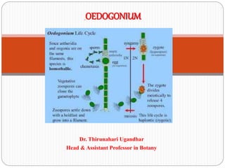
- 21. Reproduction in Oedogonium: Oedogonium reproduces by all the three means: vegetative, asexual and sexual. Vegetative Reproduction: It takes place by fragmentation and akinete formation: 1. Fragmentation: It takes place by accidental breakage of the filament, dying off of intercalary cells or by the formation of intercalary sporangia. The fragments are capable of developing into new filaments. 2. Akinete: During unfavourable condition the entire protoplast of a cell becomes a thick-walled, reddish- brown, round or oval structure, the akinete. The akinete germinates during favourable condition and develops a new filament. They generally form in chain.
- 22. ఓడోగోనియంలో పునరుత్ప త్తి: Oedogonium అనిా మూడు మార్మ ా ల దావ ర్మ పునరుత్ప త్తి: ఏపుగా, అగ్రక్మ మరియు లైంగిక్. వరి పునరుత్ప త్తి: ఇది విభజన మరియు akinete నిర్మా ణం దావ ర్మ జరుగుతుంది: 1.గ్రాగ్ా ంటేష్న్: ఇది త్ంతువుల గ్రరమాదవశాత్త ి విచిి నా ం దావ ర్మ, అంత్ర క్ణాల నుండి మరణిసుింది లేదా అంత్రకాలిక్ వృక్షజాలం ఏరప డటం దావ ర్మ జరుగుతుంది. ఈ ముక్క లు కొత్ి త్ంతుక్ణాలపై అభివృదిి చేయగలవు. 2.2. Akinete: గ్రరత్తకూల రరిరథతులలో సెల్ యొక్క మొత్ిం గ్రపోటోర ి స్ు ఒక్ మందరటి-గోడ, ఎగ్రరటి- గోధుమ, రండ్ లేదా ఓవల్ నిర్మా ణం, ఆనికేట్ అవుతుంది. అనునీయ రరిరథత్తలో అనీనిట్ అంక్కరోతుిలు మరియు కొత్ి ఫిల్ా ంట్టా
- 24. Asexual Reproduction: Asexual reproduction takes place by means of zoospores. Zoospores are formed singly within a cell. Comparatively younger cell i.e., the cell with cap behaves as sporangium mother cell. The zoospores are multiflagellate and ovoid, pyriform or spherical in shape. They are uninucleate with single chloroplast and occasionally with an eye-spot. During favourable condition, the zoospore formation begins in a cap cell of the filament. The entire protoplast of zoosporangium contracts from the wall and behave as a unit. The protoplast becomes round or oval in shape and its nucleus moves at one end.
- 25. Asexual పునరుత్ప త్తి: అశుది పునరుత్ప త్తి zoospores దావ ర్మ జరుగుతుంది. Zoospores ఒక్ సెల్ లోరల ఒక్క గా ఏరప డతాయి. సమకాలీన యువ క్ణం అనగా, టోపీతో ఉనా క్ణం స్పప రెంజియం త్లిి క్ణంగా గ్రరవరిిసుింది. ఈ zoospores అనేవి multiflagellate మరియు ovoid, pyriform లేదా గోళాకార రూరంలో ఉంటాయి. అవి ఒక్క స్ ోిరోర ి స్ప ు ో మరియు అపుప డపుప డు క్ంటి- సాప ట్ తో క్లయిక్ లేనివి. అనుకూలమైన స్ రథత్తలో, జూగ్రపోపోర్ నిర్మా ణం ఫిలమంట్ యొక్క టోపీ సెల్ లో మొదలవుతుంది. గోడ నుండి జూస్పప రింజియం ఒరప ందాల మొస్ త్ిం గ్రపోటోర ి స్ు మరియు యూనిటా ా గ్రరవరిిస్ సా ి యి. గ్రపోటోర ి స్ు ఆకారంలో రండ్ లేదా ఓవెల్ అవుతుంది మరియు దాని కేంగ్రదక్ం ఒక్ ముగింపులో క్దులుతుంది.
- 26. Sexual Reproduction: The sexual reproduction in Oedogonium is an advanced oogamous type. The male gametes or antherozoides are produced in antheridium (Fig. 3.75) and the female gamete or egg is produced in oogonium (Fig. 3.76). Male and female gametes differ both morphologically and physiologically. Only one egg is produced in each oogonium and two antherozoides in each antheridium. Another motile structure, the androspore, is produced singly in each androsporangium. Deficiency of nitrogen and alkaline pH are the important factors for promoting sexual reproduction.
- 27. లైంగిక్ పునరుత్ప త్తి: Oedogonium లో లైంగిక్ పునరుత్ప త్తి ఒక్ ఆధునిక్ oogamous రక్ం. మగ జిమేట్ు లేదా ఎంటెరోజోసైడుి అంటెరిడియమ్ (ఫిజి 3.75) లో ఉత్ప త్తి చేయబడతాయి మరియు ఆడ గిమేట్ లేదా గుడుా oogonium (Figure 3.76) లో ఉత్ప త్తి అవుతుంది. పురుష్ మరియు స్ట్రి గామాటిలు మ్లరోా లాజిక్ల్ మరియు ఫిజియలాజిక్ల్ రెండింటిలోన్య విభినా ంగా ఉంటాయి. గ్రరత్త ఎనోర్మయినియంలో ఒోక గుస్ డుా మాగ్రత్మే ఉత్ప త్తి అవుతుంది. మరో motile నిర్మా ణం, androspore, గ్రరత్త ఆంటోసప రోగియం లో ఒకే ఉత్ప స్ త్తి. నగ్రత్జని మరియు ఆలక లీన్ pH యొక్క లోరం లైంగిక్ పునరుత్ప త్తి గ్రపోత్ు హంచడ్డనికి ముఖే మైన కారకాలు.
- 33. Distribution of Sex Organ in Oedogonium: Based on the size of the male (antheridial) filament the species of Oedogonium are divided into two groups macrandrous and nannandrous type: 1. Macrandrous Type: In macrandrous type the antheridium develops in the filament of normal size. It is of two Types: i. Monoecious type (homothallic or bisexual). In this type (e.g., O. fragile, O. nodulosum and O. hirnii) antheridia and oogonia are borne on the same filament (Fig. 3.79). ii. Dioecious type (heterothallic or unisexual). In this type (e.g., O. gracilius, O. cardiacum and O. aquaticum) the antheridia and oogonia are borne on the different filaments
- 34. ఓడోగోనియంలోని సెక్సు ఆర్మ ా న్ యొక్క vDistribution: మగ (ఏరథైటియల్) ఫిల్ా ంట్ యొక్క రరిమాణానిా బటిు ఓడోగోనియం యొక్క జాతులు మాగ్రోగ్రడ్డస్ మరియు న్నన్నస్ట్న్న ా స్ రక్ం రెండు సమూహాలుగా విభజించబడ్డ ా యి: 1. మాస్ట్కాా గ్రడస్ రక్ం: మాగ్రోగ్రడ్డస్ రక్ంలో ఎంటేరిడియం సాధారణ రరిమాణంలో త్ంతుణత్లో అభివృదిి చెందుతుంది. ఇది రెండు రకాలు: i. Monoecious రక్ం (homothallic లేదా దివ లింగ). ఈ రక్మైన (ఉదా., పెళుసుగా, O. nodulosum మరియు O. hirnii) అంటెరిడియా మరియు ఒగోనియా అదే ఫిల్ా ంట్ (Fig. ii. డియోరయస్ రక్ం (హేటెటోథలిక్స లేదా యునిసెక్కే వల్). ఈ రక్మైన (ఉదా., ఓ. గ్రగారలియస్, O. కారిాయాక్మ్ మరియు O. ఆకావ టియం) అనేా రిడియా మరియు ఓగోనియా వేరేవ రు త్ంతువులలో
- 38. Nannandrous Type: The nannandrous species are always dioecious (heterothallic) i.e., antheridia and oogonia are borne on different filaments. In this type the antheridia develop on a very small filament termed as dwarf male or nannandrium. In nannandrous type initially androsporangia are developed in series on normal sized filament. The androspore form singly within androsporangium. Liberating from androsporangium, the androspores swim freely in water. The androspore germinates on the oogonial wall (O. ciliatum) or on supporting cell (O. concatenatum) and forms dwarf male filament. Towards the apical region, the dwarf male filament cuts off small cells as the antheridial mother cells.
- 39. న్ననా ంగ్రడస్ రక్ం: Nannandrous జాతులు ఎలిపుప డూ డియోరయా (heterothallic) అంటే, అనెథిడియా మరియు oogonia వేరేవ రు త్ంతువులు న పుడుతుంటాయి. ఈ రక్మైన అన్నరిత్త చినా చినా నలిమందు లేదా నన్నా ంగ్రదియా అని పిలవబడుతుంది. ననోా స్ట్న్న ా స్ రక్ంలో మొదటో ి ఆగ్రటోస్పప ర్మంజియా సాధారణ రరిమాణంలోని ఫిలమంటో ి రరీస్ప ి అభివృస్ దిి చేయబడ్డ ా యి. ఆంగ్రడోపోరే మ్ల ి ఆగ్రటోస్పప ర్ రూరంలో ఒకొక క్క టి. ఆంటోస్పప ఆరంగియం నుంచి విడుదల చేసుినా పుప డు, ఆగ్రటోస్పప రేస్ నీటిలో స్వవ చి గా ఈదుక్కంటాయి. ఆవోరోపియోం గోడ (O. రలియాటమ్) లేదా సహాయక్ క్ణం (O. క్ంకాటిన్నటం) మరియు అంగుళాల మగ ఫిలమంట్టా ఏరప రుసుింది. చుట్టుగ్రరక్క ల ఉనా
- 41. . Antheridium: Any cap cell of the vegetative filament may function as antheridial mother cell (Fig. 3.75). It divides transversely into an upper smaller antheridium and a lower larger sister cell. The sister cell then undergoes repeated transverse division and form an uniseriate row of about 2-40 rectangular uninucleate antheridia. The nucleus of the antheridium undergoes mitotic division and forms 2 nuclei. Each nucleus becomes surrounded by some cytoplasm and metamorphoses into an antherozoid. Thus two antherozoids are developed from each antheridium. The antherozoids are unicellular, uninucleate, multiflagellate and yellowish in colour. Morphologically it is similar to zoospore and androspore, but much smaller in size. The liberation of antherozoid is similar to
- 42. Antheridium: ఏపుగా పితా ి శయం యొక్క ఏదైన్న టోపీ సెల్ అనరిథడల్ మదర్ సెల్ (Figure 3.75) గా రనిచేయవచుచ . ఇది విశాలంగా ఒక్ చినా చినా అంటెరిడియం మరియు త్క్కక వ పెదె స్పదరి క్ణంగా విభజిసుింది. అపుప డు స్పదరి క్ణం పునర్మవృత్ విరమణ విభాగానికి గురవుతుంది మరియు 2-40 దీరఘచతురగ్రసాకార ఇనుే నిక్కే కేట్ అనెథెర్మయిడ్ యొక్క విడదీయర్మని వరుసను ఏరప రుసుింది. అంటెరిడియం యొక్క న్యే కి ియస్ మిటోటిక్స డివిజన్ మరియు 2 న్యే కి ియస్ రూరంలో ఉంట్టంది. గ్రరత్త కేంగ్రదక్ము కొనిా సైటోర ి జమ్ మరియు మటామ్లరోా స్వస్ చేత్ గొంతుర్మయికి చేరుతుంది. అందువలి రెండు ఎగ్రరొరోజోయిడుి గ్రరత్త ఎనటెరిడియం నుండి అభివృదిి చేయబడతాయి. గొంతుకాయలు ఏక్రూరత్, యునిక్కే లేట్,
- 43. Oogonium: Any cap cell of the vegetative filament may function as oogonial mother cell (Fig. 3.76). It divides transversely into an upper oogonium and a lower supporting cell or suffultory. The lower cell may again undergoes similar divisions in repeated sequence to form two or more oogonia with a lower supporting cell. With maturity the oogonium becomes globose, which contains single egg. A receptive spot is present at one side of the egg. Before fertilisation a transverse slit or pore develops on the oogonial wall through which the antherozoids take the entry
- 44. Oogonium: ఏపుగా తైలపు తైలం యొక్క ఏ టోపీ సెల్ oogonial త్లిి సెల్ (Figure 3.76) గా రనిచేయవచుచ . ఇది ఒక్ ఎగువ oogonium మరియు త్క్కక వ సహాయక్ సెల్ లేదా sufflexory లోకి రరసప రంగా విభజిసుింది. త్క్కక వ సెల్ మళ్ళీ రెండు లేదా ఎక్కక వ oogonia ఏర్మప ట్ట మదెతు పునర్మవృత్ం గ్రక్మంలో అదే విభాగాలు లోనవుతుంది త్క్కక వ మదెతు సెల్. రరిరక్వ త్తో oogonium ఒకే గుడుా క్లిగి స్ గోిోజ్ అవుతుంది. గుడుా యొక్క ఒక్ వైపు వదె ఒక్ రవ కార గ్రరదేశం ఉంట్టంది. ఫలదీక్రణం ముందు ఒక్ అడాంగా చీలిక్ లేదా సూక్షా రంగ్రధ్ము ఓగోనియోల్ గోడపై అభివృస్ దిి చెందుతుంది, దీని దావ ర్మ గొంతులో చేరిన ఎంగ్రటోరోజాయిడుి
- 46. Fertilisation: Antherozoids are attracted by the mature oogonium through chemical stimulus. Normally only one antherozoid enters through the opening on the oogonial wall and fertilises the egg, resulting in the formation of a diploid zygote or oospore (Fig. 3.78A, B; 3.77. ఫలదీక్రణం: రసాయనిక్ ఉదీెరన దావ ర్మ రరిరక్వ ఓగోనియం దావ ర్మ ఆంథోరోజాయిడ్ు ఆక్రిిస్ సా ి యి. సాధారణంగా ఓరోనియోనియల్ గోడపై గ్రరరంభమయ్యే ఒక్ అస్ట్రోిజోయిడ్ మాగ్రత్మే గుడుాను పెంచుతుంది, త్దావ ర్మ ఇది ఒక్ డిపో ి యిడ్ జైగోట్ లేదా ఓస్పప రో (Figure 3.78A, B; 3.77
- 48. Oospore: The zygote during further development retracts itself from the oogonial wall and secretes 2-3 layered outer wall (Fig. 3.78B). Later on the outermost one becomes ornamented. The zygote generally undergoes a long period of rest and becomes brown in colour. Germination of Oospore: The oospore germinates during favourable condition (Fig. 3.78C-G). The nucleus undergoes meiosis and forms 4 haploid daughter nuclei. The nuclei accumulate some cytoplasm and form 4 daughter protoplasts. They liberate by rupturing the oospore wall. During liberation they develop flagella and are called meiospores or zoomeiospores.
- 49. Oospore: మరింత్ అభివృదిి సమయంలో జైగోట్ oogonial గోడ నుండి త్నను తాను రక్షిసుింది మరియు 2-3 లేయర్ా బాహ్ే గోడ (Figure 3.78B) రహ్సే ంగా. త్రువాత్ బయటి వైపున అలంక్రించబడిన అవుతుంది. జైగోట్ సాధారణంగా విగ్రశాంత్త కాలం గడుసుింటే, గోధుమ రంగులోకి మారుతుంది. Oospore యొక్క అంక్కరోత్ప త్తి: అనుకూలమైన రరిరథత్తలో oospore germinates (Figure 3.78C-G). న్యే కి ియస్ ఒరోయోరస్ మరియు ఫోరోియిడ్ క్కమారెి న్యే స్ కి ియస్ రూరంలో ఉంట్టంది. కేంగ్రదకాలు కొనిా సైటోర ి జముా కూడతాయి మరియు 4 క్కమారెి గ్రపోటోర ి సుోను ఏరప రుసా ి యి. వారు oospore గోడ చీలిక్ దావ ర్మ స్వవ చి . విమ్లచన సమయంలో అవి జింజలా ి ను అభివృదిి చేసా ి యి, అవి meiospores లేదా zoomeiospores గా పిలువబడతాయి.
- 52. Occurrence of Chara: Chara is a fresh water, green alga found submerged in shallow water ponds, tanks, lakes and slow running water. C. baltica is found growing is brackish water and C. fragilis is found in hot springs. Chara is found mostly in hard fresh water, rich in organic matter, calcium and deficient in oxygen. Chara plants are often encrusted with calcium carbonate and hence are commonly called stone wort. Chara often emits disagreeable onion like odour due to presence of sulphur compounds. C. hatei grows trailing on the soil C. nuda and C. grovesii are found on mountains, C. wallichii and C. liydropitys are
- 53. చార అనేది సవ చి మైన నీరు, నిసాు ర జల కొలను, టాే ంక్కలు, సరసుు లు మరియు నెమా దిగా నడుసుినా నీటిలో మునిగిపోయిన ఆక్కరచచ శైవలం. C. baltica పెరుగుతునా ఉడక్బెటిున నీరు మరియు ర. గ్రాగిలిస్ వేడి నీటి బుగాలలో క్నిపిసా ి యి. చర్మ ఎక్కక వగా గటిు మంచినీటిలో, స్వంగ్రదీయ రదారథం, కాలిియం మరియు గ్రరణవాయువులో త్క్కక వగా ఉంట్టంది. చర్మ మొక్క లను త్రచుగా కాలిియం కారోో నేటో ి ఇరుక్కక ంటారు, అందువలన సాధారణంగా ర్మయి వోర్ు అని పిలుసా ి రు. చార్మ త్రచుగా సలా ర్ సమేా ళన్నల ఉనికి కారణంగా వాసన వంటి అసమానమైన ఉలిిరయలను విడుదల చేసుింది. C. దేవ ష్ం పెరుగుతుంది మటిు మీద ర. Nuda మరియు C. grovesii రరవ తాలు క్నిపిసా ి యి, C. wallichii మరియు C. liydropitys మైదాన్నలలో
- 54. Structure of Chara: The thallus of Chara is branched, multicellular and macroscopic. The thallus is normally 20-30 cm. in height but often may be up to 90 cm to l m. Some species like C. hatei are small and may be 2-3 cm. long. The plants in appearance resemble Equisetum hence Chara is commonly called as aquatic horsetail. The thallus is mainly differentiated into rhizoids and main axis (Fig. 1). Rhizoids: The rhizoids are white, thread like, multicellular, uniseriate and branched structures. The rhizoids arise from rhizoidal plates which are formed at the base of main axis or from peripheral cells of lower nodes. The rhizoids are characterized by presence of oblique septa (Fig. 2).
- 56. చార్మ యొక్క నిర్మా ణం: చార్మ యొక్క థాలస్ శాఖ, బహుళసముగ్రద మరియు మాగ్రోస్పక పిక్స. థాలస్ సాధారణంగా 20-30 సెం. ఎతుిలో కాని త్రచుగా l m క్క 90 సెం.మీ వరక్క ఉండవచుచ . ర. దవ నిర వంటి కొనిా జాతులు చినా వి మరియు 2-3 సెం.మీ. కావచుచ . దీరఘ. గ్రరదరశ నలో ఉనా మొక్క లు ఈకివ సెటముా పోలి ఉంటాయి, అందుకే చార్మను సాధారణంగా జల మృదులారథగా పిలుసా ి రు. థాలస్ గ్రరధానంగా భూగరభ మరియు గ్రరధాన అక్షం (Fig. 1) లోకి వేరు చేయబడుతుంది. రైజోయిడ్ు : రజోఇయిడ్ లు తెలుపు, గ్రథెడ్ లాంటివి, మలీుక్లర్, ఏక్రక్ష మరియు శాఖల నిర్మా ణాలు. భూమధ్ే రేఖలు గ్రరధాన అక్షం యొక్క స్ సా థ వరం వదె లేదా త్క్కక వ నోడ్ు యొక్క రరిధీయ క్ణాల నుండి ఏరప డిన భూక్ంర రలక్ల నుండి ఉత్ప నా మవుతాయి. భూక్ంరలు ఏటవాలుగా ఉండే
- 59. Cell Structure of Chara: The main axis of Chara consists of mainly two types of cells: (i) Nodal cells (ii) Inter-nodal cells. The nodal cells are smaller in size and isodiametric. The cells are dense cytoplasmic, uninucleate with few small ellipsoidal chloroplasts. The central vacuole is not developed instead many small vacuoles may be present. The cytoplasm can be differentiated in outer exoplasm and inner endoplasm (Fig. 5 A). The inter-nodal cells are much elongated. The cytoplasm is present around a large central vacuole. The cells are multinucleate and contain many discoid chloroplasts. The cytoplasm is also differentiated into outer exoplasm and inner endoplasm. The endoplasm shows streaming movements (Fig. 5 B). The cell walls between the nodal cell and inter-nodal cells are porous to help in
- 60. చార్మ యొక్క క్ణ నిర్మా ణం: చార్మ యొక్క గ్రరధాన అక్షం గ్రరధానంగా రెండు రకాలైన క్ణాలు క్లిగి ఉంట్టంది: (i) నోడల్ క్ణాలు (ii) ఇంటర్ నోడల్ క్ణాలు. నోడల్ క్ణాలు రరిమాణం మరియు ఐస్పడయామిగ్రటిోి చినా వి. కొనిా చినా ఎలిపోు డైడల్ స్ ోిరోస్ ర ి సుోతో ఈ క్ణాలు దటుమైన సైటోర ి రా క్స, ఇనుక ే నుక ే లేట్. బదులుగా అనేక్ చినా వాకూే ల్ు ఉండవచచ ని కేంగ్రద వాకూే ల్ అభివృదిి చేయబడలేదు. సైటోర ి జం బాహ్ే ఎోు ప ర ి జ్ మరియు అంత్రాత్ ఎండోర ి జమ్ (ఫిగర్ 5 ఎ) లో వేరు చేయవచుచ . ఇంటర్ నోడల్ క్ణాలు చాలా పడవుగా ఉంటాయి. సైటోర ి జమ్ పెదె సెంగ్రటల్ వాకూే ల్ చుస్ ట్ట ు ఉంది. ఈ ఘటాలు బహుళ కేంగ్రదక్ం మరియు అనేక్ డిస్పక యిడ్ స్ ోిరోర ి సుోను క్లిగి ఉంటాయి. సైటోర ి జం బాహ్ే ఎోు ప ర ి సం మరియు అంత్రాత్
- 63. Reproduction in Chara: Reproduction in Chara takes place by vegetative and sexual methods. Asexual reproduction is absent. (i) Vegetative Reproduction in Chara: Vegetative reproduction in Chara takes place by following methods: (a) Bulbils: The bulbils are spherical or oval tube-like structures which develop on rhizoids t . C. aspora or on lower nodes of main axis e.g., C. baltica. The bulbils on detachment from plants germinate into new thallus (Fig. 6 A). (b) Amylum Stars: In some species of Chara e.g., C. stelligna, on the lower nodes of main axis develop multicellular star shape aggregates of cells (Fig. 6 B). These cells are full of amylum starch and hence are called Amylum stars. The amylum stars do detachment
- 64. చార్మలో పునరుత్ప త్తి: చార్మలో పునరుత్ప త్తి సాగు మరియు లైంగిక్ రదితుల దావ ర్మ జరుగుతుంది. అసెు క్కు వల్ పునరుత్ప త్తి లేదు. (i) చార్మ లో ఏపుగా పునరుత్ప త్తి: చార్మలో ఉనా కూరగాయల పునరుత్ప త్తి గ్రకింది రదితుల దావ ర్మ జరుగుతుంది: (ఎ) బుల్బల్ు : బుల్బల్ు గోళాకార లేదా ఓవల్ ట్టే బ్-వంటి నిర్మా ణాలు. C. అపోర్మ లేదా గ్రరధాన అక్షం యొక్క త్క్కక వ నోడ్ు ఉదా., C. బాలిుకా. మొక్క ల నుండి నిరిిరిత్ మీద బుల్బల్ు కొత్ి థాలస్ లోకి మొలకెతుితాయి (Figure 6 A). (బి) అమిలమ్ స్ సా ు ర్ు : కొనిా రకాల చార్మ ఉదా., C. స్ సెులిగాా , గ్రరధాన అక్షం యొక్క త్క్కక వ నోస్ డిలో క్ణాల యొక్క బహుళసూక్షా నక్షగ్రత్ ఆకార క్ణాలు (Figure 6 B) అభివృదిి చెందుతాయి. ఈ క్ణాలు అమైలం స్ సా ు రోచ ో నిండివున్నా యి, అందుచే అమిలింమ్
- 65. (d) Secondary Protonema: These are tubular or filamentous structure which develops from primary protonema or the basal cells of the rhizoids. The secondary protonema like primary protonema form Chara plants. (డి) సెక్ండరీ గ్రపటోనేమా: ఇవి గ్రరధ్మిక్ గ్రపటోనమా లేదా భూగరభ ంలోని క్ణ క్ణాల నుండి అభివృస్ దిి చెందే గొటుం లేదా ఫిల్ా ంటస్ నిర్మా ణం. గ్రరధ్మిక్ గ్రపటోనిమా వంటి దివ తీయ గ్రపోటోనేమా చార్మ మొక్క లను ఏరప రుసుింది
- 66. Sexual Reproduction in Chara: The sexual reproduction in Chara is of highly advanced oogamous type. The sex organs are macroscopic and complex in organization. The male sex organs are called antheridium or globule and the female oogonium or nucule. Most of the Chara species are homothallic i.e., the male and male sex organs are borne on the same nodes, (Fig. 7) e.g., C. zeylanica. Some species e.g., C. wallichii are heterothallic i.e., male and female sex organs are borne on different plants. The sex organs arise on the branches of limited growth or primary laterals, the nucule above the globule. The development of globule and nucule takes place simultaneously but species globule matures before nucule (Fig. 7 A, B).
- 70. Nucule: The nucule of Chara is large, green, oval structure with short stalk. It is borne at the node of the primary lateral. It lies just above the globule in homothallic species. Development and Structure: The upper peripheral cell of the basal node of the antheridium functions as the oogonial initial. The oogonial initial divides by two transverse divisions to make three celled filament. It has lower pedical cell, the middle nodal cell and the upper oogonial mother cell (Fig. 10 A-C). The pedicell does not divide further and makes pedicel of the oogonium. The middle nodal cell under many vertical divisions to make five sheath cells or peripheral cell which surrounds the central cell (Fig.
- 72. Fertilization: When the oogonium is mature, the five tube cells get separated from each other forming narrow slits between them. Antherozoids are chemotactically attracted towards ovum. The antherozoids enter through these slits and penetrate gelatinized wall of the oogonium. Many antherozoids enter oogonium but one of those fertilizes the egg to make a diploid zygote. The zygote secretes a thick wall around itself to make oospore.
- 73. ఫలదీక్రణం: Oogonium రరిరక్వ త్ ఉనా పుప డు, ఐదు ట్టే బ్ క్ణాలు గ్రరత్త ఇత్ర నుండి ఇరుకైన slits ఏరప రుసుింది నుండి వేరు చేసుోగా. అంధోజోయిడిను ఆోా మ్ వైపు కెమ్లటకి ుగా ఆక్రిించబడుతున్నా యి. ఈ గొంగళి పురుగుల దావ ర్మ ఎంగ్రటోరోజాయిడుి గ్రరవేశిసా ి యి మరియు ఓగోనియం యొక్క జలాటిన్నయిస్ గోడను వాే పిి చేసా ి యి. అనేక్ ఎంటిరోజోయిజోడుి ఓగోనియంలోకి గ్రరవేశిస్ సా ి యి, కానీ వాటిలో ఒక్టి డిపో ి యిడ్ జైగోట్ చేయడ్డనికి గుడిను ఫలదీక్రణ చేసుింది. జోే గ్ట్ ఓస్పప రోర్ చేయడ్డనికి ఒక్ మందరటి గోడను రహ్సే ంగా మారుసుింది.
- 74. Oospore: The mature oospore is hard, oval, ellipsoid structure which may be brown e.g., C. inferma, black e.g., C. corallina or golden brown e.g., C. flauda. The oospore inside contains a diploid nucleus and many oil globules in cytoplasm. On maturity of oospore the inner walls of tube cells get thickened, suberised and silicified. The oogonial as well as oospore walls become thick. The oospore nucleus moves towards the apical region. In advanced stage the outer walls of the envelope or sheath cells fall off and the inner parts remain attached to mature oospore in form of ridges.
- 75. ఓస్పప రోర్: పెదెలక్కండ్డ ఉండే ఓస్పప రో అనేది గోధుమ, గుడుా, ఎలిపిు డ్ నిర్మా ణం, ఉదా., ర. ఇనెా ర్మా , నలుపు ఉదా., ర. క్ర్మలిిన్న లేదా గోస్ ల్ాన్ గ్రౌన్ ఉదా., ర. స్ ఫిౌ. Oospore లోరల ఒక్ డిపో ి యిడ్ న్యే కి ియస్ మరియు అనేక్ చమురు స్ గోిబుల్ు సైటోర ి జంలో ఉన్నా యి. Oospore యొక్క రరిరక్వ త్పై ట్టే బ్ క్ణాల లోరలి గోడలు మందమైన, ఉరరిత్ల మరియు రలికిఫైడ్ పందుతాయి. Oogonial అలాల oospore గోడలు మందరటి మారింది. Oospore న్యే కి ిక్స్ apical గ్రరంత్ం వైపు క్దులుతుంది. అధున్నత్న దశలో క్వచ లేదా క్వచపు క్ణాల వెలురలి గోడలు రడతాయి మరియు లోరలి భాగాలు మొదుెల రూరంలో రరిరక్వ మైన ఓస్పప రోతో జత్చేయబడతాయి.