More Related Content
Similar to MRobbins_Fractures of the Sacrum Poster.pptx
Similar to MRobbins_Fractures of the Sacrum Poster.pptx (20)
More from TemesgenAgegnehu1
More from TemesgenAgegnehu1 (9)
MRobbins_Fractures of the Sacrum Poster.pptx
- 1. RESEARCH POSTER PRESENTATION DESIGN © 2012
www.PosterPresentations.com
Incidence
• Rare- 0.3 per 100,0001
• Common with other injuries to pelvic ring
Mechanism
• High-energy trauma – young patients
• Motor vehicle accidents
• Car versus pedestrian
• Jump/fall from height
• Low-energy falls – elderly osteopenic patients
Prognosis
• Dependent on amount of neuro compression
Importance
• 30% missed upon initial examination2
• polytrauma or poor visualization of sacrum
• Spinopelvic dissociation increases risk of neural
injury
Introduction
5 fused vertebrae
• Makes up posterior portion of pelvic ring
• Transmits body weight from spine to
pelvis
• Nerves exit through sacral foramina
• Ala (wing) is common site for sacral fractures
• Segments fuse at puberty
Anatomy
• CT is gold standard for diagnosing sacral fracture
• 5 view pelvic films can be helpful and easier to obtain
• Traditional inlet and outlet views have been described as 45° for inlet and outlet
• Recent studies have shown 20-25° inlet and 40-60° outlet to provide superior visualization4,5
• Important for intraoperative placement of ilio-sacral screws
Imaging
Very few sacral fractures can be managed nonoperatively, but most can be fixed using either ilio-sacral screw
placement in simple fracture patterns or triangular osteosynthesis in spinoplevic dissocaition fracture
patterns.6,7 Surgical stabilization also allow earlier ambulation after injury, improving recovery times.2
Surgical Fixation
Summary
References
Contacts/Acknowledgments
Michael A. Robbins1 BS, Daniel T. O’Conner1 BS, Jonathan G. Eastman1 MD, Eric O. Klineberg1 MD
1Department of Orthopaedic Surgery, UC Davis School of Medicine, Sacramento, CA, USA
Fractures of the Sacrum
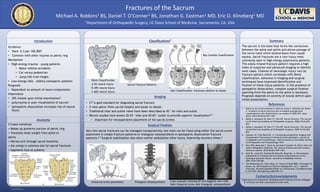
Classifications3
The sacrum is the bone that forms the connection
between the spine and pelvis and allows passage of
the nerve roots which extend down from cauda
equina. Sacral fractures are a rare injury most
commonly seen in high energy polytrauma patients.
This easily missed fracture pattern requires a high
index of suspicion and advanced imaging to identify
most cases. Chances of neurologic injury vary by
fracture pattern which correlates with Denis
classification. Advances in imaging and surgical
techniques have improved identification and
fixation of these injury patterns. In the presence of
spinopelvic dissociation, complex surgical fixation
spanning from the pelvis to the spine is necessary.
Prognosis depends on severity of neural deficit upon
initial presentation.
1. Bydon M, De la Garza-Ramos R, Macki M, Desai A, Gokaslan AK, Bydon
A. Incidence of sacral fractures and in-hospital postoperative
complications in the United States: an analysis of 2002-2011 data.
Spine. 2014;39(18):E1103-1109.
2. Mehta S, Auerbach JD, Born CT, Chin KR. Sacral fractures. The Journal
of the American Academy of Orthopaedic Surgeons. 2006;14(12):656-
665.
3. Mehta S, Auerbach JD, Born CT, Chin KR. Sacral fractures. The Journal
of the American Academy of Orthopaedic Surgeons. 2006;14(12):656-
665
4. Eastman JG, Chip Routt ML, Jr. Correlating preoperative imaging with
intraoperative fluoroscopy in iliosacral screw placement. Journal of
orthopaedics and traumatology : official journal of the Italian Society
of Orthopaedics and Traumatology. 2015
5. Ricci WM, Mamczak C, Tynan M, Streubel P, Gardner M. Pelvic inlet and
outlet radiographs redefined. The Journal of bone and joint surgery.
American volume. 2010;92(10):1947-1953.
6. Nork SE, Jones CB, Harding SP
, Mirza SK, Routt ML, Jr. Percutaneous
stabilization of U-shaped sacral fractures using iliosacral screws:
technique and early results. Journal of orthopaedic trauma.
2001;15(4):238-246.
7. Tim Pohlemann UC. Pelvic Ring. In: Thomas P Ruedi REB, Christopher G
Moran, ed. AO Principles of Fracture Management. Vol 2 - Specific
Fractures. Second Expanded Edition ed. Switzerland, Clavadelerstrasse
8, CH-7207: AO Pubishing; 2007:697-717
Thank you to my mentors Dr. Klineberg and Dr Eastman and my colleague
D. O’Conner for their contributions to this work.
Denis Classification
A-6% neural injury
B-28% neural injury
C-60% neural injury
Sacral Fracture Patterns
Roy-Camille Classification
Isler Classification- fractures relative to facets
Iliosacral screw placement Triangular Osteosynthesis Case example showing AP and Saggital view with
both iliosacral screw and triangular osteosynthesis