
Recommended
More Related Content
Similar to meckels diverticulum.pptx
Similar to meckels diverticulum.pptx (20)
Recently uploaded
Recently uploaded (20)
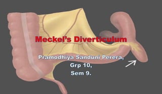
meckels diverticulum.pptx
- 2. Contents • Intro • Embryology • Epidemiology • Clinical presentation • Differential diagnosis • Diagnosis • Treatment • References
- 3. Introduction • Meckel's diverticulum is the most common congenital anomaly of the gastrointestinal tract. • It results from incomplete obliteration of the vitelline duct leading to the formation of a true diverticulum of the small intestine
- 5. Embryology • It arises from the antimesenteric surface of the middle-to-distal ileum. • The diverticulum represents a persistent remnant of the omphalomesenteric duct, which connects the midgut to the yolk sac in the fetus. • The omphalomesenteric duct normally involutes between the fifth and sixth weeks of human gestation as the bowel settles into its permanent position within the abdominal cavity. • The persistence of the omphalomesenteric duct beyond fetal development may result in a variety of anatomic patterns • The rich blood supply to the diverticulum is provided by the vitelline artery, which is a branch of the superior mesenteric artery
- 7. Epidemiology • United States data • The prevalence of Meckel diverticulum is usually noted to be approximately 2% of the population • International data • Europe and Asia have reported prevalence figures similar to those found in the United States. • In a large series of cases from 2007 to 2008, Meckel diverticulectomy was 2.3 times more common in boys, and boys accounted for 74% of the primary cases.
- 8. Clinical Presentation • The three most common symptomatic presentations are – gastrointestinal (GI) bleeding, – intestinal obstruction, and – acute inflammation of the diverticulum + intussusception.
- 9. Bleeding • When a severe bleeding episode occurs, the patient can present in hemorrhagic shock. Tachycardia is an early clinical sign of hemorrhagic shock, but pale conjunctivae and orthostatic hypotension may actually precede this.
- 10. Obstruction • Most patients with intestinal obstruction present with abdominal pain, bilious vomiting, generalized abdominal tenderness, distention, hypoactive or hyperactive bowel sounds, peritoneal signs, and rebound tenderness upon examination. Patients may develop a palpable abdominal mass. Occasionally, when patients do not present early or if the diagnosis is missed, the obstruction can progress to intestinal ischemia or infarction; the latter manifests with acute peritoneal signs and lower GI bleeding.
- 11. Inflammation • Patients with diverticulitis present with either focal or diffuse abdominal tenderness. Usually, abdominal tenderness is more marked in the periumbilical region than that from the pain of appendicitis. Children may present with abdominal guarding and rebound tenderness, in addition to abdominal tenderness. Abdominal distention and hypoactive bowel sounds are late findings. Suppurative Meckel diverticulum can present in a child with abdominal pain and periumbilical cellulitis.
- 12. Ddx Colitis Colonic Vascular Malformations Emergent Treatment of Gastroenteritis Gastrointestinal Duplications IgA Vasculitis (Henoch-Schonlein Purpura) Intestinal Polyposis Syndromes Intussusception Juvenile Polyps Necrotizing Enterocolitis Imaging Pediatric Appendicitis Pediatric Constipation Pediatric Crohn Disease Pediatric Hirschsprung Disease Pediatric Urolithiasis Peptic Ulcer Disease Peutz-Jeghers Syndrome Postoperative Adhesions Ulcerative Colitis in Children Volvulus
- 13. Diagnosis Key diagnostic factors • age <2 years • passage of bright red blood per rectum (haematochezia) • intractable constipation (obstipation) Other diagnostic factors • male sex • nausea and vomiting • abdominal cramps • lower abdominal pain • Rectal bleeding (painless) • Bloody, mucus-y stools • Co-morbid with intussusception
- 14. 1st investigations to order • FBC • technetium-99m pertechnetate scan ('Meckel's scan')- A radio-nucleotide scan called a Meckel’s scan is the most effective diagnostic test, it looks for the presence of ectopic gastric mucosa and enlarged bowel using a gamma camera for the best picture • CT scan of the abdomen and pelvis • ultrasound of the abdomen Investigations to consider • contrast enema • mesenteric angiography • endoscopic exploration of the small intestine • surgical exploration of the abdomen
- 16. Treatment • Meckel diverticulectomy If you have open surgery: • Your surgeon will make a large surgical cut in your belly to open up the area. • Your surgeon will look at the small intestine in the area where the pouch or diverticulum is located. • Your surgeon will remove the diverticulum from the wall of your intestine. • Sometimes, the surgeon may need to remove a small part of your intestine along with the diverticulum. If this is done, the open ends of your intestine will be sewn or stapled back together. This procedure is called an anastomosis. In surgery using a laparoscope: • Three to five small cuts are made in your belly. The camera and other small tools will be inserted through these cuts. • Your surgeon may also make a cut that is 2 to 3 inches (5 to 7.6 cm) long to put a hand through, if needed. • Your belly will be filled with gas to allow the surgeon to see the area and perform the surgery with more room to work. • The diverticulum is operated on as described above
- 18. Indications for surgery • Painless bleeding • Because initial bleeding from a Meckel diverticulum can be massive, it is essential that the patient be adequately resuscitated. This may require the transfusion of packed red blood cells (RBCs) to return the hematocrit level to approximately 30%. Two large-bore intravenous (IV) lines must be in place in case bleeding recurs, and crossmatched blood should be available. • Bowel obstruction • Patients present with bowel obstruction due to volvulus, intussusception, a mesodiverticular band, or incarceration of the Meckel diverticulum in a hernia (though the presence of a Meckel diverticulum in a hernia does not actually increase the risk of incarceration). • Meckel diverticulitis • The presentation of Meckel diverticulitis may be indistinguishable from that of appendicitis. As with appendicitis, the course is progressive and may result in perforation, diffuse peritoneal contamination, and septic shock. Exploration is usually performed for suspected appendicitis; an inflamed Meckel diverticulum must be sought if a normal-appearing appendix is discovered. • Umbilical drainage • Drainage of succus entericus or feculent material indicates a persistent connection between the intestine and the umbilicus. Ultrasonography (US) or contrast studies may be used to confirm the diagnosis. An exploration can then be performed to resect the fistula.
- 19. Contraindications for surgery • Because many of the operations for omphalomesenteric remnants are used in emergency situations, surgery has relatively few contraindications. • However, patients must be adequately prepared for surgery, even given short notice. • In patients who are bleeding, the blood volume must be returned to acceptable levels, and adequate IV access must be obtained. • In patients with bowel obstruction and repeated emesis, electrolyte abnormalities must be corrected while hydration is restored.
- 20. WHAT DO YOU DO BEFORE SURGERY? Provide blood transfusions to correct hypovolemia. Administer IV fluid and electrolyte replacement Provide oxygen as prescribed. Administer IV antibiotics. NPO after midnight. Maintain bed rest. Closely monitor blood loss in stools. HOW ABOUT AFTER SURGERY? Assess respiratory status and maintain airway. Provide supplemental oxygen as prescribed. Obtain vital signs. Administer analgesics for pain as prescribed. Assess surgical site for bleeding or any other abnormalities. Assess bowel sounds and bowel function. Administer IV fluids and antibiotics as prescribed. Maintain NPO status. Maintain NG tube to low continuous suction.
- 22. References • https://www.uptodate.com/contents/meckels- diverticulum • https://emedicine.medscape.com/article/931229- overview?icd=login_success_email_match_norm • https://bestpractice.bmj.com/topics/en-gb/804 • https://www.studocu.com/en-us/document/regis- college/maternal-newborn/meckels- diverticulum-fact-sheet/20409336