
Recommended
Recommended
More Related Content
What's hot
What's hot (12)
Similar to Transcranial Photobiomodulation Dosimetry: Integrating Human Data and Penetration Models
Similar to Transcranial Photobiomodulation Dosimetry: Integrating Human Data and Penetration Models (20)
Recently uploaded
Recently uploaded (20)
Transcranial Photobiomodulation Dosimetry: Integrating Human Data and Penetration Models
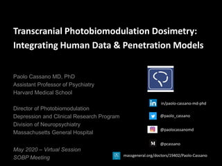
- 1. Transcranial Photobiomodulation Dosimetry: Integrating Human Data & Penetration Models May 2020 – Virtual Session SOBP Meeting Paolo Cassano MD, PhD Assistant Professor of Psychiatry Harvard Medical School Director of Photobiomodulation Depression and Clinical Research Program Division of Neuropsychiatry Massachusetts General Hospital @paolo_cassano @paolocassanomd @pcassano in/paolo-cassano-md-phd massgeneral.org/doctors/19402/Paolo-Cassano
- 2. Disclosures Niraxx Light Therapeutics Inc Advisory Board Board Member Equity Royalties LiteCure LLC Contracted Research Cerebral Sciences Inc Contracted Research Mass General Hospital – Patents No Applicable Royalties Janssen Advisory Board
- 3. Salehpour et al. 2018 Molecular Neurobiology
- 4. Salehpour et al. 2018 Molecular Neurobiology
- 7. Sharma et al. 2011 Lasers Surg Med Dose - ATP Response of PBM on Primary Cortical Neurons
- 8. Sharma et al. 2011 Lasers Surg Med Dose - Mitochondrial Membrane Potential Response of PBM on Primary Cortical Neurons
- 9. Yuan et al. 2020 Neurophotonics Simulations (MCX Model): Target Brain Regions
- 10. Simulations (MCX Model): Position of Light Sources F3/F4 position Fpz position dlPFC vmPFC Distance Yuan et al. 2020 Neurophotonics
- 11. Clinical Research Sample for Testing of Predictions Wavelength: 830 nm CW/PW: CW Irradiance (mW/cm2): 33 - 55 Fluence (J/cm2): 60 - 65 Time (min): 20 - 30 Window (cm2): 57 - 36 Total energy (kJ/session): 3.4 - 2.3 * Overall energy shed on participants’ head DOSIMETRY in MGH PBM HUMAN STUDIES * Cassano et al. 2018 - Photomed Laser Surg. Iosifescu et al. 2020 SOBP Meeting - Virtual Poster Session Spera & Sitnikova et al. https://www.biorxiv.org/content/10.1101/837591v1
- 12. Simulated Change: DOSE (Total Energy) Cassano & Tran et al. 2019 Neurophotonics
- 13. Tested with NIR-Flow Data N=10 (paired comparisons) Change: DOSE Stable: Age: young adult Condition: healthy Site: Fp1 Fp2 (F3 F4) Target: vmPFC (dlPFC)
- 14. Eyes Closed Resting State EEG: Spectogram Differences Lowering Dose (1/3) Same Sample (HS) CW: EEG effect PW: No effect Spera & Sitnikova et al. https://www.biorxiv.org/content/10.1101/837591v1
- 15. Simulated Change: AGE (Extra-Cerebral Tissue - Thickness) Yuan et al. 2020 Neurophotonics
- 16. Energy Deposition and Extra-Cerebral Tissues’ Thickness Yuan et al. 2020 Neurophotonics
- 17. Tested with NIR-Flow Data & ELATED-3 Data NIR-Flow (n=10 Healthy) ELATED-3 (n=49 MDD) Change: AGE (healthy vs. MDD) Stable: Site: Fp1 Fp2 (F3 F4) Target: vmPFC (dlPFC)
- 18. Eyes Closed Resting State EEG: Spectogram Differences Spera & Sitnikova et al. https://www.biorxiv.org/content/10.1101/837591v1
- 19. QIDS-C: Depression Scores in Overall Sample NIR – NIR Sham – NIR Sham - Sham SPCD pooled p=ns t-test n=49 phase I n=37 phase II n=36 completers 8 9 10 11 12 13 14 15 16 17 y0 y1 y3 y5 y7 y9 y11 y13 y15 y17 y19 y21 y23 y25 Visit Number QIDS-C Mean Score rx 1 rx 2 rx 3 Iosifescu et al. 2020 SOBP Meeting - Virtual Poster Session
- 20. Yuan et al. 2020 Neurophotonics Simulated Change: PROBE SITE (Target: dlPFC to vmPFC)
- 21. Simulated Change: DOSE (Total Energy) Cassano & Tran et al. 2019 Neurophotonics
- 22. Tested with ELATED-2 Data & ELATED-3 Data ELATED-2 (n=21 MDD) ELATED-3 (n=49 MDD) Change: DOSE & PROBE SITE Stable: Age: 45-50 Condition: MDD
- 23. ELATED-2: Depression Scores in Overall Sample . (Mann Whitney U-test – endpoint carried forward) p=.047 Cassano et al. 2018 - Photomed Laser Surg.
- 24. QIDS-C: Depression Scores in Overall Sample NIR – NIR Sham – NIR Sham - Sham SPCD pooled p=ns t-test n=49 phase I n=37 phase II n=36 completers 8 9 10 11 12 13 14 15 16 17 y0 y1 y3 y5 y7 y9 y11 y13 y15 y17 y19 y21 y23 y25 Visit Number QIDS-C Mean Score rx 1 rx 2 rx 3 Iosifescu et al. 2020 SOBP Meeting - Virtual Poster Session
- 26. MGH Depression Program North Eastern University Qianqian Fang Anh Phong Tran Yaoshen Yuan Nathan Kline Institute, NY Dan Iosifescu LiteCure LLC. Luis De Taboada Mass General Hospital Depression Program Sam Petrie Garrett Thomas Richard Norton David Mischoulon Cris Cusin Maurizio Fava Andy Nierenberg Anxiety Program Meredith Ward Eric Bui Tatiana Sitnikova Mari Franceschini Michael Hamblin Husam Katnani Mass Eye and Ear. Benjamin Bleier University of Pisa Vincenza Spera Marco Maiello
- 27. MGH Division of Neuropsychiatry
Editor's Notes
- “Of note, I am a co-founder of a company in the field of light therapy, however not an employee; none of the data of my presentation is from any device of this company”
- “We derive energy from NIR and visible (red) light, see the cellular membrane in the picture and the red light penetrating up to the mitochondria, which are the powerhouse of the cells”. “Even blue and green have biological properties when interfacing with our cellular membranes; but they are less likely to penetrate”. Cellular Effects of Red and NIR light Increases ATP synthesis Modulates ROS production Release of NO Increases mitochondrial membrane potential Increases Ca2+ release Activates transcription factors and signaling mediators such as NF-κB Studies have shown that 532 nm green laser can increase ATP levels and cell proliferation in vitro, likely through the modulation of the activity of the mitochondrial complex III (cytochromes b, c1, and c) (Fukuzaki et al., 2013). Irradiation with a 532 nm laser has also been shown to promote the migration of GABAergic neural stem/progenitor cells into deeper layers of the mouse neocortex (Fukuzaki et al., 2015). Moreover, it has been suggested that 420 nm blue light could effectively increase ATP synthesis, likely through the regulation of the mitochondria complex I (NADH-dehydrogenase) (Karu, 1988).
- “We derive energy from NIR and visible (red) light; see now the mitochondrial membrane and the cytochrome C oxidase; the primary photo-acceptor for the biological effects of NIR” Mechanism of Red and NIR in Mitochondria (A) Flow of electrons through the mitochondrial respiratory chain; PBM stimulates cytochrome c oxidase, improves its catalytic activity, and elevates ATP synthesis (B) Structure of cytochrome c oxidase and electrons path through its subunits PBM dissociates nitric oxide from cytochrome c oxidase, allowing oxygen to return, and facilitates electron transfer and increases proton gradient
- Sharma et al. Lasers Surg Med, 2011 https://www.ncbi.nlm.nih.gov/pmc/articles/PMC3199299/ Effect of 810-nm laser on intracellular ATP in the cultured cortical neurons. Quantification by luminescence plate reader of the relative light unit values per mg cell protein from Cell Titer Glo assay from nine wells. Error bars are SD. *P < 0.05 versus control. #P < 0.05 versus 3 J/cm2. Abstract Background and Objectives In the past four decades numerous studies have reported the efficacy of low level light (laser) therapy (LLLT) as a treatment for diverse diseases and injuries. Recent studies have shown that LLLT can biomodulate processes in the central nervous system and has been extensively studied as a stroke treatment. However there is still a lack of knowledge on the effects of LLLT at the cellular level in neurons. The present study aimed to study the effect of 810 nm laser on several cellular processes in primary cortical neurons cultured from embryonic mouse brains. Study Design/Materials and Methods Neurons were irradiated with fluences of 0.03, 0.3, 3, 10, or 30 J/cm2 of 810-nm laser delivered over varying times at 25 mW/cm2 and intracellular levels of reactive oxygen species (ROS), nitric oxide and calcium were measured using fluorescent probes within 5 minutes of the end of irradiation. The changes in mitochondrial function in response to light were studied in terms of adenosine triphosphate (ATP) and mitochondrial membrane potential (MMP). Results Light induced a significant increase in calcium, ATP and MMP at lower fluences and a decrease at higher fluences. ROS was significantly induced at low fluences, followed by a decrease and a second larger increase at 30 J/cm2. Nitric oxide levels showed a similar pattern of a double peak but values were less significant compared to ROS. Conclusions The results suggest that LLLT at lower fluences is capable of inducing mediators of cell signaling processes which in turn may be responsible for the beneficial stimulatory effects of the low level laser. At higher fluences beneficial mediators are reduced and high levels of Janus-type mediators such as ROS and NO (beneficial at low concentrations and harmful at high concentrations) may be responsible for the damaging effects of high-fluence light and the overall biphasic dose response. Keywords: low level laser therapy, LLLT, photobiomodulation, cultured cortical neurons, near-infra red laser, reactive oxygen species, nitric oxide, mitochondrial membrane potential, intracellular calcium, ATP, biphasic dose response
- Sharma et al. Lasers Surg Med, 2011 https://www.ncbi.nlm.nih.gov/pmc/articles/PMC3199299/ Effect of 810-nm laser on mitochondrial membrane potential in the cultured cortical neurons. A: JC1 non-aggregated (green), JC1 aggregated (red) and nuclear Hoechst (blue) fluorescence in control neurons. B: JC1 non-aggregated, JC1 aggregated and nuclear Hoechst fluorescence in neurons treated with 3 J/cm2 810-nm laser. Scale bar is 50 µm. C: Quantification by fluorescence plate reader of the mean red/green fluorescence ratio values from nine wells. Error bars are SD. *P < 0.05; **P < 0.05 versus control. ##P < 0.01; ###P < 0.001 versus 3 J/cm2. Abstract Background and Objectives In the past four decades numerous studies have reported the efficacy of low level light (laser) therapy (LLLT) as a treatment for diverse diseases and injuries. Recent studies have shown that LLLT can biomodulate processes in the central nervous system and has been extensively studied as a stroke treatment. However there is still a lack of knowledge on the effects of LLLT at the cellular level in neurons. The present study aimed to study the effect of 810 nm laser on several cellular processes in primary cortical neurons cultured from embryonic mouse brains. Study Design/Materials and Methods Neurons were irradiated with fluences of 0.03, 0.3, 3, 10, or 30 J/cm2 of 810-nm laser delivered over varying times at 25 mW/cm2 and intracellular levels of reactive oxygen species (ROS), nitric oxide and calcium were measured using fluorescent probes within 5 minutes of the end of irradiation. The changes in mitochondrial function in response to light were studied in terms of adenosine triphosphate (ATP) and mitochondrial membrane potential (MMP). Results Light induced a significant increase in calcium, ATP and MMP at lower fluences and a decrease at higher fluences. ROS was significantly induced at low fluences, followed by a decrease and a second larger increase at 30 J/cm2. Nitric oxide levels showed a similar pattern of a double peak but values were less significant compared to ROS. Conclusions The results suggest that LLLT at lower fluences is capable of inducing mediators of cell signaling processes which in turn may be responsible for the beneficial stimulatory effects of the low level laser. At higher fluences beneficial mediators are reduced and high levels of Janus-type mediators such as ROS and NO (beneficial at low concentrations and harmful at high concentrations) may be responsible for the damaging effects of high-fluence light and the overall biphasic dose response. Keywords: low level laser therapy, LLLT, photobiomodulation, cultured cortical neurons, near-infra red laser, reactive oxygen species, nitric oxide, mitochondrial membrane potential, intracellular calcium, ATP, biphasic dose response
- “We then teamed up with Prof. Qianqian Fang at Northeastern University in Boston to answer these questions through computerized simulations of NIR penetration. Here are the areas of interest (dlPFC in green and vmPFC in red; in blue the frontal poles); (CLICK) on the right the positions of the light sources F3, F4, Fp1, Fp2)”.
- We want to understand what causes the energy decrease over age. So, we measure ECT thickness X axis, y axis, dot size
- ELATED-2 MDD (n=21) Double-blind, randomized, parallel t-NIR vs. sham Sessions 2 x week for 8 weeks 8-week comparison ELATED-3 MDD (n=38/ SPCD) Double-blind, randomized, SPCD t-NIR vs. sham Sessions 2 x week for 12 weeks 6-week comparison Instrument: Omnilux New U (LED) TPBM-1000 (LED) Photomedex (1 W) x2 Litecure (~2 W) Sites: F3, F4 (Fp1, Fp2) F3, F4 (Fp1, Fp2) Target: dlPFC (bilateral) dlPFC & frontal poles (bilateral) Total energy (kJ/study): 45.6 28.3
- “growth and aging were correlated with progressively wider distance of the target areas from the light source; this is likely a challenge in the treatment of the elderly”
- “we found that dlPFC could be reached with sufficient energy deposition (based on effective tissue deposition in animal models); less likely to be effective was the deposition on vmPFC; also the energy deposition was inversely correlated with the thickness of extracerebral tissues; the greater the distance from the source the lower was the energy deposition on the brain target region”
- “we found that dlPFC could be reached with sufficient energy deposition (based on effective tissue deposition in animal models); less likely to be effective was the deposition on vmPFC; also the energy deposition was inversely correlated with the thickness of extracerebral tissues; the greater the distance from the source the lower was the energy deposition on the brain target region”
- Three analyses were conducted; two out of three indicated significant results despite the small sample sizes This was the baseline carried forward, which was significant for the change in severity of depression