Comparative Cytotoxic Activities of the Flavonoid-Rich Ethyl Acetate Fruit Ex...
Katie Mathewson Poster 2014.pptx
1. The Development of Folate-Targeted
Photodynamic Therapy Agents
Katherine Mathewson, Kyle Sullivan, RoJenia Jones, and K.W. Olsen
Department of Chemistry and Biochemistry Loyola University Chicago
Abstract
The goal of the project is to develop new photodynamic therapy
(PDT) agents that specifically target the rapidly dividing cells
found in cancers. PDT utilizes light to excite a photosensitizer to
produce reactive singlet oxygen species to kill the cancer cells.
There is an over-expression of folate receptors in many cancer
cells. Our proposed agent will show a double selectivity in that it
will specifically target cells that over- express folate receptors
and it will have limited-area light exposure. The proposed
research will test these hypotheses: (1) that using hemoglobin in
the PDT complex can increase singlet oxygen production by
bring additional oxygen into the cells, and (2) that HeLa cervical
cancer cell can be used to determine the effectiveness of the
PDT agents. To do this, both folate-hemoglobin-chlorin and
folate-albumin- chlorin covalent complexes will be made. The
two complexes will be tested in hypoxic HeLa cell cultures to
determine if the presence of the extra oxygen brought in by the
hemoglobin increases the compound ability to kill the cancer
cells.
Background
Photodynamic therapy (PDT) relies on oxidative damage to
cause cell death. Light excites a photosensitizer, which then
reacts with oxygen which results in the formation of a highly
reactive oxygen species, singlet oxygen. This reactive species
in turn reacts with cellular macromolecules to induce cell
death. This process requires the PDT agent to actively select
for cancer cells and enter them. Currently, mainstream
procedures rely on physical differences such as cellular pH to
differentiate between normal and cancer cells. Without an
increase in specificity, this can lead to damage in healthy cells.
In many tumor cells, the folate receptor is highly over
expressed. By using folate-photosensitizer conjugates there
would be a potential decrease in toxic side-effects to normal
cells because of its preferential uptake by cancer cells.
Future Work
• Continued development of conjugates
• More cell culture studies test
phototoxicity
References
C. Y. Ke, C. J. Mathias, M. A. Green (2003) The folate receptor
as a molecular target for tumor-selective radionuclide delivery.
Nuc. Med. & Biology 30: 811-817.
J. Khadem, A.A. Veloso Jr., F. Tolentino, T Hassan, M.R.
Hamblin (1999) Photodynamic Tissue Adhesion with Chlorin e6
Protein Conjugates. IOVS, Vol. 40, No.13: 3132-3137.
Acknowledgements
• Dr. Kenneth Olsen for directing and
advising this project.
• Doctorate candidate RoJenia Jones for
teaching and guiding me
• Kyle Sullivan for all his work on this
project
• Loyola University Chicago for supporting
this research
FA-XLHb-Ce6
Trial # of FA # of
Ce6
1 3 3
2 2 3
3 3 3
Quantification of Folate & Chlorin e6
Extraction Method
The conjugates were extracted using cold 2% acid/acetone
mixture to quantify the amount of folates and chlorin e6
molecules covalently bound to the protein. Under these harsh
conditions, the heme dissociates from the globin, and the
globin precipitates along with any covalently bound
molecules.
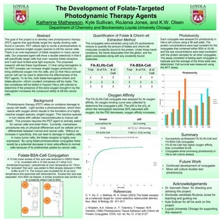
Oxygen Affinity
The FA-XLHb-Ce6 conjugate was assayed for its oxygen
affinity. An oxygen binding curve was collected to
determine the conjugate’s p50. The p50 is the pO2 at
which the hemoglobin becomes 50% saturated with
oxygen. As the P50 decreases, oxygen affinity increases.
Summary
• Successfully synthesized FA-XLHb-Ce6 and
FA-BSA-Ce6 conjugates
• FA-XLHb-Ce6 has higher oxygen affinity
than unmodified XLHb
• Conjugates show promising phototoxicity in
HeLa cell culture assays
Folate-BSA-Ce6 Conjugation
A 10 fold molar excess of folic acid was dissolved in DMSO:Water
(1:4), incubated with a 10 fold excess of 1-ethyl-3-(3-
dimethylaminopropyl) carbodiimide at room temperature for 15min.
The “activated” folic acid was added to BSA already dialyzed in PBS
buffer at pH 7.4. The mixture was incubated for 4h at room
temperature and quenched with ethanolamine. Excess folic acid was
separated from BSA via dialysis. A similar procedure was carried out
to covalently attach Ce6 to BSA.
FA-BSA-Ce6
Trial # of FA # of
Ce6
1 3 3
2 3 3
3 3 3
Sample p50
Unmodified Hb 26.5 mmHg
XLHb 10.72 (+/-.245)mmHg
FA-XLHb-Ce6 7.98 (+/-.215) mmHg
Phototoxicity
Each conjugate was assayed for phototoxicity in
HeLa cell culture using a 96 well plate. The
protein concentrations were kept constant for the
conjugates that contained either BSA or XLHb
and the dye concentrations were kept consistent
with the protein samples for all other conjugates.
The assay consisted of testing each conjugate in
triplicate and the average of the three wells was
determined. Cell survival was measured using
the MTT assay.
%Survival