1. 0
20
40
60
80
100
120
140
160
180
200
50 51
1
50
114
65
16
39
93
46
43
19
4
77
59
23
13
106
152
2
54
184
102
16
53
187
92
110
26
3
128
64
27
30
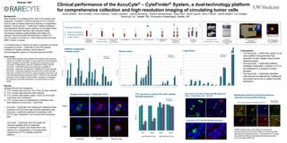
Clinical performance of the AccuCyte® – CyteFinder® System, a dual-technology platform
for comprehensive collection and high resolution imaging of circulating tumor cells
Jackie Stilwell1, Nick Drovetto1, Arturo Ramirez1, Daniel Campton1, Joshua Nordberg1, Paulina Varshavskaya1, Alisa Clein2, Steve Quarre1, Barry Friemel1, Daniel Sabath2, Eric Kaldjian1
1RareCyte, Inc., Seattle, WA; 2University of Washington, Seattle, WA
Laboratory Workflow
A. Density enrichment
B. Automated staining
C. Image analysis
A
C
B A B
Abstract 1601
Background
High numbers of circulating tumor cells (CTC) predict poor
prognosis. In addition, serial monitoring of CTC numbers
may be used to assess response to therapy or progressing
disease. The AccuCyte® – CyteFinder® system combines
comprehensive density-based collection of nucleated blood
cells with automated staining, high-resolution digital
microscopic imaging, image analysis and single-cell
retrieval. Previous studies demonstrated a model CTC
(mCTC) spike-in recovery of > 90% using various cell lines
(BMC Cancer, in press).
In a blinded study of clinical and spike-in samples we directly
compared AccuCyte - CyteFinder to the FDA-cleared
CellSearch® System (Veridex), which relies on
immunomagnetic capture of cells that express EpCAM.
Study Design
• Paired blood samples were obtained from patients with advanced
cancer (17 prostate, 24 breast, 12 lung) under the Fred Hutchinson
Cancer Research Center IRB. Samples were processed by the
University of Washington using the CellSearch® assay and the
AccuCyte - CyteFinder assay was performed in parallel by
RareCyte. RareCyte was blinded to CellSearch® counts until after
AccuCyte - CyteFinder analysis was performed and documented.
• Recovery of model CTCs (mCTCs) was compared using tumor cell
lines with high EpCAM expression (MCF7, LNCaP) or low EpCAM
expression (PC3, A549) in paired spike-in samples to investigate
the influence of EpCAM expression in the performance of each
assay.
Results
Samples fell into four categories:
1) CTC number was very low (<5) or zero by both methods
2) CTC number was similar by both methods
3) CTC number was notably higher (>50%) by AccuCyte -
CyteFinder than CellSearch
4) CTC number were not detectable by CellSearch and
were detected by AccuCyte - CyteFinder
• AccuCyte - CyteFinder and CellSearch® detected similar
numbers of mCTCs from high-EpCAM expressing cells
• AccuCyte - CyteFinder detected considerably more
mCTCs than CellSearch® from low-EpCAM expressing
cells
Platform comparison:
Prostate cancer
CTCs/7.5mL
CTC recovery in model CTCs with variable
EpCAM expression
0
10
20
30
40
50
60
70
80
90
100
%Recovery
RareCyte
CellSearch
PC3 A549 MCF7 LnCAP
Breast cancer
0
10
20
30
40
50
60
70
80
90
100
1 2
4
0 0 0 0 0 0
58
0
283
0
17
13
15
5
8
3
0
28
56
7
680
0
100
200
300
400
500
600
700
800
900
0 5 0 1 0
24
391
0
135
195
273
565
886
0 0 9 5
37 39
112
3 2
216
01 10 0 3 2
17
603
4
231 231
317
740
812
6 0 10 11
104
51
132
4 2
275
5
Lung Cancer
CTCs/7.5mL
CTCs/7.5mL
Images of AccuCyte – CyteFinder CTCs
RareCyte
CellSearch
Lung cancer CTC with low EpCAM expression
Cytokeratin EpCAM Merged image
RareCyte
CellSearch
CellSearch
RareCyte
Breast
Lung
PC3 model CTCs with variable EpCAM expression
(high – arrowheads, low – arrows)
AccuCyte – CyteFinder also has single cell
retrieval capability with its integrated
CytePicker® module. See Poster #30 in this
session for a presentation of next generation
sequencing of CTCs isolated using this
platform.
Low EpCAM cell lines High EpCAM cell lines
Conclusions
• The AccuCyte – CyteFinder system is an
open platform that generates high
resolution 6-color images using custom
antibody panels.
• The AccuCyte – CyteFinder platform
identifies comparable numbers of CTCs
as CellSearch in a subset of cancer
samples.
• The AccuCyte – CyteFinder identifies
cells that are not detected by CellSearch
most likely due to low or absent EpCAM
expression.
Cytokeratin - AF647
Her2 - AF488
EGFR - QD605
CD45 - BV421
Nucleus
Sytox-Orange
EpCAM - QD800
(Cy7Filter) Vimentin - QD605
CD15 - AF488
Multiplexed staining using 6 fluorescence
channels and sequential staining
SKBR3 breast cancer cells spiked into blood and
processed by AccuCyte were imaged on CyteFinder after
staining with 6 fluorescent dyes. A second round of 2 dyes
was performed after an automated de-staining process.
Arrow indicates SKBR cell.
Second round
in 2 channelsFirst round of staining in 6 fluorescence channels
Cytokeratin EpCAM
Cytokeratin EpCAM