Gowtham's 2nd ca cervix anatomy
•Download as PPTX, PDF•
2 likes•109 views

Basic of Anatomy - ca cervix

Recommended
More Related Content
What's hot
What's hot (20)
Similar to Gowtham's 2nd ca cervix anatomy
Similar to Gowtham's 2nd ca cervix anatomy (20)
More from Gowtham Manimaran
Recently uploaded
Recently uploaded (20)
Gowtham's 2nd ca cervix anatomy
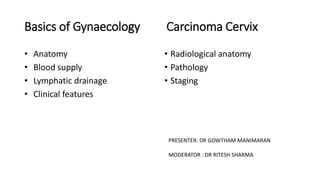
- 1. Basics of Gynaecology Carcinoma Cervix • Anatomy • Blood supply • Lymphatic drainage • Clinical features • Radiological anatomy • Pathology • Staging PRESENTER: DR GOWTHAM MANIMARAN MODERATOR : DR RITESH SHARMA
- 2. Anatomyof female genitaltract • Externalgenitalia/Vulva • Internalgenitalia
- 3. Mons Pubis External genital organs: • Mons pubis • Labia Majora • Labia Minora • Clitoris • Vestibule • Vestibular bulb • The greater vestibular glands
- 4. • Bloodsupply – Arteries • Br.of internal pudendalartery(br of In.iliac artery) – LabialA., – TransverseperinealA., – A to vestibularbulb, – deep& dorsalarteries toclitoris • Br.of FemoralA – Superficial& deepexternalpudendalA – Veins • Internal ,external pudendalvein • Vesical/vaginalvenousplexuses • UltimatelyintoLongsaphenousvein
- 5. • Nervesupply – Ant-suppart • Ilioinguinal& genitalbr.of genitofemoralN(L1,L2) – Post-inf part • Pudendalbr.of post.cut. Nof thigh(S123) – Inbetween • Labial& perinealbr.of PudendalN(S234) • Lymphatics – Superf.Inguinalnodes – Glandof Cloquet(deepinguinalnode) – Int. Iliacnodes – Rectallymphaticplexus
- 6. Vagina ovries Internal genital organs: Vagina Uterus Fallopian tubes Ovaries
- 7. Vagina : • Walls – Ant : 7.5 cm – Post : 9cm – 2 lateral walls • Fornices : – Ant : shallow – 2 lateral – Post : deep
- 8. • BloodSupply – Arteries( from internal iliac artery) • Cervicovaginalbr.Ofuterine A • VaginalA • Middle rectalA • Internalpudendal • UterineandvaginalbranchesAnastomose -2azygosarteries/median longitudinal vessels – Veinsdrain into: • Vaginal veins(from lateral plexus)Internal iliacV • Internal pudendal V • Lymphaticsdrain into • Upper1/3rd : internal iliacnodesandexternaliliacgroup • Middle 1/3rd: internal iliacnodes • Lower1/3rd (below hymen): superficial inguinalgp. • Nervesupply • Parasympathetic:S234 • Sympathetic: hypogastricplexus • Lowerend : pudendal N(sensory)
- 9. Uterus : • Hollow, pyriform muscular organ in pelvis • Position: Anteversion and Anteflexion • Measurements: 7.5 cm long 5 cm wide 3 cm thick • Parts: Body Isthmus cervix
- 10. – Anterior • Above int.os : uterovesical pouch • Below int.os: separated from UB by loose areolar tissue – Posterior • Pouch of Douglas with coils on intestine – Lateral • Broad ligament • Mackenrodt’s ligament • Uterine A &ureter Relations of Uterus
- 11. Ligaments of uterus • Uterosacral ligament • Transverse cervical/ Mackenrodt's ligament • Pubovesicocervical lig • Round ligament • Broad ligament o Mesovarium o Mesosalpinx o Mesometrium • Suspensory lig of ovary/ infundibulopelvic ligament
- 13. • Blood supply – Arteries: • Uterine A • Ovarian & Vaginal As. – Veins drain into • Internal iliac veins • Nerves – Sympathetic • Motor : T5 & T6 • Sensory : T10 – L1 – Parasympathetic •Pelvic N ( S2,3,4) : both motor & sensory : ends in ganglia of Frankenhauser
- 15. Fallopian tubes : • 10-14 cm • Lies within the superior border of broad ligament • 2 openings – Medially into cornua – Laterally into abdominal cavity
- 16. • BloodSupply – Arteries • UterineA • OvarianA – Veins • Throughpampiniforn plexusinto ovarianveins • Lymphatics • Para-aorticnodes • Nervesupply – Uterine & ovariannerves
- 17. The Ovary • Paired, situated on either side of uterus • Close to lateral pelvic wall • In ovarian fossa of Waldeyer • Size: 4*3*2 cm • Only intra-abdominal structure not covered by peritoneum
- 18. Cortex: • Lined single layer of germinal epithelium of Waldeyer (cuboidal epi.) • Tunica albuginea : stromal cells thickened beneath germinal epithelium • Contain primordial follicles • Corpus albicans/ atretic follicles Medulla: • Loose connective tissues, blood vessels,nerves, muscles • Hilus cells : homologous to interstitial cells of testes
- 19. • Blood Supply – Arterial : Ovarian A – Veins : • Through pampiniform plexus ovarian V Lt. Renal Vein IVC • Lymphatics • Para-aortic nodes Nerve supply : • Sympathetic supply from T10 along ovarian A
- 20. Lymphatics of Female genital tract
- 22. Innervation Of Female genital tract
- 23. Clinical Features of Gynaecologic malignancies • Cervical CA: 1. MC during routine gynecologic(before symptoms appear) cytologic smears Pap smear, colposcopy and biopsies, and HPV testing 2. Visible lesions present with an exophytic mass or a barrel-shaped cervix because of an endocervical lesion 3. Metrorrhagia (intermenstrual bleeding), menorrhagia (heavier menstrual flow), or postcoital bleedingsymptoms related to anemia 4. Advanced diseasebowel obstruction, renal failure, foulsmelling serosanguinous or yellowish vaginal discharge, pelvic pain, flank and/or leg pain, rectal bleeding, obstipation, dysuria, hematuria, or persistent edema of lower extremities because of lymphatic/venous blockade by pelvic sidewall disease may occur. 5. Pain in the pelvis or hypogastrium may be caused by tumor necrosis or associated pelvic inflammatory disease. 6. Pain in the lumbosacral area the possibility of PALN involvement with extension into the lumbosacral roots or hydronephrosis should be considered.
- 24. • Endometrial CA: 1. The most common presentation for endometrial cancer is postmenopausal vaginal bleeding, 80%-90% • Ovarian CA: 1. Ovarian cancer has insidious growth and is asymptomatic in early-stage disease. Vague gastrointestinal complaints of dyspepsia, nausea, early satiety, bloating, constipation, or obstipation are common presenting symptoms, as are genitourinary symptoms including frequency, urgency, or incontinence. Other ill-defined symptoms include fatigue, back pain, pain with intercourse, and menstrual irregularities 2. Germ cell and stromal cell malignancies abdominal discomfort or symptoms of excessive estrogen or androgen production. 3. Granulosa cell tumorsprecocious puberty. 4. Sertoli-Leydig cellvirilization. • Fallopian Tube CA: 1. Two Triads (a)pelvic pain,pelvic mass, and leukorrhea and (b) vaginal bleeding, vaginal discharge, and lower abdominal pain. (11%) 2. Hydrops Tubae Profluensis a sudden emptying of accumulated fluid in the distended fallopian tube that causes profuse, watery, serosanguineous vaginal discharge.(9%)
- 25. • Vaginal CA: Rare malignancy, constituting 1% to 2% of all gynecologic malignancies. • Urethral CA: 1. Irritative or obstructive urethral symptoms. Bleeding (hematuria) or spotting is the prevailing presenting sign in 50% to 60% of patients. 2. 30% to 50% of patients experience pain or irritative symptoms, difficulty urinating, and frequent micturition.Urinary retention and overflow incontinence may occur in advanced cases. 3. 10% to 20% patients, dyspareunia, perineal pain, and inguinal lymphadenopathy. Urethrovaginal and vesicovaginal fistulas may develop in advanced, neglected cases. • Vulval CA: 1. Vulvar pruritus, bleeding, pain, discharge.
- 26. CT images
- 38. MRI of uterus and vagina • On T2-weighted magnetic resonance imaging (MRI), the uterus displays a zonal anatomy, with three distinct zones: the endometrium, junctional zone and myometrium. • The endometrium and uterine cavity appear as a high-signal stripe; the thickness varies with the stage of the menstrual cycle. In the early proliferative phase, it measures up to 5 mm, and widens to up to 1 cm in the mid-secretory phase. • A band of low signal, the junctional zone, borders the endometrium. It represents the inner myometrium and is of constant thickness and signal throughout the menstrual cycle; it usually measures 5 mm. • The outer myometrium is of medium-signal intensity in the proliferative phase, and of high-signal intensity in the mid- secretory phase as a result of the increased vascularity and prominence of the arcuate vessels.