
Recommended
More Related Content
More from Cultura Arte Sociedad (Cuarso)
More from Cultura Arte Sociedad (Cuarso) (20)
Recently uploaded
Recently uploaded (20)
Supporting information: Kaolin Alleviates Graphene Oxide Toxicity
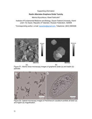
- 1. S1 Supporting information Kaolin Alleviates Graphene Oxide Toxicity Marina Kryuchkova, Rawil Fakhrullin* Institute of Fundamental Medicine and Biology, Kazan Federal University, Kreml uramı 18, Kazan, Republic of Tatarstan, Russian Federation, 420008 *Corresponding author, e-mail: kazanbio@gmail.com, Telephone: (843) 5905506 (a) (b) Figure S1. Atomic force microscopy images of graphene oxide (a) and kaolin (b) particles Figure S2. Optical microscopy images of Paramecium caudatum protists at lower (a) and higher (b) magnification.
- 2. S2 Figure S3. Investigation of chemotaxis in P. caudatum. The distribution of P. caudatum cells in media and nanoparticle-doped droplets within 1 hour after inoculation: a) pure graphene oxide; b) – graphene oxide with added kaolin (at equal concentrations) Figure S4. Optical microscopy images of Congo Red-stained food vacuoles in P. caudatum: a) – intact cell; b) – graphene oxide at 100 µg mL-1 ; c) – graphene oxide with kaolin at 100 µg mL-1 Figure S5. Galvanotaxis assay: remediation of anodic aggregation of P. caudatum cells (disrupted by graphene oxide) by kaolin (at 1 mg mL-1 ).