
Recommended
More Related Content
What's hot
What's hot (20)
Similar to Diagnostic Outcomes for Women with Postcoital Bleeding
Similar to Diagnostic Outcomes for Women with Postcoital Bleeding (20)
More from AymanEwies
More from AymanEwies (20)
Recently uploaded
Recently uploaded (20)
Diagnostic Outcomes for Women with Postcoital Bleeding
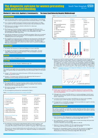
- 1. The diagnostic outcome for women presenting with postcoital bleeding x Postcoital bleeding (PCB) is defined as bleeding occurring during or immediately after sexual intercourse at a time separated from menstruation (Fraser et al, 1996). x It is a common multifactorial gynaecological symptom (Rosenthal et al, 2001) and could be the first presentation of cervical cancer (Jha and Sabharwal, 2002). x PCB alone is not currently an absolute indication for colposcopy (Jha and Sabharwal, 2002). x The frequency of cervical cancer in women presenting with PCB varies from 3.8 to 5.5% (Rosenthal et al, 2001; Rajaram et al, 1998, respectively) and cervical intraepithelial neoplasia (CIN) from 17% to 17.8% (Rosenthal et al, 2001; Jha and Sabharwal, 2002 respectively). x Currently the management of women with PCB is inconsistent. There are no standard NHS guidelines to ensure good practice. The investigations offered depend on individual preferences and expertise. x The Guidelines for Suspected Cancer published by the Department of Health (DoH, April 2000) in the United Kingdom set criteria for referral only. Urgent referral (within 2 weeks) for PCB more than four weeks in women > 35 years of age and early referral (within 4-6 weeks) in any other cases of repeated unexplained PCB was recommended. 1. To measure the frequency of abnormal findings in women with PCB. 2. To compare the frequency of abnormal findings in women aged > 35 years with PCB for more than 4 weeks with those ≤ 35 years aiming to assess the validity of DoH referral guidelines. x The majority (89.3%) of women in group 1 had a routine referral by their general practitioners, while the majority (94.1%) in group 2 were referred directly from the cytology department. x All women in group 2 and only 39% of group 1 were referred to the colposcopy clinic. x The investigations performed are summarised in Table 1. Table 1: Investigations performed for women with PCB Group 1 Group 2 Cervical Check-up: 99 (59.6) 118 (100%) 1. Cervical smear 82 (49.4%) 59 (50%) 2. Colposcopy 84 (50.6%) 109 (92.4%) 3. Cervical biopsy 26 (15.6%) 52 (44%) 4. LLETZ 15 (9%) 52 (44%) Infection Screen: 50 (30.1%) 10 (8.5%) 1. Chlamydial Swab 44 (26.5%) 7 (6%) 2. High Vaginal Swab 39 (23.5%) 2 (1.7%) 3. Endocervical Swab 10 (23.5%) 2 (1.7%) Endometrial Check-up: 69 (41.6%) 2(1.7%) 1.Hysteroscopy 59 (35.5%) 1 (0.8%) 2. Endometrial Biopsy 36 (21.7%) 1 (0.8%) 3. Ultrasound Scanning 25 (15%) 0 x No pathology was detected in approximately half of women in group 1 and 17% of group 2. Nevertheless, 6 cases of cervical cancer were diagnosed in each group. The incidence of CIN was 9% and 66.1% in group 1 and 2, respectively. There were no cases of endometrial cancer in this series. Out of 51 women tested for Chlamydia, 4 (7.8%) had a positive test. Diagnostic findings are summarised in Table 2 and Figure 1 Group 1 Group 2 Cervical Cancer 6 (3.6%) 6 (5%) CIN 15 (9%) 78 (66.1%) Infection/Inflammation 16 (9.6%) 6 (5%) 1. Chlamydia 4 (2.4%) 0 2. Bacterial vaginosis 2 (1.2%) 0 3. HPV 0 3 (2.5%) 4. Chronic cervicitis 10 (6%) 3 (2.5%) Cervical Polyp 13 (7.8%) 3 (2.5%) Ectropion 38 (23%) 6 (5%) DUB 18 (22.9%) 0 NAD 81 (48.8%) 20 (17%) x Seventy two (43.4%) women in group 1 were more than 35 years of age and had PCB for more than 4 weeks. Figure 2 shows a comparison between diagnostic findings in these 72 women versus the 94 women aged ≤ 35 years in group 1. The incidence of benign cervical polyp was statistically significantly higher (p= 0.005) and that of ectropion was statistically significantly lower (p=0.0002) in women older than 35 years of age. There was no significant difference in the frequency of cervical cancer or CIN between the two subgroups. x Neither age nor duration of PCB is a reliable indication for serious pathology, and our data, therefore, does not support the distinction within the two week rule referral guidelines. x Although most women with PCB will have no serious pathology, cervical cancer and CIN occurred in 3.6% and 9% of women referred with PCB (and normal last smear), respectively. x A normal cervical smear in women with PCB does not rule out the possibility of cervical cancer or CIN. Therefore, prompt referral is indicated. x It is imperative to standardise assessment and care providers are urged to develop guidelines for the management of PCB. Khattab A.F., Ewies A.A.A., Appleby D., Cruickshank D.J. The James Cook University Hospital, Middlesbrough BACKGROUND OBJECTIVES Retrospective review of hospital case notes. DESIGN SETTING The James Cook University Hospital, Middlesbrough, UK. POPULATION Two hundred and eighty four non-pregnant, non-hysterectomised women with PCB, seen in the gynaecology and colposcopy clinics from first January 1996 to 31 December 2003. x Group-1: 166 women were referred because of PCB with normal last smear (within the previous 3 years). x Group-2: 118 women were referred to the colposcopy clinic because of abnormal cervical cytology associated with PCB. RESULTS Table 2: Diagnostic findings in women with PCB Figure 1: Diagnostic findings in women with PCB CONCLUSION REFERENCE 1. Fraser IS, Petrucco MO. Management of Intermenstrual and Postcoital Bleeding. Aust.and N.Z. Journal of Obstetrics and Gynaecology 1996; 36: 67-73. 2. Jha S, Sabharwal S. outcome of colposcopy in women presenting with postcoital bleeding and negative or no cytology. Journal of Obstetrics and Gynaecology 2002; 22: 299-301 3. Rajaram S, Suneja A, Mahishee AN. How alarming is postcoital bleeding. Gynecology and Obstetric investigation 1998; 45: 205-208. 4. Rosenthal AN, Panoskaltsis T, Smith T, Soutter WP. The frequency of significant pathology in women attending a general gynaecological service for PCB. British journal of Obstetrics and Gynaecology 2001 108; 103-106 ACKNOWLEDGEMENT The authors would like to thank Mrs. Caroline Marshall and Mrs. Sharon Poulter, clinical audit facilitators, The James Cook University Hospital for their help in collecting and processing the data. under 35 over 35 Figure 2: Comparison between the diagnostic findings in women >35 years of age and those <35 years *= Statistically significant 60 50 40 30 20 10 0 Cancer CINInfection Polyp*Ectropion* D U B N A D Group 1 Group 2 Cancer CINInfection Polyp Ectropion D U B N A D 90 80 70 60 50 40 30 20 10 0