Mystus catapogon, a new catfish (Siluriformes: Bagridae) species from Kerala,...
SICB Poster
1. Functional morphology of cephalic protuberances
in the oyster toadfish, Opsanus tau
AN Marranzino 1,7 MM Frank 2,7, SD Lindemann 3,7, BA Guiffrida 4,7, K Sipper 5,7, JF Webb 6,7 and AF Mensinger 3,7
1Regis University, Denver, CO; 2St. Olaf College, Northfield, MN; 3University of Minnesota, Duluth; 4Wareham Middle School, MA; 5Northern Michigan University, Marquette, MI;
6University of Rhode Island; 7Marine Biological Laboratory, Woods Hole, MA
Morphology of Cirri A B
Conclusions
Introduction
• Superficial neuromasts were found between pairs of papillae. The
hair cell orientation in neuromasts was perpendicular to the axis
between the papillae. This is the same as the hair cell orientation
relative to the canal axis found in canal neuromasts of other
teleosts. The relative lack of canal neuromasts in the cephalic region
of O. tau may be due to the benthic nature of the fish, which could
cause canals neuromasts to become clogged with sediment. Papillae
may allow superficial neuromasts to function as canal neuromasts
(responding to water accelerations) by channeling the water
through the papillae perpendicular to the axis of the papillae, while
shielding neuromasts from sedimentation.
• There appears to be a positive correlation between fish size and the
surface area of the cirri. Multiple lobes on the cirri are also more
pronounced in larger cirri, indicating that cirri may grow and
develop throughout the fish’s life.
• The sensory cells found on the cirri are similar in appearance to
other extra-oral taste buds in teleosts with characteristic microvillar
projections as well as bulbiform cell shape. The cells found appear
to be possible type II taste buds, consistent with the hypothesis that
cirri function in gustatory detection.
Acknowledgements
The oyster toadfish, Opsanus tau, is a demersal fish species indigenous
to the inshore waters of the Eastern United States. It is commonly found
in the waters near Woods Hole, MA. Its large size and robust physiology
has made it a model organism for biomedical studies of muscle and
sensory physiology at the Marine Biological Laboratory since 1888.
Despite >100 years of study, the function of these prominent anatomical
features on the head remain unknown. These include large, fleshy,
multi-lobed protuberances, called cirri, which project from the mandible
and above the eye, and smaller paired projections, termed papillae,
which sit on either side of superficial neuromasts.
We would like to give special thanks to the following people for their aid in this
research: Dr. Paul Forlano (Brooklyn College and MBL), Dr. Joe Sisneros (University
of Washington and MBL), Louis Kerr (MBL), Ryan Stephansky (MBL), Dan Keeley
(MBL).
This research was funded by NSF DBI-1005378 (Biological Discovery in Woods Hole
REU Program) to AFM.
Purpose
The purpose of this study was to examine the functional morphology of
the papillae and cirri. It was hypothesized that the papillae serve to
channel water over the superficial neuromasts and allow them to
function as canal neuromasts. It was also hypothesized that the sensory
cells located on the cirri function in gustatory detection.
Toadfish were obtained from the Marine Resources Center at the Marine
Biological Laboratory (Woods Hole). All experiments conformed to
institutional animal care protocols. Toadfish (13.5 to 28.5 cm) were split
into three size groups and digital images of cirri were taken using a Zeiss
SteREO Discovery z12 dissecting scope to calculate surface area. Cirri were
numbered consecutively starting at the mandibular symphysis progressing
along the length of the mandible. For histological examination, cirri were
transected from additional adult toadfish that were transcardially perfused
with 3% paraformaldehyde and 1% glutaraldehyde in 0.1 M PBS and placed
overnight in the fixative solution. Alternatively, cirri were removed from
toadfish (prior to fixation) and pinned in a Sylgard lined petri dish in 3.7%
formalin in seawater at 4°C. Tissue was cryo-protected by immersion in a
20% sucrose solution in 0.1% PBS and then sectioned (10-25 μm
increment) with a Zeiss cryostat. Sectioned tissue was kept in PBS until
staining with a standard hematoxylin and eosin protocol.The surface of the
cirri and the paired papillae with superficial neuromasts were examined
using scanning electron microscopy. Cirri and sections of skin containing
papillae and superficial neuromasts flanked by papillae from the dorsal
portion of the head, infraorbital, pre operculum, and the trunk were
dissected. Skin with papillae/neuromasts were pinned to expose the
neuromasts. Cirri and skin tissue was placed in a 0.1% solution of S-carboxymethyl-
L-cysteine in PBS for 7 minutes with agitation to remove
mucus. Cirri and skin samples were then fixed in cold 3.7% formalin in PBS
for at least 2 hours. Tissue was dehydrated in an ascending ethanol series
(with some were sonicated for 30 seconds to remove the cupulae from
neuromasts) and critical point dried. Tissue was mounted on stubs;
papillae were pushed open to reveal superficial neuromasts. Samples were
sputter coated with 15 nm of platinum and imaged at 3KV with a Zeiss NTS
Supra 40VP SEM.
Results
The lateral line system is a mechanosensory system found in all fishes. It
is composed of a series of neuromasts, which are comprised of hair cells
that sense water motion. Neuromasts can either be enclosed in canals
or sit superficially on the surface of the skin. O. tau has relatively few
canal neuromasts on the head, and the numerous superficial
neuromasts in this region are flanked by paired papillae. These papillae
are not common in other fish and their function is unknown.
Methods
Results
B
C D
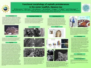
Figure 1. A - Paired papillae closed over a superficial neuromast. B- Superficial
neuromast where papillae have been separated to reveal underlying neuromast
(solid red arrow indicates hair cell orientation and dashed red arrow indicates
papillae axis). C and D - Superficial neuromast with sensory strip of hair cells
visible (white arrow indicates hair cell orientation). E and F – Superficial
neuromasts showing line hair cells where cupula has been removed. The longer
structures are kinocilia and the shorter are stereocilia of the superfical
neuromast. Hair cell orientation is perpendicular to the axis between the
papillae. Solid red arrow indicates hair cell orientation.
A
C D
Figure 2. A - ventral view of a small O. tau with an enlargement showing
submandibular cirri (left). B - Surface area of mandibular cirri versus fish length (n= 18;
error bars = 1 SD). Cirri are numbered consecutively from caudal to rostral. Images on
right show mandibular cirri from a large (upper) and a small (lower) fish. C - SEM
images of Type I (upper image) and Type II (lower image) taste buds in the
oropharyngeal cavity of rainbow trout, Salmo gairdenari (from Ezeasor, 1982) D - SEM
image of sensory cell found on a cirrus of O. tau.
F
F
References
Ezeasor D. (1982) Distribution and ultrastructure of taste buds in the
oropharyngeal cavity of the rainbow trout, Salmo gairdenari Richardson. J. Fish
Biol. 20: 53-68.
Hara TJ. (2006) Gustation. Fish Physiology. 25: 45-96
The multi-lobed cirri in O. tau have been hypothesized to serve as
camouflage and/or function in either mechanosensory or gustatory
detection. While camouflage cannot be ruled out by this study, no
neuromasts were found on the surface of the cirri, indicating that they
do not function as a part of the lateral line system. Other sensory cells
were found on the cirri which appear to be taste buds. Fish are known
to possess a variety of extra-oral taste buds on various parts of their
bodies including lips, skin, and barbels. Three distinct types of taste
buds have been distinguished among fishes. All three are characterized
by a bulbiform shape and microvillar projections.
Figure 3. A - Schematic drawing of a taste bud (TB) typical for teleosts.
Dark cells (Cd) with microvilli, light cells (Cl) with a single rodlike
protrusion, and basal cells (Cb) constitute a pear‐ or onion‐shaped TB,
which sits on a dermal papilla (DP). Marginal cells (Cm), not belonging to
the TB proper, form the interface between TB and the epithelial cells
(Ce). The TB nerve (TBN) reaches into the TB to form the nerve fiber
plexus (NFP). BL, basal lamina; RA, receptive area; and VC, capillary
vessel. (from Hara, 2006). B – Cross section of cirrus showing putative
taste bud on cirrus of O. tau.
A
E
B
Morphology of Papillae