
Recommended
More Related Content
Similar to FACIAL NERVE.pptx
Similar to FACIAL NERVE.pptx (20)
Recently uploaded
Recently uploaded (20)
FACIAL NERVE.pptx
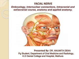
- 1. FACIAL NERVE Embryology, Internuclear connections, Intracranial and extracranial course, anatomy and applied anatomy. Presented By- DR. ANUMITA DEKA Pg Student, Department of Oral Medicine and Radiology, K.D Dental College and Hospital, Mathura
- 2. Introduction: The facial nerve is one of a group of nerves called the cranial nerves, twelve pairs of nerves that, with the exception of the spinal accessory nerve, originate in the brain and contribute to the peripheral nervous system. They are referred to as “ cranial” because they arise from the brain and upper spinal cord and supply structures of the head and neck. The 12 paired cranial nerves include: * the olfactory nerve * the optic nerve * the occulomotor nerve * the trochlear nerve * the trigeminal nerve * the abducens nerve * the facial nerve * the vestibulocochlear nerve * the glossopharyngeal nerve * the vagus nerve * the accessory nerve * the hypoglossal nerve
- 3. Organization of the cranial nerves: The olfactory, optic and vestibulocochlear nerves are entirely sensory. The oculomotor, trochlear, abducent, accessory and hypoglossal nerves are entirely motor. The trigeminal, facial, glossopharyngeal and vagus nerves are both sensory and motor nerves. The cranial nerves have central motor and/ or sensory nuclei within the brain and peripheral nerve fibres that emerge from the brain and exit from the skull to reach their effector or sensory organs. The functions of the cranial nerves are summarized shortly as follows:
- 5. The facial nerve The facial nerve ( also known as the VII cranial nerve) is one of the twelve cranial nerves. It is predominantly a motor nerve. However, sensory and vegetative ( parasympathetic) functions are also involved in its complex role. Facial expressions as the result of activity of craniofacial muscles, contraction of the muscles derived from the second pharyngeal arch such as stapedius, platysma, stylohyoid and posterior belly of digastric, and secretory participation through general visceral efferent fibres ( salivary, lacrimal, nasal and palatal glands) are some examples that illustrate the physiological importance of the VII nerve.
- 6. Facial nerve embryology *In the 3rd week of intrauterine development appears the facio- acoustic primordium, this structure originates from the rhombencephalon and develops in the rostral sense to the otic placode. The facial nerve becomes the nerve of the second branchial arch, and for this reason it will supply all the elements that derive from it; the stapes, the styloid process of the temporal bone, the stylohyoid ligament, the minor horn and the upper portion from the body of the hyoid, also to the muscles of the stapes, stylohyoid, posterior belly of the digastric, auricular and facial expression.
- 7. * In the fourth week of embryogenesis the first branch of the facial nerve appears in the rostral aspect of the embryo. By the fifth week, the intermediate nerve becomes evident and two weeks later will be recognized as an independent nerve. *Towards the seventh week the tympanic cord will be derived from the first branch of the facial nerve, joining elements of the first branchial arch and the lingual nerve (branch of the V part). By the middle of this week, the trunk of the facial nerve, which is already formed, bifurcates into the temporofacial and cervicofacial branches. Around the end of this week different fascicles that originate from these branches are recognized clearly. The temporal, zygomatic, oral, mandibular and cervical regions will give origin to the five main (terminal branches) of the facial nerve.
- 8. * In the eighth week of pregnancy the cartilaginous capsule from the previously formed ear vesicle forms a groove around the facial nerve, the stapedial artery and the stapes muscle. This groove will become the facial canal. By week 12 all the muscles of the face are formed, and all are innervated by one of the branches of the facial nerve, if the innervation is not present, the affected muscle fibers will sustain an adipose involution, a finding described in the Moebius syndrome, in which there is agenesis or destruction of the nuclei of cranial nerves VI and VII.
- 9. * In the twenty-first week, the ossification of the facial canal begins, which will only end when the collateral branches of the facial nerve are found in their definitive location and when the artery has been reversed,4 in many cases, the ossification ends in the early childhood. *With respect to the trajectory of the facial nerve during intrauterine life, literature reports that initially the nerve presents itself as a rectilinear structure. However, by the sixth week, due to the mesencephalic growth, there is a change of direction in acute angle, which will become the knee of the facial nerve.
- 10. * Towards the fourth month of formation, a second change of direction appears, secondary to the development of the tympanic cavity, which originates from the first branchial pouch; this second tier will be the elbow of the seventh pair. *Towards the end of pregnancy, the tympanic bone and the mastoid process are not fully developed, so the petrous portion of the facial nerve does not exist and will not be formed until between 2 and 4 years of age.
- 11. Facial Nerve Nuclei The facial nerve has three nuclei: (1) the main motor nucleus,(2) the parasympathetic nuclei, and (3) the sensory nucleus. Main Motor Nucleus The main motor nucleus lies deep in the reticular formation of the lower part of the pons. The part of the nucleus that supplies the muscles of the upper part of the face receives corticonuclear fibers from both cerebral hemispheres. The part of the nucleus that supplies the muscles of the lower part of the face receives only corticonuclear fibers from the opposite cerebral hemisphere. These pathways explain the voluntary control of facial muscles. However, another involuntary pathway exists; it is separate and controls mimetic or emotional changes in facial expression. This other pathway forms part of the reticular formation.
- 12. Parasympathetic Nuclei Parasympathetic nuclei lie posterolateral to the main motor nucleus. They are the superior salivatory and lacrimal nuclei. The superior salivatory nucleus receives afferent fibers from the hypothalamus through the descending autonomic pathways. Information concerning taste also is received from the nucleus of the solitary tract from the mouth cavity. The lacrimal nucleus receives afferent fibers from the hypothalamus for emotional responses and from the sensory nuclei of the trigeminal nerve for reflex lacrimation secondary to irritation of the cornea or conjunctiva.
- 13. Sensory Nucleus The sensory nucleus is the upper part of the nucleus of the tractus solitarius and lies close to the motor nucleus. Sensations of taste travel through the peripheral axons of nerve cells situated in the geniculate ganglion on the seventh cranial nerve. The central processes of these cells synapse on nerve cells in the nucleus. Efferent fibers cross the median plane and ascend to the ventral posterior medial nucleus of the opposite thalamus and to a number of hypothalamic nuclei. From the thalamus, the axons of the thalamic cells pass through the internal capsule and corona radiata to end in the taste area of the cortex in the lower part of the postcentral gyrus.
- 15. Course of the Facial Nerve *The facial nerve consists of a motor and a sensory root. The fibers of the motor root first travel posteriorly around the medial side of the abducent nucleus. They then pass around the nucleus beneath the colliculus facialis in the floor of the fourth ventricle and, finally pass anteriorly to emerge from the brainstem. *The sensory root (nervus intermedius) is formed of the central processes of the unipolar cells of the geniculate ganglion. It also contains the efferent preganglionic parasympathetic fibers from the parasympathetic nuclei. The two roots of the facial nerve emerge from the anterior surface of the brain between the pons and the medulla oblongata.They pass laterally in the posterior cranial fossa with the vestibulocochlear nerve and enter the internal acoustic meatus in the petrous part of the temporal bone.
- 16. At the bottom of the meatus, the nerve enters the facial canal and runs laterally through the inner ear. On reaching the medial wall of the tympanic cavity, the nerve expands to form the sensory geniculate ganglion and turns sharply backward above the promontory. At the posterior wall of the tympanic cavity, the facial nerve turns downward on the medial side of the aditus of the mastoid antrum, descends behind the pyramid, and emerges from the stylomastoid foramen.
- 17. The course of the facial nerve is very complex. There are many branches, which transmit a combination of sensory, motor and parasympathetic fibers. Anatomically, the course of the facial nerve can be divided into two parts: Intracranial – the course of the nerve through the cranial cavity, and the cranium itself. Extracranial – the course of the nerve outside the cranium, through the face and neck. Intracranial The nerve arises in the pons, an area of the brainstem. It begins as two roots; a large motor root, and a small sensory root (the part of the facial nerve that arises from the sensory root is sometimes known as the intermediate nerve). The two roots travel through the internal acoustic meatus, a 1cm long opening in the petrous part of the temporal bone. Here, they are in very close proximity to the inner ear. Still within the temporal bone, the roots leave the internal acoustic meatus, and enter into the facial canal. The canal is a ‘Z’ shaped structure.
- 18. Within the facial canal, three important events occur: Firstly the two roots fuse to form the facial nerve. Next, the nerve forms the geniculate ganglion (a ganglion is a collection of nerve cell bodies). Lastly, the nerve gives rise to: Greater petrosal nerve – parasympathetic fibers to mucous glands and lacrimal gland. Nerve to stapedius – motor fibers to stapedius muscle of the middle ear. Chorda tympani – special sensory fibers to the anterior 2/3 tongue and parasympathetic fibers to the submandibular and sublingual glands. The facial nerve then exits the facial canal (and the cranium) via the stylomastoid foramen. This is an exit located just posterior to the styloid process of the temporal bone.
- 19. Branches of facial nerve
- 20. Extracranial After exiting the skull, the facial nerve turns superiorly to run just anterior to the outer ear. The first extracranial branch to arise is the posterior auricular nerve. It provides motor innervation to the some of the muscles around the ear. Immediately distal to this, motor branches are sent to the posterior belly of the digastric muscle and to the stylohyoid muscle. The main trunk of the nerve, now termed the motor root of the facial nerve, continues anteriorly and inferiorly into the parotid gland (note – the facial nerve does not contribute towards the innervation of the parotid gland, which is innervated by the glossopharyngeal nerve).
- 21. Within the parotid gland, the nerve terminates by splitting into five branches: *Temporal branch- innervating the frontalis and orbicularis oris muscles and the muscles in the upper part of the face. *Zygomatic branch- innervating the middle part of the face. *Buccal branch- innervating the cheek muscles, including the buccinator muscle. *Marginal mandibular branch- innervating muscles of the lower part of the face. *Cervical branch- innervating the muscles below the chin and, among others, the platysma muscle. These branches are responsible for innervating the muscles of facial expression.
- 22. Terminal branches of the facial nerve:
- 23. Functions: Motor Functions Branches of the facial nerve are responsible for innervating many of the muscles of the head and neck. All these muscles are derivatives of the second pharyngeal arch. The first motor branch arises within the facial canal; the nerve to stapedius. The nerve passes through the pyramidal eminence to supply the stapedius muscle in the middle ear. Between the stylomastoid foramen, and the parotid gland, three more motor branches are given off: Posterior auricular nerve – Ascends in front of the mastoid process, and innervates the intrinsic and extrinsic muscles of the outer ear. It also supplies the occipital part of the occipitofrontalis muscle. Nerve to the posterior belly of the digastric muscle – Innervates the posterior belly of the digastric muscle (a suprahyoid muscle of the neck). It is responsible for raising the hyoid bone. Nerve to the stylohyoid muscle – Innervates the stylohyoid muscle (a suprahyoid muscle of the neck). It is responsible for raising the hyoid bone.
- 24. Special Sensory Functions The chorda tympani branch of the facial nerve is responsible for innervating the anterior 2/3 of the tongue with the special sense of taste. The nerve arises in the facial canal, and travels across the bones of the middle ear, exiting via the petrotympanic fissure, and entering the infratemporal fossa. Here, the chorda tympani ‘hitchhikes’ with the lingual nerve. The parasympathetic fibres of the chorda tympani stay with the lingual nerve, but the main body of the nerve leaves to innervate the anterior 2/3 of the tongue.
- 25. Parasympathetic Functions The parasympathetic fibres of the facial nerve are carried by the greater petrosal and chorda tympani branches. Greater Petrosal Nerve The greater petrosal nerve arises immediately distal to the geniculate ganglion within the facial canal. It then moves in anteromedial direction, exiting the temporal bone into the middle cranial fossa. From here, its travels across (but not through) the foramen lacerum, combining with the deep petrosal nerve to form the nerve of the pterygoid canal. The nerve of pterygoid canal then passes through the pterygoid canal (Vidian canal) to enter the pterygopalatine fossa, and synapses with the pterygopalatine ganglion. Branches from this ganglion then go on to provide parasympathetic innervation to the mucous glands of the oral cavity, nose and pharynx, and the lacrimal gland.
- 26. Chorda Tympani The chorda tympani also carries some parasympathetic fibres. These combine with the lingual nerve (a branch of the trigeminal nerve) in the infratemporal fossa and form the submandibular ganglion. Branches from this ganglion travel to the submandibular and sublingual salivary glands.
- 27. Image demonstrating branches of the facial nerve:
- 28. Applied Anatomy of the facial nerve: Facial paralysis, often unilateral, may be due to: (1) Supranuclear lesions in the cortico nuclear fibers from the frontal lobe, variably combined with numerous other descending fibers converging in the facial nucleus. (2) Nuclear or infranuclear lesions involving lower motor neurons.
- 29. Supranuclear facial paralysis involving “upper motor neuron” pathways is usually a part of hemiplegia. Movements in the lower part of the face are usually more severely affected, voluntary movements being weak or absent though emotional expression is little affected. Electrical reaction of affected muscles are unaltered. Occasionally supranuclear lesions may abolish or weaken emotional movements but not voluntary movements. This dissociation shows that the supranuclear control of expressive movements is separate from the corticonuclear path for voluntary movements.
- 30. Nuclear or infranuclear lesions vary in their effects according to the lesion’s site. If the nucleus or facial pontine fibres are involved, neighboring structures are inevitably also involved. Facial muscles are represented in cell groups within the nucleus; their degree of involvement governs the extent of paralysis, which is ipsilateral. Lesions due to adjacent damage include paralysis of the lateral rectus because of involvement of the abducent nucleus around which the facial nerve loops.
- 31. Bilateral facial paralysis are caused by (1) Bilateral infranuclear lesion’s : • Acute idiopathic polyneuritis • Leprosy • Leukemia • Syphilitic or meningococcal meningitis • Double otitis media • Rheumatic • Post diphtheritic • Bell’s Palsy • Uveoparotid paralysis (2) Muscle diseases: • Myasthenia gravis/ myotonic dystrophy • Facio-scapulo humeral dystrophy Signs: Flattening of all normal folds, sagging of corners of mouth, fixed expression less mask like face, no voluntary movements of facial muscles. White of eye seen when patient attempts to close them. Patient talks as if he had severe stomatitis.
- 32. BELL’S PALSY Is an acute apparently isolated facial palsy for which no cause can be found. Aetiology: (a) Associated known clinical condition – Diabetes Mellitus, severe hypertension in last trimester of pregnancy, dental anesthesia. (b) Causes – (i) Exposure to cold, oedema and subsequent compression of the nerve trunk within rigid fallopian canal causes circulatory disturbance, (ii) other important causes of acute facial palsy include suppurative otitis media, herpes zoster, head injury, Guillian-Barre syndrome, sarcoidosis and multiple sclerosis.
- 34. Symptoms: sudden following exposure to chill or without any apparent precipitating cause, maximum paralysis in twenty-four hours. Post auricular pain is common and many precede paralysis by two days. There may be spontaneous loss of sense of taste, hyperacusis and watering of eyes. Sweating is less on affected side. Signs: Forehead cannot be wrinkled, frowning lost, eyes cannot be closed. On attempting closure eye ball turns upwards and outwards, known as Bell’s phenomenon. On showing teeth, the lips do not separate on affected side. Whistling not possible. Articulation of labial components difficult. Nasolabial fold flattened out. Angle of affected side droops with dribbling of saliva. Cheeks puffs out with expiration because of buccinator paralysis. Food collects between teeth and paralysed cheek. Fluid runs out while drinking. Base of tongue lowered.Vesicles within the external auditory meatus and ear drum in Ramsay Hunt Syndrome. Pain may precede facial weakness. Deafness may result. Investigations: Electromyography. Management: Local heat. Local treatment of muscles. Protection of eye
- 35. REFERENCES: * Snell- Neuroanatomy- 7th- Edition, Chapter 11 “ The Cranial Nerve Nuclei and Their Central Connections and Distribution.” * Anatomy, descriptive and surgical by Henry Gray, H.V. Carter. * Danilo Alejandro Garcia, “ Facial nerve: embryology and anatomy of its nucleus.”; Mini Review Article Med Crave; MOJ Anatomy & Physiology. 2018; 5(3): 164- 166. * Terence M. Myckatyn, M.D. And Susan E. Mackinnon, M.D. “ A review of Facial Nerve Anatomy.” Seminars in Plastic Surgery/ Volume 18, Number 1 2004. * Liam Curry, “ The Facial Nerve ( CN VII).”; August 16, 2020, 66th Revision.
- 36. THANK YOU