
Recommended
More Related Content
Similar to ADRENAL DISORDER.pptx
Similar to ADRENAL DISORDER.pptx (20)
Recently uploaded
Recently uploaded (20)
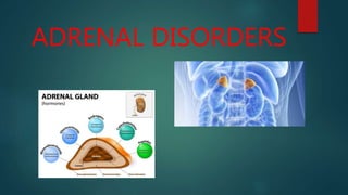
ADRENAL DISORDER.pptx
- 2. ADDISON’S DISEASE : Primary hypoadrenalism There is Destruction of entire adrenal cortex. Glucocorticoids, Mineralocorticoid and sex steroid pdtn are all reduced. Reduced cortisol → feedback→ Increased CRH and ACTH production. Rare disorder Incidence : 3-4/million per year Prevalence : 40-60/million Marked female preponderance
- 3. CAUSES Autoimmune disease(autoimmune adrenalitis) Tuberculosis Surgical removal Hemorrhage/infarction Meningococcal septicaemia Venography Infiltration Malignant destruction Amyloid Schilders disease(adrenal leukodystrophy)
- 4. CLINICAL FEATURES SYMPTOMS SIGNS Weight loss Malaise Weakness Fever Anorexia Nausea Diarrhea Abdominal pain Constipation Depression Confusion Myalgia Joint/back pain Impotence/amenorrhea sycope Loss of weight General wasting Pigmentation(specially new scars and palmar creases) Buccal pigmentation Postural hypotension Loss of body hair Dehydration
- 5. INVESTIGATIONS Single cortisol measurements random cortisol below 100mmol/L during day-highly suggestive Short ACTH Stimulation test absent or impaired cortisol response confirms the presence of hypoadrenalism. 9:00 hrs. plasma ACTH level high level with low or low normal cortisol confirm primary hypoadrenalism electrolytes and urea show hyponatremia, hyperkalemia and high urea. Blood glucose – low Adrenal antibodies –present Chest and abdominal X-rays –show evidence of TB and calcification Plasma renin activity – high ←high aldosterone hypercalcemia and anaemia
- 6. MANAGEMENT ACUTE CASES LONG TERM Assuming normal CVS Activity • 1L of 0.9% saline given over 30-60 min with 100mg of i.v. bolus hydrocortisone. • Subsq req. of several litres of saline within 24h + hydrocortisone 100mg IM 6 hourly , until until patient is stable • Oral replacement Medication hydrocortisone 20mg 8 hourly reducing 20- 30 mg in divided doses over few days . • long term therapy with replacement glucocorticoid and mineralocorticoid.
- 7. CONGENITAL ADRENAL HYPERPLASIA Autosomal recessive deficiency of an enzyme in the cortisol synthetic pathways. Six major types ; most common is 21-hydroxylase deficiency 1 in 15000 live birth Cortisol lvls are reduced → feedback → increased ACTH secretion → adrenal hyperplasia Diversion of steroid precursors into androgenic steroid pathways occurs. 17-OH progesterone ,androstenedione and testosterone lvs are increased → virilization. Aldosterone synthesis may be impaired → salt wasting .
- 8. CLINICAL FEATURES Presents at birth Cortisol deficiency – Hypoglycemia ,inability to withstand stress ,vasomotor collapse, hyperpigmentation, apneic spells, muscle weakness & fatigue Aldosterone deficiency- hyponatremia, hyperkalemia, vomiting, urinary sodium wasting, salt craving, acidosis, failure to thrive, volume depletion, hypotension, dehydration, shock , diarrhea. Androgen excess – Ambiguous gentilia, virilization of external genitilia, hirsutism, early appearance of pubic hair, penile enlargement, excessive height gain and skeletal advance. Late onset CAH – normal genitilia , have acne , hirsutism, irregular menses/ amenorrhoea.
- 9. INVESTIGATIONS Profile of adrenocorticoid hormones drawn up before and 1hr after ACTH Administration 17-OH progesterone levels – increased Urinary pregnanetriol excretion – increased Androstenedione levels – raised Basal ACTH Levels – raised
- 10. PRIMARY HYPERALDOSTERONISM Disorder of adrenal cortex Increased mineralocorticoid secretion from the adrenal cortex. Excess aldosterone production → sodium retention , potassium loss + combination of hypokalemia and hypertension. Account for 5-10% of all hypertension. Highest chances of detecting in patients : Under 35 yrs. especially without a family history of HTN With accelerated HTN With hypokalemia before diuretic therapy Resistant to conventional antihypertensive therapy With unusual symptoms.
- 11. ENDOCRINE CASES OF HYPERTENSION Excessive renin , and thus angiotensin II pdtn. Renal artery stenosis Renin secreting tumors Excessive production of catecholamines pheochromocytoma Excessive growth hormone(GH) production Acromegaly excessive aldosterone production Adrenal adenoma (Conn Syndrome) Idiopathic adrenal hyperplasia Excessive production other mineralocorticoids cushing syndrome Congenital adrenal hyperplasia
- 12. CLINICAL FEATURES Hypertension Hypokalemia Lack of edema Metabolic alkalosis Mild hypernatremia, hypomagnesmia ↑ GFR , polyuria , proteinuria, CRF Muscle weakness & Cramps LVH, MI, CVA, AF
- 13. MANAGEMENT INVESTIGATIONS • Plasma Aldosterone: Renin ratio(ARR) : screening test raised ARR • Plasma aldosteronone levels : elevated , not suppressed with 0.9% saline infusion • Plasma renin activity: suppressed • Hypokalemia • Urinary potassium loss > 30 mmol daily.
- 14. PHAEOCHROMOCYTOMA Very rare tumors of sympathetic nervous system. Secrete catecholamines, noradrenaline , adrenaline and their metabolites arise in adrenal medulla Maye be associated with MEN 2 syndromes and the von Hippel – Lindau syndrome. Oval groups of cells occur in clusters and stain for chromogranin A. 25% are multiple and 10% malignant
- 15. CLINICAL FEATURES SYMPTOMS SIGNS Anxiety/panic attacks Palpitations Tremor Sweating Headache Flushing Nausea and/or Vomitting Weight loss Constipation/ constipation Raynaud’s phenomenon Chest pain polyuria Hypertension Tachycardia/arrhythmia Bradycardia Orthstatic hypotension Pallor or flushing Glycosuria fever
- 16. INVESTIGATIONS Measurement of urinary catecholamines and metabolites useful screening test, normal levels on three 24h collections of metanephrines virtually exclude the diagnosis. Resting plasma metanephrines – raised Plasma chromogranin A – raised Clonidine suppression test CT/MRI Scans – localize the tumours Scanning with MIBG (meta –iodobenzylguanidine) Gentic testing – MEN2, VHL, SDHB and SDHD mutations should be performed in all people with confirmed cases.
- 17. MANAGEMENT