SlideShare a Scribd company logo
1 of 9
Download to read offline
cervical color of metal ceramic crowns
             Gianluca Paniz, DDS, MS,a Yongjeong Kim, DDS, DMD, MS,b
             Haythem Abualsaud, BDT, MS,c and Hiroshi Hirayama, DDS,
             DMD, MSd
             School of Dental Medicine, Tufts University, Boston, Mass;
             University of Padova, Padova, Italy; College of Dentistry, King
             Saud University, Riyadh, Saudi Arabia
Statement of problem. The replication of natural teeth, especially for single tooth restorations in patients with high
esthetic needs, represents a challenge.

Purpose. The purpose of the study was to analyze the color of the cervical portion of single metal ceramic crowns
fabricated with different metal framework designs.

Material and methods. The color, as measured on the CIELAB color scale, of 3 different groups of restorations
(n=10) fabricated with a high noble metal alloy (V-Deltaloy) and feldspathic porcelain (Noritake Super Porcelain) was
analyzed with a colorimeter. Conventional metal ceramic crowns with metal facial margins were compared to metal
ceramic crowns with porcelain facial margins and a horizontal reduction of the metal framework (1.0 mm reduction)
or an additional vertical reduction (1.0 mm reduction). In all specimens, the finish line was positioned at a subgingi-
val or equigingival level. The 6 groups obtained by the combination of the levels of the 2 factors (framework extension
and finish line location) were examined with an ANOVA Fisher’s F-test and a post hoc Tukey’s HSD test ( =.05).

Results. The mean color difference for all the groups was clinically acceptable (∆ <3.7). Conventional metal ceramic
crowns showed higher differences in relation to finish line location (∆ =2.34), while a vertical reduction of the frame-
work was related to lower values (∆ =0.96). Mean Lab* values were reported for all the groups of crowns. Statistically
significant differences were present for L*, a*, and b* values when related to framework extension. Considering each
value in relation to the interaction between framework extension and finish line location factors, significant differ-
ences were present only for L* and a* values.

Conclusions. No significant differences in base shade were present among the investigated crowns. Nevertheless metal
ceramic crowns with vertical cut-back and porcelain facial margins presented more consistent results that tended to
be closer to those of natural teeth. (J Prosthet Dent 2011;106:310-318)


     If metal ceramic is selected as the restorative material for a crown in the esthetic zone, a
     vertical cut-back of approximately 1 mm is recommended on the facial metal framework/
     porcelain facial margin to achieve an adequate esthetic result. If a conventional metal frame-
     work design is selected, deep subgingival margins might reduce the color impairment.


Presented as a poster at the Greater New York Academy of Prosthodontics Annual Meeting, December 2006, New York, NY.

a
  Adjunct Assistant Professor, Graduate & Postgraduate Prosthodontics, Department of Prosthodontics and Operative Dentistry,
Tufts University; Adjunct Professor, Department of Prosthodontics, University of Padova, Dental School.
b
  Associate Professor, Graduate and Postgraduate Prosthodontics, Department of Prosthodontics and Operative Dentistry.
c
 Senior Specialist, College of Dentistry, King Saud University.
d
  Professor, Division Head of Postgraduate Prosthodontics, Director of Graduate and Postgraduate Prosthodontics, Director of
Advanced Education in Esthetic Dentistry, and Director of Advanced Dental Technology and Research program, Tufts University,
School of Dental Medicine.

The Journal of Prosthetic Dentistry                                                                              Paniz et al
November 2011                                                                                                                     311
    The replication of natural teeth         metal framework reduction of 2 mm          timate goal in achieving an accurate
with single tooth restorations in pa-        coronally from the buccocervical line      color match is to achieve the smallest
tients with high esthetic needs and          angle. With this design, metal was not     possible ∆E value.34 The correlation
expectations represents a challenging        visible at the external crown surface,     between ∆E and clinical observation
task in restorative dentistry.1-4 An im-     light transmission was increased, and      was analyzed in an in vivo study of
portant consideration is color repro-        the opaquing effect of metal and           subjects treated with composite resin
duction in the cervical portion, where       opaque porcelain was eliminated in         veneers. Within the limitation of the
a thinner layer of restorative material      the cervical region. Recent studies        study, 3.7 was the average color dif-
must be used to prevent harm to the          have confirmed that the metal frame-       ference among teeth rated as a match
pulpal tissue or interference with the       work should be cut back a minimum          in the oral environment.44 When ana-
proper emergence profile.5-7                 of 2 mm away from the shoulder to          lyzing metal ceramic crowns, different
    Different materials and techniques       avoid a color mismatch.33 However,         results were obtained. Thresholds for
have been proposed to achieve an             others show similar esthetic results       acceptability were reduced to ∆E=1.7,
improved esthetic result. The metal          with less reduction of the metal           while perceptibility was reduced to
ceramic crown is one of the most             framework, which also provides im-         ∆E=0.4. In addition, observers were
popular restorations since it com-           proved porcelain support.27                more sensitive to and critical of
bines good esthetics with adequate               One of the objective methods           crowns whose color differed in red-
strength, accurate fit, and long-term        for evaluating color in dentistry is       ness (∆a*) than crowns whose color
survival.8 Esthetic problems with this       through colorimetric or spectropho-        differed to the same extent in yellow-
type of restoration are related to the       tometric analysis.34-40 These instru-      ness (∆b*).45 More recently, different
opaque porcelain layer used to mask          ments use the CIELAB color scale,          thresholds for perceptibility (∆E<2.6)
the dark color of the underlying met-        which identifies tooth color through       and acceptability (∆E<5.5) of shade
al framework. Acting as a barrier to         the black/white (L* value), green/red      mismatch have been described in a
light transmission, this opaque layer        (a* value) and yellow/blue dimen-          clinical setting and used in experimen-
causes an impairment of color and            sions (b* value).41 In an analysis of      tal research.46,47
translucency, which is more evident          the color of natural, maxillary central        The purpose of this study was to
in the cervical portion, where the por-      incisor teeth, Hasegawa et al42 found      compare, through spectrophotomet-
celain is thinner.2, 9-11 Furthermore, the   significant variations in Lab* values      ric digital technology, the color of
presence of a facial, marginal metal         along the axis of the surface of the       the cervical portion of single metal
collar thick enough to support the           teeth: L* was higher in the center; a*     ceramic crowns fabricated with dif-
overlying layers of porcelain and resist     was higher in the cervical region and      ferent metal framework designs. Con-
metal deformation during porcelain           significantly lower when approaching       ventional metal ceramic crowns with
firing12,13 often becomes an esthetic        the incisal edge; and b* was higher in     metal margins (metal extended to the
issue.14 To fulfill the esthetic require-    the cervical area but with gradual and     finish line) were compared with col-
ments, collarless metal ceramic resto-       significant reduction toward the in-       larless metal ceramic crowns fabri-
rations have been suggested, limiting        cisal area. Older subjects had darker      cated with different metal extensions.
the extension of the metal framework         and more yellowish color at the cen-       Subgingival and equigingival margin
to a certain distance from the margin        ter of the natural tooth. Both reddish     positions were evaluated. The null hy-
of the preparation. Several techniques       and yellowish colors of natural teeth      pothesis was that there would be no
have been proposed to fabricate col-         tended to increase from the incisal to     color differences present in the cer-
larless metal ceramic restorations, in-      the cervical area, whereas translucen-     vical portion of single metal ceramic
cluding platinum foil techniques,14,15       cy decreased. In an in vitro study, the    crowns fabricated with different met-
the direct-lift technique,16 and the         distribution of color was identified for   al framework design and tested with
wax technique.17-20 Research has con-        3 regions with respective mean Lab*        equigingival and subgingival margin
firmed adequate marginal adapta-             values of 71.4, 0.9, and 12.8 for the      placement.
tion21-24 and fracture resistance25-31 for   incisal portion, 72.4, 1.2, and 16.2
this type of restoration. In particular,     for the middle portion, and 72.6, 1.5,        MATERIAL AND METHODS
to achieve adequate strength, a deep         and 18.4 for the gingival portion.43
chamfer preparation and no more                  The most commonly used method             The color of 3 different margin
than 1 mm of unsupported ceramic             for evaluating color differences among     design groups of restorations (n=10)
are recommended.27                           specimens is through ∆L*, ∆a*, and         fabricated with high noble metal alloy
    Analyzing the esthetic effect of         ∆b*. Their combination is described        (V-Deltaloy; Metalor Dental AG, Biel-
metal ceramic restorations through           as color difference (∆E), which is de-     Bienne, Switzerland) and feldspathic
transillumination and photography,           termined by the following equation:        porcelain, (Noritake Super Porcelain;
Geller and Kwiatkowski32 proposed a          ∆E = (∆L*2+ ∆a*2+∆b*2)1/2.34 The ul-       Noritake Dental Supply Co, Nagoya,
Paniz et al
312
      Japan) was measured with a color-
      imeter (ShadeVision System; X-Rite,
      Inc, Grand Rapids, Mich). The sam-
      ple size was arbitrary as no a priori
      power analysis was performed, and
      no reliable estimate of within-group
      standard deviation for the population
      could be obtained. The conventional
      metal ceramic crowns with metal
      margins (group C) were compared
      with metal ceramic crowns with 2
      different designs of porcelain facial
      margin. One group had a horizontal
      reduction of the metal framework                 1 Different designs of metal framework tested. Conventional metal
      (group H: 1.0 mm reduction), and                ceramic crowns (Cmc), metal ceramic crowns with horizontal re-
      the second had an additional vertical           duction of metal framework (Hmc), and metal ceramic crowns with
      reduction (group V: 1.0 mm vertical             additional vertical reduction of metal framework (Vmc).
      reduction) (Fig. 1).
          One extracted, intact, human                              Type and thickness of materials used for crowns
      maxillary central incisor was prepared
      to receive the crowns. The facial re-                                        Materials                  Cervical
      duction was 1.5 mm following the                    Crown Layers           (Manufacturer)              Thickness
      depth of a rounded shoulder finishing
      line. The margin was positioned at the              Metal framework       High noble metal alloy         0.3 mm
                                                                                (V-Delta Alloy, Metalor)
      level of the cement-enamel junction
      and 2.0 mm of incisal reduction was
                                                          Opaque layer          Porcelain opaque               0.1 mm
      performed. Throughout the study, the                                      (POA1, Noritake Porcelain)
      tooth was stored in a 0.05 % thymol
      and distilled water solution. The pre-              Dentin porcelain      Feldspathic porcelain          1.0 mm
      pared tooth was duplicated with a                                         (A1B, Noritake Porcelain)
      vinyl polysiloxane impression mate-
      rial (CapSil, Aquatrols Corporation of              Enamel porcelain      Feldspathic porcelain          0.1 mm
      America, Paulsboro, NJ). Type IV den-                                     (A1B, Noritake Porcelain)
      tal stone (New Fuji-Rock, GC Corp,
                                                          Porcelain for         Feldspathic porcelain          Minimal
      Tokyo, Japan) was used to fabricate
                                                          butt margin           (MA1, Noritake Porcelain)     achievable
      the 30 dies, each corresponding to a
      single crown.                                       Cement medium         Translucent cement            Thin layer
          A standardized framework was                                          (Variolink II, trial-base,
      cast for each die in a high noble metal                                   Ivoclar Vivadent AG)
      alloy (V-Delta; Metalor Dental AG).
      After the heat treatment process,          lingual side of the crown. For group          applications of margin porcelain were
      the intaglio surfaces were finished to     V crowns, an additional 1.0 mm was            applied to achieve adequate marginal
      a uniform facial and interproximal         added to the vertical reduction of the        integrity. The thickness of the margin
      thickness of 0.3 mm. For the conven-       metal framework.                              porcelain was reduced as much as
      tional margin design, group C, the             Dental porcelain (Noritake Super          possible. Two layers of dentin porce-
      metal framework remained extended          Porcelain; Noritake Dental Supply             lain were applied to all 3 groups to
      to the preparation finish line (Fig.       Co) was applied to all groups with            achieve proper contour and reduced
      1). For group H crowns, the cervi-         a brush-on technique according to             to a cervical thickness of 1.4 mm.
      cal margin of the metal framework          the manufacturer’s recommenda-                Enamel porcelain was then applied
      was reduced at the axiocervical line       tions and fired under vacuum in a             over the dentin porcelain to achieve a
      angle at a distance of 1.5 mm from         porcelain furnace (Programat P300;            proper final crown contour with a 1.5
      the preparation finishing line; this re-   Ivoclar Vivadent AG, Schaan, Liech-           mm thickness at the cervical area. All
      duction was made on the facial and         tenstein). Three layers of opaque por-        specimens were autoglazed according
      interproximal margin, leaving 150 de-      celain were applied to all of the metal       to the manufacturer’s recommenda-
      grees of metal collar exposure on the      frameworks. For groups V and H, 2             tions. A caliper (Iwanson Decimal
      The Journal of Prosthetic Dentistry                                                                                  Paniz et al
November 2011                                                                                                                       313
                                                                                         conditions for each of the experimen-
                                                                                         tal margins. Six experimental groups
                                                                                         were evaluated in the study.
                                                                                             On the computer screen, the cervi-
                                                                                         cal portion of the teeth was selected
                                                                                         by using a transparent custom-made
                                                                                         plastic template, which was fabricat-
                                                                                         ed to fit the computer screen to better
                                                                                         identify the different tooth portions. A
                                                                                         4 mm length was considered enough
                                                                                         to represent the cervical portion since
       2 Selected cervical portion in equigingival (left) and subgingival                the initial length of the tooth before
      (right) groups of metal ceramic crowns. Note that subgingival                      preparation was approximately 12
      crowns have margin positioned 0.5 mm apical to gingival level.                     mm, and a standardized area, extend-
                                                                                         ing 2 mm from the gingival level, was
           Groups analyzed. Variables included framework extension (3                    selected in the cervical portion (Fig.
groups of specimens) and finish line location (total 6 study groups)                     2). The measurements were recorded
                                                                                         in CIELAB coordinates.
   Groups of                Framework
                                                                                             The color difference among the
   Specimens                 Extension                  Study         Finish Line
                                                                                         experimental groups was determined
    (n=30)                   (C, H, V)                 Groups       Location (S, E)
                                                                                         with the equation: ∆E= [(∆L*)2 +
       Cmc                    Finish line                 CST
                                                                                         (∆a*)2 + (∆b*)2]1/2,33,34 calculated
                                                                                         from the mean values of L*, a*, and
       Hmc          1.5 mm horizontal reduction,         HST                             b*. To determine clinical significance,
                                                                       Subgingival       the ∆E values were related to those
                      (buccocervical line angle)
                                                                        (0.5mm)          present in the literature, which range
       Vmc             1 mm additional vertical           VST
                                                                                         from 1.7 to 3.7. 44,45
                                                                                             A 2-way ANOVA was calculated
                              reduction
                                                                                         for each CIELAB coordinate (L*, a*,
                                                                                         and b* values) to show which of the 2
       Cmc                    Finish line                CET
                                                                                         factors (framework extension or finish
       Hmc          1.5 mm horizontal reduction,         HET
                                                                                         line location) had a significant impact
                                                                       Equigingival      on the values of each coordinate. A
                       (buccocervical line angle)
                                                                                         post hoc Tukey’s Honestly Significant
                                                                                         Difference (HSD) test was performed
       Vmc            1 mm additional vertical            VET
                                                                                         to determine statistically significant
                               reduction
                                                                                         differences among the group means.

Caliper; Asa Dental s.p.a., Lucca,            cally (Fig. 2). To achieve repeatability      RESULTS
Italy) with an accuracy of 0.05 mm            of measurements, the position of the
was used to measure and adjust the            tooth and of the measuring instru-             The mean L*, a*, and b* values
final thicknesses, which are reported         ment was standardized for all the          are presented in Table III. When con-
in Table I.                                   measurements with custom-position-         sidering L* values through the analy-
    The color of the cervical region of       ing indices. Each measurement was          sis of variance (2-way ANOVA), there
the crowns, fitted on a prepared max-         performed 5 times to reduce mea-           were significant differences (P=.012)
illary central incisor, was measured          surement error. Since natural teeth        among the different groups of crowns,
with a colorimeter (ShadeVision Sys-          are usually observed in a wet environ-     with framework extension having the
tem; X-Rite, Inc). The measurements           ment, all measurements were made           main effect (Table IV). The mean L*
were made in a dental mannequin               with the specimens in a slightly moist     value for the Vmc group (78.8) was
(KaVo Dental, Biberach/Riss, Germa-           condition. All the crowns were mea-        significantly lower (P=.027) than
ny) with the margin of the restoration        sured with the application of trans-       the mean value for the Cmc group
positioned either 0.5 mm subgingi-            parent cement medium try-in paste          (79.4) and also significantly lower
vally (subgroup S) or at the gingival         (Variolink II; Ivoclar Vivadent AG). Ta-   (P=.023) than the value for the Hmc
level (subgroup E) by moving the pre-         bles I and II summarize the porcelain      group (79.4) (Fig. 3). Considering L*
pared tooth 0.5 mm more or less api-          layer thicknesses and measurement          values in relation to framework ex-
Paniz et al
314

                                Mean L* values, a* values, and b* values for each single group of crowns (n=6)
                                                               L*                       a*                         b*
                                                   Mean                    Mean                          Mean
                                  Group            Values           SD     Values            SD          Values         SD

                                    CST                78.48        0.93   3.77              0.34         12.26         0.65


                                    HST                79.96        0.81   3.06              0.47         11.87         1.00


                                    VST                78.89        0.55   3.64              0.07         13.33         0.60


                                    CET                80.37        0.84   2.53              0.27         11.65         0.58


                                    HET                78.92        0.68   2.31              0.23         11.78         1.15


                                    VET                78.71        0.57   2.69              0.25         13.30         0.82


                                        Two-way ANOVA table for L* value (main effects and interaction term)

                                                               Sum of                         Mean
                                Source                         Squares      df               Square            F               P

                                Framework extension              5.39       2                  2.69          4.81              .12


                                Finish line location             0.74       1                  0.74          1.33              .25


                                Framework extension ×            22.5       2                 11.25          20.07        <.001
                                Finish line location


                                Error                            30.3       54                 0.56


                         Framework Extension; LS Means                                              Framework Extension* Finishing Line; LS Means
                       Current effect: (F2, 54) = 4.82, P=.012                                          Current effect: (F2, 54) = 20.0, P<.001
           80.0                                                                  81.5

           79.8                                                                  81.0                                                    Sub-gingival
           79.6                                                                                                                          Equi-gingival
                                                                                 80.5
           79.4
                                                                                 80.0
           79.2
      L*


                                                                           L*


                                                                                 79.5
           79.0
                                                                                 79.0
           78.8

           78.6                                                                  78.5

           78.4                                                                  78.0

           78.2                                                                  77.5
                       Cmc               Hmc              Vmc                                            Cmc                   Hmc          Vmc
                               Framework Extension                                                                 Framework Extension

      3 L* values in relation to framework extension factor.                4 L* values in relation to framework extension and fin-
                                                                           ishing line location factors.


      The Journal of Prosthetic Dentistry                                                                                                     Paniz et al
November 2011
tension and finish line location fac-           with framework extension having the            measured subgingivally and 2.7 when
tors, significant differences (P<.001)          main effect (Table V). Hmc crowns              measured equigingivally (Fig. 6).
were present (Table IV). The post               showed lower a* values (2.69) than                 When considering b* values, there
hoc Tukey test revealed no significant          Cmc crowns (3.17) and Vmc crowns               were significant differences (P<.001)
differences (P=.95) between subgin-             (3.17) (Fig. 5). Considering a* values         among the different groups of crowns,
gival (78.9) and equigingival (78.7)            in relation to framework extension             with framework extension having the
measurements within the Vmc group.              and finish line location factors, signif-      main effect (Table VI). Vmc crowns
However, significant differences were           icant differences (P=.043) were pres-          show higher values (13.3) than Cmc
present within the Cmc (P<.001) and             ent (Table V). The post hoc Tukey’s            (12, P<.001) and Hmc (11.9, P<.001)
Hmc (P=.035) groups. Conventional               test showed that, for all of the groups        crowns (Fig. 7). Considering b* val-
metal ceramic crowns had an L* value            of crowns, mean values were signifi-           ues in relation to framework exten-
of 78.4 when measured subgingivally             cantly higher when measured subgin-            sion and finish line location factors,
and 80.4 when measured equigingi-               givally. Cmc crowns (P<.001) showed            no significant differences (P=.488)
vally. Specimens from the Hmc group             a value of 3.78 when measured sub-             were present (Table VI and Fig. 8).
had a mean L* value of 80 when mea-             gingivally and 2.54 when measured                  The ∆E values among the groups
sured subgingivally and 78.9 when               equigingivally. Hmc crowns (P<.001)            of crowns are represented in Table
measured equigingivally (Fig. 4).               showed a value of 3.06 when mea-               VII. Evaluating subgingival groups of
    When considering a* values, there           sured subgingivally and 2.31 when              crowns, the base shade comparison
were significant differences (P<.001)           measured equigingivally. Vmc crowns            between Cmc crowns (CST) and Vmc
among the different groups of crowns,           (P<.001) showed a value of 3.65 when           crowns (VST) showed ∆ =1.15. The


                  Table V. Two-way ANOVA table for a* value (main effects and interaction term)
                                                     Sum of                      Mean
                         Source                      Squares         df         Square             F           P

                         Framework extension               3.03      2           1.51         16.85          <.001


                         Finish line location              14.4      1           14.4         159.4          <.001


                         Framework extension ×             0.60      2           0.30             3.34        .04
                         Finish line location


                         Error                             4.86      54          0.09


                   Framework Extension; LS Means                                   Framework Extension* Finishing Line; LS Means
                  Current effect: (F2, 54) = 16.9 P<.001                                Current effect: (F2, 54) = 3.3, P=.043
     3.4                                                                  4.2
     3.3                                                                  4.0
                                                                          3.8
     3.2
                                                                          3.6
     3.1
                                                                          3.4
     3.0                                                                  3.2      Sub-gingival
b*


     2.9
                                                                    L*


                                                                          3.0      Equi-gingival
     2.8                                                                  2.8
                                                                          2.6
     2.7
                                                                          2.4
     2.6
                                                                          2.2
     2.5                                                                  2.0
     2.4                                                                  1.8
                  Cmc              Hmc              Vmc                                    Cmc                Hmc            Vmc
                          Framework Extension                                                          Framework Extension
5 a* values in relation to framework extension factor.               6 a* values in relation to framework extension and fin-
                                                                    ishing line location factors.


Paniz et al
316

                                         Two-way ANOVA for b* value (main effects and interaction term)

                                                              Sum of                          Mean
                               Source                         Squares          df            Square           F           P

                               Framework extension              27.1           2              13.6       19.53           <.001


                               Finish line location             0.89           1              0.89          1.28          .26


                               Framework extension ×            1.01           2              0.50          0.72          .48
                               Finish line location


                               Error                            37.5           54             0.69


                        Framework Extension; LS Means                                           Framework Extension* Finishing Line; LS Means
                       Current effect: (F2, 54) = 19.5 P<.001                                       Current effect: (F2, 54) = 0.73, P=.488
           14.0                                                                     14.5

                                                                                    14.0              Sub-gingival
           13.5
                                                                                    13.5              Equi-gingival
           13.0
                                                                                    13.0
      b*


                                                                           b*


           12.5                                                                     12.5

                                                                                    12.0
           12.0
                                                                                    11.5
           11.5
                                                                                    11.0

           11.0                                                                     10.5
                       Cmc              Hmc               Vmc                                         Cmc                Hmc            Vmc
                               Framework Extension                                                                Framework Extension

      7 b* values in relation to framework extension factor.                8 b* values in relation to framework extension and fin-
                                                                           ishing line location factors.

                                                        ∆E values among different groups of crowns

                                              ---      CST       HST    VST           CET      HET     VET


                                              CST       ---      1.68   1.15          2.34     1.59    1.51


                                              HST     1.68        ---   1.90          0.70     1.28    1.93


                                              VST     1.15       1.90    ---          2.49     2.04    0.96


                                              CET     2.34       0.70   2.49           ---     1.47    2.34


                                              HET     1.59       1.28   2.04          1.47      ---    1.58


                                              VET     1.51       1.93   0.96          2.34     1.58     ---


      The Journal of Prosthetic Dentistry                                                                                                Paniz et al
November 2011                                                                                                                       317
same comparison performed equig-             strated ∆E to be lower than 1.7 (Table     crowns (Hmc) showed lower values,
ingivally (CET vs. VET), found higher        VII). Evaluating subgingival groups of     indicating a color closer to that of
mean values (∆ =2.34). Consider-             crowns, excluding the first 0.5 mm         natural teeth.43
ing the color of each specific group         coronal to the finish line (Fig. 2), the       Considering b* values, equigingival
of specimens, Vmc crowns showed              base shade comparison between Cmc          and subgingival crowns showed simi-
∆ =0.96 when comparing equigin-              crowns (CST) and Vmc crowns (VST)          lar b* values, except for the analysis of
gival and subgingival groups, Hmc            showed clinically acceptable results       Cmc crowns, which showed lower val-
crowns showed ∆ =1.28, and Cmc               (∆ =1.15). The same comparison             ues when equigingival (Figs. 7, 8). In
crowns showed ∆ =2.34 (Table VII).           performed equigingivally, including        general, vertical cut-back crowns (Vmc)
                                             the most apical 0.5 mm (CET vs VET),       showed higher values, indicating a color
   DISCUSSION                                found mean values above the limit          closer to that of natural teeth.43
                                             of acceptability (∆ =2.34). This re-           The use of a dental mannequin
    This study was designed to achieve       sult suggests that, when the shade of      with a plastic gingival substitute al-
a standardized evaluation of 3 differ-       conventional metal ceramic crowns is       lowed exclusion of portions of the
ent metal framework designs for the          analyzed, there is significant color im-   crowns, creating 2 different groups of
color of the cervical area of metal          pairment in the most apical 0.5 mm         specimens, subgingival (S) and equig-
ceramic restorations. The goal was           of the restoration. Furthermore, the       ingival (E). However, the plastic gingi-
to understand whether the technical          color of the cervical portion of Vmc       va did not replicate a clinical situation
challenges of fabricating and man-           crowns is most consistent between          since it eliminated the impact of the
aging a collarless metal ceramic res-        the equigingival and subgingival           crown on the soft tissue, whose color
toration19, 21-24, 26-32 were justified to   groups (∆ =0.96), while that of the        influences the final esthetic outcome.
achieve an improved esthetic result.         Hmc crowns is moderate (∆ =1.28),          Similarly, this study did not consider
The data support rejecting the null          and that of the Cmc crowns is the          the possibility of short-term or long-
hypothesis that no color differences         worst (∆ =2.34) (Table VII). The pos-      term tissue recession. The use of a dif-
would be present in the cervical por-        sible positive esthetic implication of     ferent gingival substitute could help
tion of single metal ceramic crowns          the use of an additional millimeter of     overcome these shortcomings in pos-
fabricated with different metal frame-       metal framework reduction was not          sible future research. Furthermore,
work designs.                                tested in this study because of the in-    considering the development of ce-
    In analyzing tooth color, the pri-       creased fracture risk present with this    ramic restorations, a similar study
mary concern was to obtain clinically        type of restoration.25,27,28               design could be used to evaluate core
relevant results, and for this reason,           Through the analysis of Lab* val-      ceramic restorations.
the data obtained through the colo-          ues, a deeper understanding of the
rimetric measurement were related to         color differences and of influenc-            CONCLUSION
the values reported in the literature.       ing factors can be drawn (Figs. 3-8).
A natural tooth was not selected as a        Considering L* values, Cmc crowns             Within the limitations of this
standard since previously completed          showed the highest value when equi-        study, the following conclusions were
in vitro research clearly showed signif-     gingival (CET), the lowest when sub-       drawn:
icant color differences when compar-         gingival (CST), and the highest differ-       1. No significant differences in
ing tooth structure to a metal ceramic       ence of values between equigingival        base shade were present among the
restoration. 33 According to Johnston        and subgingival specimens (Figs. 3,        investigated crowns.
and Kao,44 a ∆E > 3.7 indicates vi-          4). As discussed in the ∆E analysis,          2. Vertical cut-back crowns (Vmc)
sually perceivable color differences         this confirmed that significant color      had a cervical shade which was more
which are clinically unacceptable. By        impairment occurs in the most apical       consistent and appeared to be more
using this threshold and comparing           0.5 mm of the ceramic of metal ce-         similar to that of a natural tooth.
the groups with the most clinical rel-       ramic crowns. In contrast, when ana-          3. Conventional metal ceramic
evance, no clinical differences were         lyzing Vmc crowns, the results were        crowns (Cmc) were characterized by
noted (Table VII). A similar compari-        more consistent and the color also         significant color impairment in the
son can be made to other recent clini-       closer to that of natural teeth.43         most apical 0.5 mm of the ceramic.
cal references.47 Even if Johnston and           Considering a* values, consis-            4. Vertical cut-back crowns (Vmc)
Kao44 is often cited in the literature,      tent differences were present be-          tended to have mean L* and b* values
other values were considered for this        tween equigingival (E) and subgingival     closer to those of natural teeth, while
in vitro study. According to Douglas         (S) crowns. In general, equigingival       horizontal cut-back crowns (Hmc)
and Brewer,45 the threshold for accept-      crowns demonstrated lower values           tended to have closer mean a* values.
ability is ∆E=1.7 and for perceptibil-       (Figs. 5 and 6). Among the different
ity, 0.4. Several comparisons demon-         types of design, horizontal cutback
Paniz et al
318
          REFERENCES                                        20.McLean JW, Jeansonne EE, Bruggers H,            34.Chu SJ, Devigus A, Mieleszko AJ. Funda-
                                                               Lynn DB. A new metal-ceramic crown. J              mentals of color: Shade matching and com-
      1. Mayekar SM. Shades of a color. Illusion or            Prosthet Dent 1978;40:273-87.                      munication in esthetic dentistry. Chicago:
         reality? Dent Clin North Am 2001;45:155-72.        21.Belser UC, MacEntee MI, Richter WA. Fit            Quintessence; 2004.
      2. McLean JW. The science and art of dental              of three porcelain-fused-to-metal marginal      35.Berns RS. Billmeyer and Saltzman’s prin-
         ceramics. Volume I: the nature of dental              designs in vivo: a scanning electron micro-        ciples of color technology. New York: John
         ceramics and their clinical use. Chicago:             scope study. J Prosthet Dent 1985;53:24-9.         Wiley & Sons; 2000. p. 75-105.
         Quintessence Publishing Co Inc; 1979. p.           22.Belles DM, Cronin RJ Jr, Duke ES. Effect        36.Al-Wahadni A, Ajlouni R, Al-Omari Q,
         102-10.                                               of metal design and technique on the               Cobb D, Dawson D. Shade-match percep-
      3. Joiner A. Tooth colour: a review of the               marginal characteristics of the collarless         tion of porcelain-fused-to-metal restora-
         literature. J Dent 2004;32:3-12.                      metal ceramic restoration. J Prosthet Dent         tions: a comparison between dentist and
      4. Kelly JR, Nishimura I, Campbell SD. Ceramics          1991;65:611-9.                                     patient. J Am Dent Assoc 2002;133:1220-5.
         in dentistry: historical roots and current per-    23.Omar R. Scanning electron microscopy of         37.van der Burgt TP, ten Bosch JJ, Borsboom PC,
         spectives. J Prosthet Dent 1996;75:18-32.             the marginal fit of ceramometal restora-           Kortsmit WJ. A comparison of new and con-
      5. Croll BM. Emergence profiles in natural               tions with facially butted porcelain margins.      ventional methods for quantification of tooth
         tooth contour. Part I: Photographic obser-            J Prosthet Dent 1987;58:13-9.                      color. J Prosthet Dent 1990;63:155-62.
         vations. J Prosthet Dent 1989;62:4-10.             24.Morris HF. Department of Veterans Affairs       38.Ishikawa-Nagai S, Sato RR, Shiraishi A,
      6. Croll BM. Emergence profiles in natural               Cooperative Studies Project No. 242.               Ishibashi K. Using a computer color-
         tooth contour. Part II: Clinical consider-            Quantitative and qualitative evaluation of         matching system in color reproduction
         ations. J Prosthet Dent 1990; 63:374-9.               the marginal fit of cast ceramic, porcelain-       of porcelain restorations. Part 3: A newly
      7. Felton DA, Bergenholtz G, Kanoy BE.                   shoulder, and cast metal full crowns               developed spectrophotometer designed
         Evaluation of the desensitizing effect of             margins. Participants of CSP No. 147/242.          for clinical application. Int J Prosthodont
         Gluma Dentin Bond on teeth prepared                   J Prosthet Dent 1992;67:198-204.                   1994;7:50-5.
         for complete-coverage restorations. Int J          25.Lehner CR, Männchen R, Schärer P. Vari-         39.Tung FF, Goldstein GR, Jang S, Hittelman
         Prosthodont 1991;4: 292-8.                            able reduced metal support for collarless          E. The repeatability of an intraoral dental
      8. Walton JN, Gardner FM, Agar JR. A survey              metal ceramic crowns: a new model for              colorimeter. J Prosthet Dent 2002;88:585-90.
         of crown and fixed partial denture failures:          strength evaluation. Int J Prosthodont          40.Douglas RD. Precision of in vivo colorimet-
         length of service and reasons for replace-            1995;8:337-45.                                     ric assessments of teeth. J Prosthet Dent
         ment. J Prosthet Dent 1986;56:416-21.              26.Anusavice KJ, Hojjatie B. Stress distribution      1997;77:464-70.
      9. Douglas RD, Przybylska M. Predicting por-             in metal-ceramic crowns with a facial por-      41.Wee AG, Monaghan P, Johnston WM. Vari-
         celain thickness required for dental shade            celain margin. J Dent Res 1987;66:1493-8.          ation in color between intended matched
         matches. J Prosthet Dent 1999;82:143-9.            27.O’Boyle KH, Norling BK, Cagna DR,                  shade and fabricated shade of dental por-
      10.Terada Y, Maeyama S, Hirayasu R. The                  Phoenix RD. An investigation of new metal          celain. J Prosthet Dent. 2002;87:657-66.
         influence of different thicknesses of dentin          framework design for metal ceramic resto-       42.Hasegawa A, Ikeda I, Kawaguchi S. Color
         porcelain on the color reflected from thin            rations. J Prosthet Dent 1997;78:295-301.          and translucency of in vivo natural central
         opaque porcelain fused to metal. Int J             28.Gardner FM, Tillman-McCombs KW, Gas-               incisors. J Prosthet Dent 2000;83:418-23.
         Prosthodont 1989;2:352-6.                             ton ML, Runyan DA. In vitro failure load        43.O’Brien WJ, Hemmendinger H, Boenke
      11.Jacobs SH, Goodacre CJ, Moore BK,                     of metal-collar margins compared with              KM, Linger JB, Groh CL. Color distribution
         Dykema RW. Effects of porcelain thickness             porcelain facial margins of metal-ceramic          of three regions of extracted human teeth.
         and type of metal-ceramic alloy on color. J           crowns. J Prosthet Dent 1997;78:1-4.               Dent Mater 1997;13:179-85.
         Prosthet Dent 1987;57:138-45.                      29.Bishop K, Briggs P, Kelleher M. Margin          44.Johnston WM, Kao EC. Assessment of
      12.Campbell SD, Pelletier LB. Thermal cycling            design for porcelain fused to metal restora-       appearance match by visual observa-
         distortion of metal ceramics: Part I--Metal col-      tions which extend onto the root. Br Dent J        tion and clinical colorimetry. J Dent Res
         lar width. J Prosthet Dent 1992;67:603-8.             1996;180:177-84.                                   1989;68:819-22.
      13.Campbell SD, Sirakian A, Pelletier LB, Gior-       30.Dalvit DL, Jackson RA, Hawkins MC,              45.Douglas RD, Brewer JD. Acceptability of
         dano RA. Effects of firing cycle and surface          Parker MH. Mathematical derivation of the          shade differences in metal ceramic crowns.
         finishing on distortion of metal ceramic              minimally acceptable all-porcelain margin          J Prosthet Dent 1998;79:254-60.
         castings. J Prosthet Dent 1995;74:476-81.             angle. J Prosthet Dent 2005;93:467-72.          46.Yilmaz B, Ozçelik TB, Wee AG. Effect of
      14.Goodacre CJ, Van Roekel NB, Dykema RW,             31.Michalakis KX, Stratos A, Hirayama H,              repeated firings on the color of opaque
         Ullman RB. The collarless metal-ceramic               Kang K, Touloumi F, Oishi Y. Fracture re-          porcelain applied on different dental alloys.
         crown. J Prosthet Dent 1977;38:615-22.                sistance of metal ceramic restorations with        J Prosthet Dent 2009;101:395-404.
      15.Sozio RB, Riley DJ. A precision ceramic-              two different margin designs after exposure     47.Douglas RD, Steinhauer TJ, Wee AG.
         metal restoration with a facial butted mar-           to masticatory simulation. J Prosthet Dent         Intraoral determination of the tolerance
         gin. J Prosthet Dent 1977;37:517-21.                  2009;102:172-8.                                    of dentists for perceptibility and accept-
      16.Toogood GD, Archibald JF. Technique for            32.Geller W, Kwiatkowski SJ. The Willi’s glas         ability of shade mismatch. J Prosthet Dent
         establishing porcelain margins. J Prosthet            crown: a new solution in the dark and              2007;97;200-8.
         Dent 1978;40:464-6.                                   shadowed zones of esthetic porcelain
      17.Hagen WHB. A combination gold and porce-              restorations. Quintessence Dent Technol         Corresponding author:
         lain crown. J Prosthet Dent 1960;10:325-9.            1987;11:233-42.                                 Dr Gianluca Paniz
      18.Vickery RC, Badinelli LA, Waltke RW. The           33.Swain VL, Pesun IJ, Hodges JS. The effect       Via Cesarotti 31
         direct fabrication of restorations without            of metal ceramic restoration framework          35100 Padova
         foil on a refractory die. J Prosthet Dent             design on tooth color. J Prosthet Dent          ITALY
         1969;21:227-34.                                       2008;99:468-76.                                 Fax: +39-049-8776791
      19.Prince J, Donovan TE, Presswood RG. The                                                               E-mail: panizg@hotmail.com
         all-porcelain labial margin for ceramometal
         restorations: a new concept. J Prosthet                                                               Copyright © 2011 by the Editorial Council for
         Dent 1983;50:793-6.                                                                                     The Journal of Prosthetic Dentistry.


      The Journal of Prosthetic Dentistry                                                                                                     Paniz et al

More Related Content

What's hot

Repair of teeth with cracks in crowns and roots: An observational clinical study
Repair of teeth with cracks in crowns and roots: An observational clinical studyRepair of teeth with cracks in crowns and roots: An observational clinical study
Repair of teeth with cracks in crowns and roots: An observational clinical studyDR.AJAY BABU GUTTI M.D.S
 
Comparative evaluation of treatment of noncarious cervical hypersensitivity b...
Comparative evaluation of treatment of noncarious cervical hypersensitivity b...Comparative evaluation of treatment of noncarious cervical hypersensitivity b...
Comparative evaluation of treatment of noncarious cervical hypersensitivity b...DR.AJAY BABU GUTTI M.D.S
 
Complementary remineralizing effect of self-assembling peptide (P11-4) with C...
Complementary remineralizing effect of self-assembling peptide (P11-4) with C...Complementary remineralizing effect of self-assembling peptide (P11-4) with C...
Complementary remineralizing effect of self-assembling peptide (P11-4) with C...DR.AJAY BABU GUTTI M.D.S
 
Methods of detecting microleakage/ orthodontic course by indian dental academy
Methods of detecting microleakage/ orthodontic course by indian dental academyMethods of detecting microleakage/ orthodontic course by indian dental academy
Methods of detecting microleakage/ orthodontic course by indian dental academyIndian dental academy
 
Article presentation: enamel repair with amorphous ceramics
Article presentation: enamel repair with amorphous ceramicsArticle presentation: enamel repair with amorphous ceramics
Article presentation: enamel repair with amorphous ceramicsDr. Ritu Gupta
 
Single Visit Replacement of Central Maxillary Using Fiber-Reinforced Composi...
 Single Visit Replacement of Central Maxillary Using Fiber-Reinforced Composi... Single Visit Replacement of Central Maxillary Using Fiber-Reinforced Composi...
Single Visit Replacement of Central Maxillary Using Fiber-Reinforced Composi...Abu-Hussein Muhamad
 
Denture base resins ./ cosmetic dentistry training
Denture base resins ./ cosmetic dentistry trainingDenture base resins ./ cosmetic dentistry training
Denture base resins ./ cosmetic dentistry trainingIndian dental academy
 
ACID BASE RESISTANT ZONE
ACID BASE RESISTANT ZONEACID BASE RESISTANT ZONE
ACID BASE RESISTANT ZONEDeepa jinan
 
Bioactive materials Operative dentistry
Bioactive materials Operative dentistry Bioactive materials Operative dentistry
Bioactive materials Operative dentistry Lama K Banna
 
Bioceramic in dentistry ( Endodontics)
Bioceramic in dentistry ( Endodontics)Bioceramic in dentistry ( Endodontics)
Bioceramic in dentistry ( Endodontics)Nakul Patidar
 
Monoblock
MonoblockMonoblock
Monoblocklacho24
 
color stability of orthodontic esthetic arch wires
color stability of orthodontic esthetic arch wirescolor stability of orthodontic esthetic arch wires
color stability of orthodontic esthetic arch wiresDr.Sai kiran Kovoor
 
Esthethic And Functional Consideration in Restoring Endodontically
Esthethic And Functional Consideration in Restoring Endodontically Esthethic And Functional Consideration in Restoring Endodontically
Esthethic And Functional Consideration in Restoring Endodontically Andres Cardona
 
Fiber reinforced composite fixed prosthesis
Fiber reinforced composite fixed prosthesisFiber reinforced composite fixed prosthesis
Fiber reinforced composite fixed prosthesisPriyam Javed
 
JOURNAL CLUB: Diffusion of hydrogen ion and hydroxyl ion from various sources...
JOURNAL CLUB: Diffusion of hydrogen ion and hydroxyl ion from varioussources...JOURNAL CLUB: Diffusion of hydrogen ion and hydroxyl ion from varioussources...
JOURNAL CLUB: Diffusion of hydrogen ion and hydroxyl ion from various sources...Urvashi Sodvadiya
 

What's hot (18)

Adhesives in endodontics
Adhesives in endodonticsAdhesives in endodontics
Adhesives in endodontics
 
Repair of teeth with cracks in crowns and roots: An observational clinical study
Repair of teeth with cracks in crowns and roots: An observational clinical studyRepair of teeth with cracks in crowns and roots: An observational clinical study
Repair of teeth with cracks in crowns and roots: An observational clinical study
 
Comparative evaluation of treatment of noncarious cervical hypersensitivity b...
Comparative evaluation of treatment of noncarious cervical hypersensitivity b...Comparative evaluation of treatment of noncarious cervical hypersensitivity b...
Comparative evaluation of treatment of noncarious cervical hypersensitivity b...
 
Complementary remineralizing effect of self-assembling peptide (P11-4) with C...
Complementary remineralizing effect of self-assembling peptide (P11-4) with C...Complementary remineralizing effect of self-assembling peptide (P11-4) with C...
Complementary remineralizing effect of self-assembling peptide (P11-4) with C...
 
Methods of detecting microleakage/ orthodontic course by indian dental academy
Methods of detecting microleakage/ orthodontic course by indian dental academyMethods of detecting microleakage/ orthodontic course by indian dental academy
Methods of detecting microleakage/ orthodontic course by indian dental academy
 
Article presentation: enamel repair with amorphous ceramics
Article presentation: enamel repair with amorphous ceramicsArticle presentation: enamel repair with amorphous ceramics
Article presentation: enamel repair with amorphous ceramics
 
Single Visit Replacement of Central Maxillary Using Fiber-Reinforced Composi...
 Single Visit Replacement of Central Maxillary Using Fiber-Reinforced Composi... Single Visit Replacement of Central Maxillary Using Fiber-Reinforced Composi...
Single Visit Replacement of Central Maxillary Using Fiber-Reinforced Composi...
 
Denture base resins ./ cosmetic dentistry training
Denture base resins ./ cosmetic dentistry trainingDenture base resins ./ cosmetic dentistry training
Denture base resins ./ cosmetic dentistry training
 
ACID BASE RESISTANT ZONE
ACID BASE RESISTANT ZONEACID BASE RESISTANT ZONE
ACID BASE RESISTANT ZONE
 
Bioceramic materials
Bioceramic materialsBioceramic materials
Bioceramic materials
 
Bioactive materials Operative dentistry
Bioactive materials Operative dentistry Bioactive materials Operative dentistry
Bioactive materials Operative dentistry
 
Bioceramic in dentistry ( Endodontics)
Bioceramic in dentistry ( Endodontics)Bioceramic in dentistry ( Endodontics)
Bioceramic in dentistry ( Endodontics)
 
Monoblock
MonoblockMonoblock
Monoblock
 
color stability of orthodontic esthetic arch wires
color stability of orthodontic esthetic arch wirescolor stability of orthodontic esthetic arch wires
color stability of orthodontic esthetic arch wires
 
ibrahim ahsen dikici
ibrahim ahsen dikiciibrahim ahsen dikici
ibrahim ahsen dikici
 
Esthethic And Functional Consideration in Restoring Endodontically
Esthethic And Functional Consideration in Restoring Endodontically Esthethic And Functional Consideration in Restoring Endodontically
Esthethic And Functional Consideration in Restoring Endodontically
 
Fiber reinforced composite fixed prosthesis
Fiber reinforced composite fixed prosthesisFiber reinforced composite fixed prosthesis
Fiber reinforced composite fixed prosthesis
 
JOURNAL CLUB: Diffusion of hydrogen ion and hydroxyl ion from various sources...
JOURNAL CLUB: Diffusion of hydrogen ion and hydroxyl ion from varioussources...JOURNAL CLUB: Diffusion of hydrogen ion and hydroxyl ion from varioussources...
JOURNAL CLUB: Diffusion of hydrogen ion and hydroxyl ion from various sources...
 

Viewers also liked

Influence of abutment material on the gingival color of implant-supported all...
Influence of abutment material on the gingival color of implant-supported all...Influence of abutment material on the gingival color of implant-supported all...
Influence of abutment material on the gingival color of implant-supported all...elenaAbcdental
 
Comparison of 2 Techniques of Subepithelial Connective Tissue Graft in the Tr...
Comparison of 2 Techniques of Subepithelial Connective Tissue Graft in the Tr...Comparison of 2 Techniques of Subepithelial Connective Tissue Graft in the Tr...
Comparison of 2 Techniques of Subepithelial Connective Tissue Graft in the Tr...elenaAbcdental
 
Genetics,study designs- Dr Harshavardhan Patwal
Genetics,study designs- Dr Harshavardhan PatwalGenetics,study designs- Dr Harshavardhan Patwal
Genetics,study designs- Dr Harshavardhan PatwalDr Harshavardhan Patwal
 
JOURNAL CLUB ON INTERLEUKIN‑17 AND INTERLEUKIN‑18 LEVELS IN DIFFERENT STAGES ...
JOURNAL CLUB ON INTERLEUKIN‑17 AND INTERLEUKIN‑18LEVELS IN DIFFERENT STAGES ...JOURNAL CLUB ON INTERLEUKIN‑17 AND INTERLEUKIN‑18LEVELS IN DIFFERENT STAGES ...
JOURNAL CLUB ON INTERLEUKIN‑17 AND INTERLEUKIN‑18 LEVELS IN DIFFERENT STAGES ...Shilpa Shiv
 
Journal club on Surgical treatment of periiMplantitis using a bone substitute...
Journal club on Surgical treatment of periiMplantitis using a bone substitute...Journal club on Surgical treatment of periiMplantitis using a bone substitute...
Journal club on Surgical treatment of periiMplantitis using a bone substitute...Shilpa Shiv
 
journal club on Full Mouth Disinfection As A Non Surgical Treatment Approach ...
journal club on Full Mouth Disinfection As A Non Surgical Treatment Approach ...journal club on Full Mouth Disinfection As A Non Surgical Treatment Approach ...
journal club on Full Mouth Disinfection As A Non Surgical Treatment Approach ...Shilpa Shiv
 
Journal club on Minimally Invasive Single Implant Treatment (M.I.S.I.T.) base...
Journal club on Minimally Invasive Single Implant Treatment (M.I.S.I.T.) base...Journal club on Minimally Invasive Single Implant Treatment (M.I.S.I.T.) base...
Journal club on Minimally Invasive Single Implant Treatment (M.I.S.I.T.) base...Shilpa Shiv
 
JOURNAL CLUB ON CORONALLY ADVANCED FLAP vs THE POUCH TECHNIQUE COMBINED WITH ...
JOURNAL CLUB ON CORONALLY ADVANCED FLAP vs THE POUCH TECHNIQUE COMBINED WITH ...JOURNAL CLUB ON CORONALLY ADVANCED FLAP vs THE POUCH TECHNIQUE COMBINED WITH ...
JOURNAL CLUB ON CORONALLY ADVANCED FLAP vs THE POUCH TECHNIQUE COMBINED WITH ...Shilpa Shiv
 

Viewers also liked (14)

Influence of abutment material on the gingival color of implant-supported all...
Influence of abutment material on the gingival color of implant-supported all...Influence of abutment material on the gingival color of implant-supported all...
Influence of abutment material on the gingival color of implant-supported all...
 
Comparison of 2 Techniques of Subepithelial Connective Tissue Graft in the Tr...
Comparison of 2 Techniques of Subepithelial Connective Tissue Graft in the Tr...Comparison of 2 Techniques of Subepithelial Connective Tissue Graft in the Tr...
Comparison of 2 Techniques of Subepithelial Connective Tissue Graft in the Tr...
 
Genetics,study designs- Dr Harshavardhan Patwal
Genetics,study designs- Dr Harshavardhan PatwalGenetics,study designs- Dr Harshavardhan Patwal
Genetics,study designs- Dr Harshavardhan Patwal
 
Host modulation therapy
Host modulation therapyHost modulation therapy
Host modulation therapy
 
Calculus Ppt
Calculus PptCalculus Ppt
Calculus Ppt
 
Treatment plan
Treatment planTreatment plan
Treatment plan
 
Peridontal pocket
Peridontal pocketPeridontal pocket
Peridontal pocket
 
Periodontal Disease
Periodontal DiseasePeriodontal Disease
Periodontal Disease
 
JOURNAL CLUB ON INTERLEUKIN‑17 AND INTERLEUKIN‑18 LEVELS IN DIFFERENT STAGES ...
JOURNAL CLUB ON INTERLEUKIN‑17 AND INTERLEUKIN‑18LEVELS IN DIFFERENT STAGES ...JOURNAL CLUB ON INTERLEUKIN‑17 AND INTERLEUKIN‑18LEVELS IN DIFFERENT STAGES ...
JOURNAL CLUB ON INTERLEUKIN‑17 AND INTERLEUKIN‑18 LEVELS IN DIFFERENT STAGES ...
 
Journal club on Surgical treatment of periiMplantitis using a bone substitute...
Journal club on Surgical treatment of periiMplantitis using a bone substitute...Journal club on Surgical treatment of periiMplantitis using a bone substitute...
Journal club on Surgical treatment of periiMplantitis using a bone substitute...
 
journal club on Full Mouth Disinfection As A Non Surgical Treatment Approach ...
journal club on Full Mouth Disinfection As A Non Surgical Treatment Approach ...journal club on Full Mouth Disinfection As A Non Surgical Treatment Approach ...
journal club on Full Mouth Disinfection As A Non Surgical Treatment Approach ...
 
Journal club on Minimally Invasive Single Implant Treatment (M.I.S.I.T.) base...
Journal club on Minimally Invasive Single Implant Treatment (M.I.S.I.T.) base...Journal club on Minimally Invasive Single Implant Treatment (M.I.S.I.T.) base...
Journal club on Minimally Invasive Single Implant Treatment (M.I.S.I.T.) base...
 
JOURNAL CLUB ON CORONALLY ADVANCED FLAP vs THE POUCH TECHNIQUE COMBINED WITH ...
JOURNAL CLUB ON CORONALLY ADVANCED FLAP vs THE POUCH TECHNIQUE COMBINED WITH ...JOURNAL CLUB ON CORONALLY ADVANCED FLAP vs THE POUCH TECHNIQUE COMBINED WITH ...
JOURNAL CLUB ON CORONALLY ADVANCED FLAP vs THE POUCH TECHNIQUE COMBINED WITH ...
 
Periimplantitis
PeriimplantitisPeriimplantitis
Periimplantitis
 

Similar to Influence of framework design on the cervical color of metal ceramic crowns

Fiber-reinforced composite posts.pdf
Fiber-reinforced composite posts.pdfFiber-reinforced composite posts.pdf
Fiber-reinforced composite posts.pdfpedrampishgahi2
 
Jc12-08/2016-subharti dental college, Meerut
Jc12-08/2016-subharti dental college, MeerutJc12-08/2016-subharti dental college, Meerut
Jc12-08/2016-subharti dental college, Meerutdeekshasaxena12
 
Porcelain Layering On Zirconia Coping
Porcelain Layering On Zirconia CopingPorcelain Layering On Zirconia Coping
Porcelain Layering On Zirconia CopingDental Evo
 
JOURNAL CLUB: Impact of Access Cavity Design and Root Canal Taper on Fracture...
JOURNAL CLUB: Impact of Access Cavity Design and Root Canal Taper on Fracture...JOURNAL CLUB: Impact of Access Cavity Design and Root Canal Taper on Fracture...
JOURNAL CLUB: Impact of Access Cavity Design and Root Canal Taper on Fracture...Urvashi Sodvadiya
 
indirect inlay restoration.pptx
indirect inlay restoration.pptxindirect inlay restoration.pptx
indirect inlay restoration.pptxAmmar Al-Kazan
 
Journal club presentaion on zirconia fixed partial dentured on endodonticaloy...
Journal club presentaion on zirconia fixed partial dentured on endodonticaloy...Journal club presentaion on zirconia fixed partial dentured on endodonticaloy...
Journal club presentaion on zirconia fixed partial dentured on endodonticaloy...NAMITHA ANAND
 
Ppt for the conferance effect of postoperative bleaching on clinical perfor...
Ppt for the conferance   effect of postoperative bleaching on clinical perfor...Ppt for the conferance   effect of postoperative bleaching on clinical perfor...
Ppt for the conferance effect of postoperative bleaching on clinical perfor...Ameer Al-Ameedee
 
Investigation of the Remineralization Effect Tnrough Scanning Electron Micros...
Investigation of the Remineralization Effect Tnrough Scanning Electron Micros...Investigation of the Remineralization Effect Tnrough Scanning Electron Micros...
Investigation of the Remineralization Effect Tnrough Scanning Electron Micros...IJERA Editor
 
Ceramic inlays and onlays
Ceramic inlays  and onlaysCeramic inlays  and onlays
Ceramic inlays and onlaysPooja Jayan
 
A simple method for reconstruction of severely damaged primary anterior teeth
A simple method for reconstruction of severely damaged primary anterior teethA simple method for reconstruction of severely damaged primary anterior teeth
A simple method for reconstruction of severely damaged primary anterior teethDR KARUNA SHARMA
 
Lasers in Medical Science - Bond strength LED
Lasers in Medical Science - Bond strength LEDLasers in Medical Science - Bond strength LED
Lasers in Medical Science - Bond strength LEDEduardo Souza-Junior
 
1. estudio clinico de 10 años de inlays direcra e indirecto de composite y ce...
1. estudio clinico de 10 años de inlays direcra e indirecto de composite y ce...1. estudio clinico de 10 años de inlays direcra e indirecto de composite y ce...
1. estudio clinico de 10 años de inlays direcra e indirecto de composite y ce...Lady viviana panduro monteiro
 
Evaluation of shear bond strength of veneering
Evaluation of shear bond strength of veneeringEvaluation of shear bond strength of veneering
Evaluation of shear bond strength of veneeringdentalid
 

Similar to Influence of framework design on the cervical color of metal ceramic crowns (20)

Fiber-reinforced composite posts.pdf
Fiber-reinforced composite posts.pdfFiber-reinforced composite posts.pdf
Fiber-reinforced composite posts.pdf
 
Jc12-08/2016-subharti dental college, Meerut
Jc12-08/2016-subharti dental college, MeerutJc12-08/2016-subharti dental college, Meerut
Jc12-08/2016-subharti dental college, Meerut
 
Porcelain Layering On Zirconia Coping
Porcelain Layering On Zirconia CopingPorcelain Layering On Zirconia Coping
Porcelain Layering On Zirconia Coping
 
JOURNAL CLUB: Impact of Access Cavity Design and Root Canal Taper on Fracture...
JOURNAL CLUB: Impact of Access Cavity Design and Root Canal Taper on Fracture...JOURNAL CLUB: Impact of Access Cavity Design and Root Canal Taper on Fracture...
JOURNAL CLUB: Impact of Access Cavity Design and Root Canal Taper on Fracture...
 
indirect inlay restoration.pptx
indirect inlay restoration.pptxindirect inlay restoration.pptx
indirect inlay restoration.pptx
 
Jada doktora
Jada doktoraJada doktora
Jada doktora
 
Journal club presentaion on zirconia fixed partial dentured on endodonticaloy...
Journal club presentaion on zirconia fixed partial dentured on endodonticaloy...Journal club presentaion on zirconia fixed partial dentured on endodonticaloy...
Journal club presentaion on zirconia fixed partial dentured on endodonticaloy...
 
Ppt for the conferance effect of postoperative bleaching on clinical perfor...
Ppt for the conferance   effect of postoperative bleaching on clinical perfor...Ppt for the conferance   effect of postoperative bleaching on clinical perfor...
Ppt for the conferance effect of postoperative bleaching on clinical perfor...
 
Ceramic inlays and onlays
Ceramic inlays and onlaysCeramic inlays and onlays
Ceramic inlays and onlays
 
Investigation of the Remineralization Effect Tnrough Scanning Electron Micros...
Investigation of the Remineralization Effect Tnrough Scanning Electron Micros...Investigation of the Remineralization Effect Tnrough Scanning Electron Micros...
Investigation of the Remineralization Effect Tnrough Scanning Electron Micros...
 
Veneers /prosthodontic courses
Veneers /prosthodontic coursesVeneers /prosthodontic courses
Veneers /prosthodontic courses
 
Veneers/ fixed orthodontics courses
Veneers/ fixed orthodontics coursesVeneers/ fixed orthodontics courses
Veneers/ fixed orthodontics courses
 
Principles Of Tooth Preparation.pptx
Principles Of Tooth Preparation.pptxPrinciples Of Tooth Preparation.pptx
Principles Of Tooth Preparation.pptx
 
Ceramic inlays and onlays
Ceramic inlays  and onlaysCeramic inlays  and onlays
Ceramic inlays and onlays
 
A simple method for reconstruction of severely damaged primary anterior teeth
A simple method for reconstruction of severely damaged primary anterior teethA simple method for reconstruction of severely damaged primary anterior teeth
A simple method for reconstruction of severely damaged primary anterior teeth
 
2008 all ceramics at a glance
2008 all ceramics at a glance2008 all ceramics at a glance
2008 all ceramics at a glance
 
Lasers in Medical Science - Bond strength LED
Lasers in Medical Science - Bond strength LEDLasers in Medical Science - Bond strength LED
Lasers in Medical Science - Bond strength LED
 
1. estudio clinico de 10 años de inlays direcra e indirecto de composite y ce...
1. estudio clinico de 10 años de inlays direcra e indirecto de composite y ce...1. estudio clinico de 10 años de inlays direcra e indirecto de composite y ce...
1. estudio clinico de 10 años de inlays direcra e indirecto de composite y ce...
 
Evaluation of shear bond strength of veneering
Evaluation of shear bond strength of veneeringEvaluation of shear bond strength of veneering
Evaluation of shear bond strength of veneering
 
Sub1563
Sub1563Sub1563
Sub1563
 

Influence of framework design on the cervical color of metal ceramic crowns

  • 1. cervical color of metal ceramic crowns Gianluca Paniz, DDS, MS,a Yongjeong Kim, DDS, DMD, MS,b Haythem Abualsaud, BDT, MS,c and Hiroshi Hirayama, DDS, DMD, MSd School of Dental Medicine, Tufts University, Boston, Mass; University of Padova, Padova, Italy; College of Dentistry, King Saud University, Riyadh, Saudi Arabia Statement of problem. The replication of natural teeth, especially for single tooth restorations in patients with high esthetic needs, represents a challenge. Purpose. The purpose of the study was to analyze the color of the cervical portion of single metal ceramic crowns fabricated with different metal framework designs. Material and methods. The color, as measured on the CIELAB color scale, of 3 different groups of restorations (n=10) fabricated with a high noble metal alloy (V-Deltaloy) and feldspathic porcelain (Noritake Super Porcelain) was analyzed with a colorimeter. Conventional metal ceramic crowns with metal facial margins were compared to metal ceramic crowns with porcelain facial margins and a horizontal reduction of the metal framework (1.0 mm reduction) or an additional vertical reduction (1.0 mm reduction). In all specimens, the finish line was positioned at a subgingi- val or equigingival level. The 6 groups obtained by the combination of the levels of the 2 factors (framework extension and finish line location) were examined with an ANOVA Fisher’s F-test and a post hoc Tukey’s HSD test ( =.05). Results. The mean color difference for all the groups was clinically acceptable (∆ <3.7). Conventional metal ceramic crowns showed higher differences in relation to finish line location (∆ =2.34), while a vertical reduction of the frame- work was related to lower values (∆ =0.96). Mean Lab* values were reported for all the groups of crowns. Statistically significant differences were present for L*, a*, and b* values when related to framework extension. Considering each value in relation to the interaction between framework extension and finish line location factors, significant differ- ences were present only for L* and a* values. Conclusions. No significant differences in base shade were present among the investigated crowns. Nevertheless metal ceramic crowns with vertical cut-back and porcelain facial margins presented more consistent results that tended to be closer to those of natural teeth. (J Prosthet Dent 2011;106:310-318) If metal ceramic is selected as the restorative material for a crown in the esthetic zone, a vertical cut-back of approximately 1 mm is recommended on the facial metal framework/ porcelain facial margin to achieve an adequate esthetic result. If a conventional metal frame- work design is selected, deep subgingival margins might reduce the color impairment. Presented as a poster at the Greater New York Academy of Prosthodontics Annual Meeting, December 2006, New York, NY. a Adjunct Assistant Professor, Graduate & Postgraduate Prosthodontics, Department of Prosthodontics and Operative Dentistry, Tufts University; Adjunct Professor, Department of Prosthodontics, University of Padova, Dental School. b Associate Professor, Graduate and Postgraduate Prosthodontics, Department of Prosthodontics and Operative Dentistry. c Senior Specialist, College of Dentistry, King Saud University. d Professor, Division Head of Postgraduate Prosthodontics, Director of Graduate and Postgraduate Prosthodontics, Director of Advanced Education in Esthetic Dentistry, and Director of Advanced Dental Technology and Research program, Tufts University, School of Dental Medicine. The Journal of Prosthetic Dentistry Paniz et al
  • 2. November 2011 311 The replication of natural teeth metal framework reduction of 2 mm timate goal in achieving an accurate with single tooth restorations in pa- coronally from the buccocervical line color match is to achieve the smallest tients with high esthetic needs and angle. With this design, metal was not possible ∆E value.34 The correlation expectations represents a challenging visible at the external crown surface, between ∆E and clinical observation task in restorative dentistry.1-4 An im- light transmission was increased, and was analyzed in an in vivo study of portant consideration is color repro- the opaquing effect of metal and subjects treated with composite resin duction in the cervical portion, where opaque porcelain was eliminated in veneers. Within the limitation of the a thinner layer of restorative material the cervical region. Recent studies study, 3.7 was the average color dif- must be used to prevent harm to the have confirmed that the metal frame- ference among teeth rated as a match pulpal tissue or interference with the work should be cut back a minimum in the oral environment.44 When ana- proper emergence profile.5-7 of 2 mm away from the shoulder to lyzing metal ceramic crowns, different Different materials and techniques avoid a color mismatch.33 However, results were obtained. Thresholds for have been proposed to achieve an others show similar esthetic results acceptability were reduced to ∆E=1.7, improved esthetic result. The metal with less reduction of the metal while perceptibility was reduced to ceramic crown is one of the most framework, which also provides im- ∆E=0.4. In addition, observers were popular restorations since it com- proved porcelain support.27 more sensitive to and critical of bines good esthetics with adequate One of the objective methods crowns whose color differed in red- strength, accurate fit, and long-term for evaluating color in dentistry is ness (∆a*) than crowns whose color survival.8 Esthetic problems with this through colorimetric or spectropho- differed to the same extent in yellow- type of restoration are related to the tometric analysis.34-40 These instru- ness (∆b*).45 More recently, different opaque porcelain layer used to mask ments use the CIELAB color scale, thresholds for perceptibility (∆E<2.6) the dark color of the underlying met- which identifies tooth color through and acceptability (∆E<5.5) of shade al framework. Acting as a barrier to the black/white (L* value), green/red mismatch have been described in a light transmission, this opaque layer (a* value) and yellow/blue dimen- clinical setting and used in experimen- causes an impairment of color and sions (b* value).41 In an analysis of tal research.46,47 translucency, which is more evident the color of natural, maxillary central The purpose of this study was to in the cervical portion, where the por- incisor teeth, Hasegawa et al42 found compare, through spectrophotomet- celain is thinner.2, 9-11 Furthermore, the significant variations in Lab* values ric digital technology, the color of presence of a facial, marginal metal along the axis of the surface of the the cervical portion of single metal collar thick enough to support the teeth: L* was higher in the center; a* ceramic crowns fabricated with dif- overlying layers of porcelain and resist was higher in the cervical region and ferent metal framework designs. Con- metal deformation during porcelain significantly lower when approaching ventional metal ceramic crowns with firing12,13 often becomes an esthetic the incisal edge; and b* was higher in metal margins (metal extended to the issue.14 To fulfill the esthetic require- the cervical area but with gradual and finish line) were compared with col- ments, collarless metal ceramic resto- significant reduction toward the in- larless metal ceramic crowns fabri- rations have been suggested, limiting cisal area. Older subjects had darker cated with different metal extensions. the extension of the metal framework and more yellowish color at the cen- Subgingival and equigingival margin to a certain distance from the margin ter of the natural tooth. Both reddish positions were evaluated. The null hy- of the preparation. Several techniques and yellowish colors of natural teeth pothesis was that there would be no have been proposed to fabricate col- tended to increase from the incisal to color differences present in the cer- larless metal ceramic restorations, in- the cervical area, whereas translucen- vical portion of single metal ceramic cluding platinum foil techniques,14,15 cy decreased. In an in vitro study, the crowns fabricated with different met- the direct-lift technique,16 and the distribution of color was identified for al framework design and tested with wax technique.17-20 Research has con- 3 regions with respective mean Lab* equigingival and subgingival margin firmed adequate marginal adapta- values of 71.4, 0.9, and 12.8 for the placement. tion21-24 and fracture resistance25-31 for incisal portion, 72.4, 1.2, and 16.2 this type of restoration. In particular, for the middle portion, and 72.6, 1.5, MATERIAL AND METHODS to achieve adequate strength, a deep and 18.4 for the gingival portion.43 chamfer preparation and no more The most commonly used method The color of 3 different margin than 1 mm of unsupported ceramic for evaluating color differences among design groups of restorations (n=10) are recommended.27 specimens is through ∆L*, ∆a*, and fabricated with high noble metal alloy Analyzing the esthetic effect of ∆b*. Their combination is described (V-Deltaloy; Metalor Dental AG, Biel- metal ceramic restorations through as color difference (∆E), which is de- Bienne, Switzerland) and feldspathic transillumination and photography, termined by the following equation: porcelain, (Noritake Super Porcelain; Geller and Kwiatkowski32 proposed a ∆E = (∆L*2+ ∆a*2+∆b*2)1/2.34 The ul- Noritake Dental Supply Co, Nagoya, Paniz et al
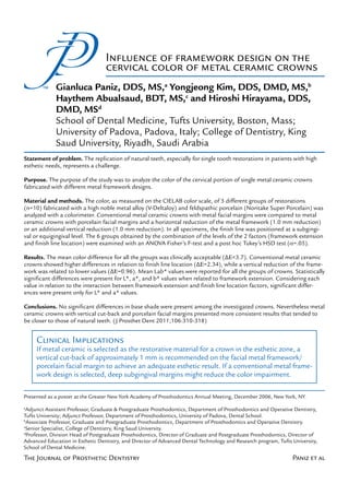
  • 3. 312 Japan) was measured with a color- imeter (ShadeVision System; X-Rite, Inc, Grand Rapids, Mich). The sam- ple size was arbitrary as no a priori power analysis was performed, and no reliable estimate of within-group standard deviation for the population could be obtained. The conventional metal ceramic crowns with metal margins (group C) were compared with metal ceramic crowns with 2 different designs of porcelain facial margin. One group had a horizontal reduction of the metal framework 1 Different designs of metal framework tested. Conventional metal (group H: 1.0 mm reduction), and ceramic crowns (Cmc), metal ceramic crowns with horizontal re- the second had an additional vertical duction of metal framework (Hmc), and metal ceramic crowns with reduction (group V: 1.0 mm vertical additional vertical reduction of metal framework (Vmc). reduction) (Fig. 1). One extracted, intact, human Type and thickness of materials used for crowns maxillary central incisor was prepared to receive the crowns. The facial re- Materials Cervical duction was 1.5 mm following the Crown Layers (Manufacturer) Thickness depth of a rounded shoulder finishing line. The margin was positioned at the Metal framework High noble metal alloy 0.3 mm (V-Delta Alloy, Metalor) level of the cement-enamel junction and 2.0 mm of incisal reduction was Opaque layer Porcelain opaque 0.1 mm performed. Throughout the study, the (POA1, Noritake Porcelain) tooth was stored in a 0.05 % thymol and distilled water solution. The pre- Dentin porcelain Feldspathic porcelain 1.0 mm pared tooth was duplicated with a (A1B, Noritake Porcelain) vinyl polysiloxane impression mate- rial (CapSil, Aquatrols Corporation of Enamel porcelain Feldspathic porcelain 0.1 mm America, Paulsboro, NJ). Type IV den- (A1B, Noritake Porcelain) tal stone (New Fuji-Rock, GC Corp, Porcelain for Feldspathic porcelain Minimal Tokyo, Japan) was used to fabricate butt margin (MA1, Noritake Porcelain) achievable the 30 dies, each corresponding to a single crown. Cement medium Translucent cement Thin layer A standardized framework was (Variolink II, trial-base, cast for each die in a high noble metal Ivoclar Vivadent AG) alloy (V-Delta; Metalor Dental AG). After the heat treatment process, lingual side of the crown. For group applications of margin porcelain were the intaglio surfaces were finished to V crowns, an additional 1.0 mm was applied to achieve adequate marginal a uniform facial and interproximal added to the vertical reduction of the integrity. The thickness of the margin thickness of 0.3 mm. For the conven- metal framework. porcelain was reduced as much as tional margin design, group C, the Dental porcelain (Noritake Super possible. Two layers of dentin porce- metal framework remained extended Porcelain; Noritake Dental Supply lain were applied to all 3 groups to to the preparation finish line (Fig. Co) was applied to all groups with achieve proper contour and reduced 1). For group H crowns, the cervi- a brush-on technique according to to a cervical thickness of 1.4 mm. cal margin of the metal framework the manufacturer’s recommenda- Enamel porcelain was then applied was reduced at the axiocervical line tions and fired under vacuum in a over the dentin porcelain to achieve a angle at a distance of 1.5 mm from porcelain furnace (Programat P300; proper final crown contour with a 1.5 the preparation finishing line; this re- Ivoclar Vivadent AG, Schaan, Liech- mm thickness at the cervical area. All duction was made on the facial and tenstein). Three layers of opaque por- specimens were autoglazed according interproximal margin, leaving 150 de- celain were applied to all of the metal to the manufacturer’s recommenda- grees of metal collar exposure on the frameworks. For groups V and H, 2 tions. A caliper (Iwanson Decimal The Journal of Prosthetic Dentistry Paniz et al
  • 4. November 2011 313 conditions for each of the experimen- tal margins. Six experimental groups were evaluated in the study. On the computer screen, the cervi- cal portion of the teeth was selected by using a transparent custom-made plastic template, which was fabricat- ed to fit the computer screen to better identify the different tooth portions. A 4 mm length was considered enough to represent the cervical portion since 2 Selected cervical portion in equigingival (left) and subgingival the initial length of the tooth before (right) groups of metal ceramic crowns. Note that subgingival preparation was approximately 12 crowns have margin positioned 0.5 mm apical to gingival level. mm, and a standardized area, extend- ing 2 mm from the gingival level, was Groups analyzed. Variables included framework extension (3 selected in the cervical portion (Fig. groups of specimens) and finish line location (total 6 study groups) 2). The measurements were recorded in CIELAB coordinates. Groups of Framework The color difference among the Specimens Extension Study Finish Line experimental groups was determined (n=30) (C, H, V) Groups Location (S, E) with the equation: ∆E= [(∆L*)2 + Cmc Finish line CST (∆a*)2 + (∆b*)2]1/2,33,34 calculated from the mean values of L*, a*, and Hmc 1.5 mm horizontal reduction, HST b*. To determine clinical significance, Subgingival the ∆E values were related to those (buccocervical line angle) (0.5mm) present in the literature, which range Vmc 1 mm additional vertical VST from 1.7 to 3.7. 44,45 A 2-way ANOVA was calculated reduction for each CIELAB coordinate (L*, a*, and b* values) to show which of the 2 Cmc Finish line CET factors (framework extension or finish Hmc 1.5 mm horizontal reduction, HET line location) had a significant impact Equigingival on the values of each coordinate. A (buccocervical line angle) post hoc Tukey’s Honestly Significant Difference (HSD) test was performed Vmc 1 mm additional vertical VET to determine statistically significant reduction differences among the group means. Caliper; Asa Dental s.p.a., Lucca, cally (Fig. 2). To achieve repeatability RESULTS Italy) with an accuracy of 0.05 mm of measurements, the position of the was used to measure and adjust the tooth and of the measuring instru- The mean L*, a*, and b* values final thicknesses, which are reported ment was standardized for all the are presented in Table III. When con- in Table I. measurements with custom-position- sidering L* values through the analy- The color of the cervical region of ing indices. Each measurement was sis of variance (2-way ANOVA), there the crowns, fitted on a prepared max- performed 5 times to reduce mea- were significant differences (P=.012) illary central incisor, was measured surement error. Since natural teeth among the different groups of crowns, with a colorimeter (ShadeVision Sys- are usually observed in a wet environ- with framework extension having the tem; X-Rite, Inc). The measurements ment, all measurements were made main effect (Table IV). The mean L* were made in a dental mannequin with the specimens in a slightly moist value for the Vmc group (78.8) was (KaVo Dental, Biberach/Riss, Germa- condition. All the crowns were mea- significantly lower (P=.027) than ny) with the margin of the restoration sured with the application of trans- the mean value for the Cmc group positioned either 0.5 mm subgingi- parent cement medium try-in paste (79.4) and also significantly lower vally (subgroup S) or at the gingival (Variolink II; Ivoclar Vivadent AG). Ta- (P=.023) than the value for the Hmc level (subgroup E) by moving the pre- bles I and II summarize the porcelain group (79.4) (Fig. 3). Considering L* pared tooth 0.5 mm more or less api- layer thicknesses and measurement values in relation to framework ex- Paniz et al
  • 5. 314 Mean L* values, a* values, and b* values for each single group of crowns (n=6) L* a* b* Mean Mean Mean Group Values SD Values SD Values SD CST 78.48 0.93 3.77 0.34 12.26 0.65 HST 79.96 0.81 3.06 0.47 11.87 1.00 VST 78.89 0.55 3.64 0.07 13.33 0.60 CET 80.37 0.84 2.53 0.27 11.65 0.58 HET 78.92 0.68 2.31 0.23 11.78 1.15 VET 78.71 0.57 2.69 0.25 13.30 0.82 Two-way ANOVA table for L* value (main effects and interaction term) Sum of Mean Source Squares df Square F P Framework extension 5.39 2 2.69 4.81 .12 Finish line location 0.74 1 0.74 1.33 .25 Framework extension × 22.5 2 11.25 20.07 <.001 Finish line location Error 30.3 54 0.56 Framework Extension; LS Means Framework Extension* Finishing Line; LS Means Current effect: (F2, 54) = 4.82, P=.012 Current effect: (F2, 54) = 20.0, P<.001 80.0 81.5 79.8 81.0 Sub-gingival 79.6 Equi-gingival 80.5 79.4 80.0 79.2 L* L* 79.5 79.0 79.0 78.8 78.6 78.5 78.4 78.0 78.2 77.5 Cmc Hmc Vmc Cmc Hmc Vmc Framework Extension Framework Extension 3 L* values in relation to framework extension factor. 4 L* values in relation to framework extension and fin- ishing line location factors. The Journal of Prosthetic Dentistry Paniz et al
  • 6. November 2011 tension and finish line location fac- with framework extension having the measured subgingivally and 2.7 when tors, significant differences (P<.001) main effect (Table V). Hmc crowns measured equigingivally (Fig. 6). were present (Table IV). The post showed lower a* values (2.69) than When considering b* values, there hoc Tukey test revealed no significant Cmc crowns (3.17) and Vmc crowns were significant differences (P<.001) differences (P=.95) between subgin- (3.17) (Fig. 5). Considering a* values among the different groups of crowns, gival (78.9) and equigingival (78.7) in relation to framework extension with framework extension having the measurements within the Vmc group. and finish line location factors, signif- main effect (Table VI). Vmc crowns However, significant differences were icant differences (P=.043) were pres- show higher values (13.3) than Cmc present within the Cmc (P<.001) and ent (Table V). The post hoc Tukey’s (12, P<.001) and Hmc (11.9, P<.001) Hmc (P=.035) groups. Conventional test showed that, for all of the groups crowns (Fig. 7). Considering b* val- metal ceramic crowns had an L* value of crowns, mean values were signifi- ues in relation to framework exten- of 78.4 when measured subgingivally cantly higher when measured subgin- sion and finish line location factors, and 80.4 when measured equigingi- givally. Cmc crowns (P<.001) showed no significant differences (P=.488) vally. Specimens from the Hmc group a value of 3.78 when measured sub- were present (Table VI and Fig. 8). had a mean L* value of 80 when mea- gingivally and 2.54 when measured The ∆E values among the groups sured subgingivally and 78.9 when equigingivally. Hmc crowns (P<.001) of crowns are represented in Table measured equigingivally (Fig. 4). showed a value of 3.06 when mea- VII. Evaluating subgingival groups of When considering a* values, there sured subgingivally and 2.31 when crowns, the base shade comparison were significant differences (P<.001) measured equigingivally. Vmc crowns between Cmc crowns (CST) and Vmc among the different groups of crowns, (P<.001) showed a value of 3.65 when crowns (VST) showed ∆ =1.15. The Table V. Two-way ANOVA table for a* value (main effects and interaction term) Sum of Mean Source Squares df Square F P Framework extension 3.03 2 1.51 16.85 <.001 Finish line location 14.4 1 14.4 159.4 <.001 Framework extension × 0.60 2 0.30 3.34 .04 Finish line location Error 4.86 54 0.09 Framework Extension; LS Means Framework Extension* Finishing Line; LS Means Current effect: (F2, 54) = 16.9 P<.001 Current effect: (F2, 54) = 3.3, P=.043 3.4 4.2 3.3 4.0 3.8 3.2 3.6 3.1 3.4 3.0 3.2 Sub-gingival b* 2.9 L* 3.0 Equi-gingival 2.8 2.8 2.6 2.7 2.4 2.6 2.2 2.5 2.0 2.4 1.8 Cmc Hmc Vmc Cmc Hmc Vmc Framework Extension Framework Extension 5 a* values in relation to framework extension factor. 6 a* values in relation to framework extension and fin- ishing line location factors. Paniz et al
  • 7. 316 Two-way ANOVA for b* value (main effects and interaction term) Sum of Mean Source Squares df Square F P Framework extension 27.1 2 13.6 19.53 <.001 Finish line location 0.89 1 0.89 1.28 .26 Framework extension × 1.01 2 0.50 0.72 .48 Finish line location Error 37.5 54 0.69 Framework Extension; LS Means Framework Extension* Finishing Line; LS Means Current effect: (F2, 54) = 19.5 P<.001 Current effect: (F2, 54) = 0.73, P=.488 14.0 14.5 14.0 Sub-gingival 13.5 13.5 Equi-gingival 13.0 13.0 b* b* 12.5 12.5 12.0 12.0 11.5 11.5 11.0 11.0 10.5 Cmc Hmc Vmc Cmc Hmc Vmc Framework Extension Framework Extension 7 b* values in relation to framework extension factor. 8 b* values in relation to framework extension and fin- ishing line location factors. ∆E values among different groups of crowns --- CST HST VST CET HET VET CST --- 1.68 1.15 2.34 1.59 1.51 HST 1.68 --- 1.90 0.70 1.28 1.93 VST 1.15 1.90 --- 2.49 2.04 0.96 CET 2.34 0.70 2.49 --- 1.47 2.34 HET 1.59 1.28 2.04 1.47 --- 1.58 VET 1.51 1.93 0.96 2.34 1.58 --- The Journal of Prosthetic Dentistry Paniz et al
  • 8. November 2011 317 same comparison performed equig- strated ∆E to be lower than 1.7 (Table crowns (Hmc) showed lower values, ingivally (CET vs. VET), found higher VII). Evaluating subgingival groups of indicating a color closer to that of mean values (∆ =2.34). Consider- crowns, excluding the first 0.5 mm natural teeth.43 ing the color of each specific group coronal to the finish line (Fig. 2), the Considering b* values, equigingival of specimens, Vmc crowns showed base shade comparison between Cmc and subgingival crowns showed simi- ∆ =0.96 when comparing equigin- crowns (CST) and Vmc crowns (VST) lar b* values, except for the analysis of gival and subgingival groups, Hmc showed clinically acceptable results Cmc crowns, which showed lower val- crowns showed ∆ =1.28, and Cmc (∆ =1.15). The same comparison ues when equigingival (Figs. 7, 8). In crowns showed ∆ =2.34 (Table VII). performed equigingivally, including general, vertical cut-back crowns (Vmc) the most apical 0.5 mm (CET vs VET), showed higher values, indicating a color DISCUSSION found mean values above the limit closer to that of natural teeth.43 of acceptability (∆ =2.34). This re- The use of a dental mannequin This study was designed to achieve sult suggests that, when the shade of with a plastic gingival substitute al- a standardized evaluation of 3 differ- conventional metal ceramic crowns is lowed exclusion of portions of the ent metal framework designs for the analyzed, there is significant color im- crowns, creating 2 different groups of color of the cervical area of metal pairment in the most apical 0.5 mm specimens, subgingival (S) and equig- ceramic restorations. The goal was of the restoration. Furthermore, the ingival (E). However, the plastic gingi- to understand whether the technical color of the cervical portion of Vmc va did not replicate a clinical situation challenges of fabricating and man- crowns is most consistent between since it eliminated the impact of the aging a collarless metal ceramic res- the equigingival and subgingival crown on the soft tissue, whose color toration19, 21-24, 26-32 were justified to groups (∆ =0.96), while that of the influences the final esthetic outcome. achieve an improved esthetic result. Hmc crowns is moderate (∆ =1.28), Similarly, this study did not consider The data support rejecting the null and that of the Cmc crowns is the the possibility of short-term or long- hypothesis that no color differences worst (∆ =2.34) (Table VII). The pos- term tissue recession. The use of a dif- would be present in the cervical por- sible positive esthetic implication of ferent gingival substitute could help tion of single metal ceramic crowns the use of an additional millimeter of overcome these shortcomings in pos- fabricated with different metal frame- metal framework reduction was not sible future research. Furthermore, work designs. tested in this study because of the in- considering the development of ce- In analyzing tooth color, the pri- creased fracture risk present with this ramic restorations, a similar study mary concern was to obtain clinically type of restoration.25,27,28 design could be used to evaluate core relevant results, and for this reason, Through the analysis of Lab* val- ceramic restorations. the data obtained through the colo- ues, a deeper understanding of the rimetric measurement were related to color differences and of influenc- CONCLUSION the values reported in the literature. ing factors can be drawn (Figs. 3-8). A natural tooth was not selected as a Considering L* values, Cmc crowns Within the limitations of this standard since previously completed showed the highest value when equi- study, the following conclusions were in vitro research clearly showed signif- gingival (CET), the lowest when sub- drawn: icant color differences when compar- gingival (CST), and the highest differ- 1. No significant differences in ing tooth structure to a metal ceramic ence of values between equigingival base shade were present among the restoration. 33 According to Johnston and subgingival specimens (Figs. 3, investigated crowns. and Kao,44 a ∆E > 3.7 indicates vi- 4). As discussed in the ∆E analysis, 2. Vertical cut-back crowns (Vmc) sually perceivable color differences this confirmed that significant color had a cervical shade which was more which are clinically unacceptable. By impairment occurs in the most apical consistent and appeared to be more using this threshold and comparing 0.5 mm of the ceramic of metal ce- similar to that of a natural tooth. the groups with the most clinical rel- ramic crowns. In contrast, when ana- 3. Conventional metal ceramic evance, no clinical differences were lyzing Vmc crowns, the results were crowns (Cmc) were characterized by noted (Table VII). A similar compari- more consistent and the color also significant color impairment in the son can be made to other recent clini- closer to that of natural teeth.43 most apical 0.5 mm of the ceramic. cal references.47 Even if Johnston and Considering a* values, consis- 4. Vertical cut-back crowns (Vmc) Kao44 is often cited in the literature, tent differences were present be- tended to have mean L* and b* values other values were considered for this tween equigingival (E) and subgingival closer to those of natural teeth, while in vitro study. According to Douglas (S) crowns. In general, equigingival horizontal cut-back crowns (Hmc) and Brewer,45 the threshold for accept- crowns demonstrated lower values tended to have closer mean a* values. ability is ∆E=1.7 and for perceptibil- (Figs. 5 and 6). Among the different ity, 0.4. Several comparisons demon- types of design, horizontal cutback Paniz et al
  • 9. 318 REFERENCES 20.McLean JW, Jeansonne EE, Bruggers H, 34.Chu SJ, Devigus A, Mieleszko AJ. Funda- Lynn DB. A new metal-ceramic crown. J mentals of color: Shade matching and com- 1. Mayekar SM. Shades of a color. Illusion or Prosthet Dent 1978;40:273-87. munication in esthetic dentistry. Chicago: reality? Dent Clin North Am 2001;45:155-72. 21.Belser UC, MacEntee MI, Richter WA. Fit Quintessence; 2004. 2. McLean JW. The science and art of dental of three porcelain-fused-to-metal marginal 35.Berns RS. Billmeyer and Saltzman’s prin- ceramics. Volume I: the nature of dental designs in vivo: a scanning electron micro- ciples of color technology. New York: John ceramics and their clinical use. Chicago: scope study. J Prosthet Dent 1985;53:24-9. Wiley & Sons; 2000. p. 75-105. Quintessence Publishing Co Inc; 1979. p. 22.Belles DM, Cronin RJ Jr, Duke ES. Effect 36.Al-Wahadni A, Ajlouni R, Al-Omari Q, 102-10. of metal design and technique on the Cobb D, Dawson D. Shade-match percep- 3. Joiner A. Tooth colour: a review of the marginal characteristics of the collarless tion of porcelain-fused-to-metal restora- literature. J Dent 2004;32:3-12. metal ceramic restoration. J Prosthet Dent tions: a comparison between dentist and 4. Kelly JR, Nishimura I, Campbell SD. Ceramics 1991;65:611-9. patient. J Am Dent Assoc 2002;133:1220-5. in dentistry: historical roots and current per- 23.Omar R. Scanning electron microscopy of 37.van der Burgt TP, ten Bosch JJ, Borsboom PC, spectives. J Prosthet Dent 1996;75:18-32. the marginal fit of ceramometal restora- Kortsmit WJ. A comparison of new and con- 5. Croll BM. Emergence profiles in natural tions with facially butted porcelain margins. ventional methods for quantification of tooth tooth contour. Part I: Photographic obser- J Prosthet Dent 1987;58:13-9. color. J Prosthet Dent 1990;63:155-62. vations. J Prosthet Dent 1989;62:4-10. 24.Morris HF. Department of Veterans Affairs 38.Ishikawa-Nagai S, Sato RR, Shiraishi A, 6. Croll BM. Emergence profiles in natural Cooperative Studies Project No. 242. Ishibashi K. Using a computer color- tooth contour. Part II: Clinical consider- Quantitative and qualitative evaluation of matching system in color reproduction ations. J Prosthet Dent 1990; 63:374-9. the marginal fit of cast ceramic, porcelain- of porcelain restorations. Part 3: A newly 7. Felton DA, Bergenholtz G, Kanoy BE. shoulder, and cast metal full crowns developed spectrophotometer designed Evaluation of the desensitizing effect of margins. Participants of CSP No. 147/242. for clinical application. Int J Prosthodont Gluma Dentin Bond on teeth prepared J Prosthet Dent 1992;67:198-204. 1994;7:50-5. for complete-coverage restorations. Int J 25.Lehner CR, Männchen R, Schärer P. Vari- 39.Tung FF, Goldstein GR, Jang S, Hittelman Prosthodont 1991;4: 292-8. able reduced metal support for collarless E. The repeatability of an intraoral dental 8. Walton JN, Gardner FM, Agar JR. A survey metal ceramic crowns: a new model for colorimeter. J Prosthet Dent 2002;88:585-90. of crown and fixed partial denture failures: strength evaluation. Int J Prosthodont 40.Douglas RD. Precision of in vivo colorimet- length of service and reasons for replace- 1995;8:337-45. ric assessments of teeth. J Prosthet Dent ment. J Prosthet Dent 1986;56:416-21. 26.Anusavice KJ, Hojjatie B. Stress distribution 1997;77:464-70. 9. Douglas RD, Przybylska M. Predicting por- in metal-ceramic crowns with a facial por- 41.Wee AG, Monaghan P, Johnston WM. Vari- celain thickness required for dental shade celain margin. J Dent Res 1987;66:1493-8. ation in color between intended matched matches. J Prosthet Dent 1999;82:143-9. 27.O’Boyle KH, Norling BK, Cagna DR, shade and fabricated shade of dental por- 10.Terada Y, Maeyama S, Hirayasu R. The Phoenix RD. An investigation of new metal celain. J Prosthet Dent. 2002;87:657-66. influence of different thicknesses of dentin framework design for metal ceramic resto- 42.Hasegawa A, Ikeda I, Kawaguchi S. Color porcelain on the color reflected from thin rations. J Prosthet Dent 1997;78:295-301. and translucency of in vivo natural central opaque porcelain fused to metal. Int J 28.Gardner FM, Tillman-McCombs KW, Gas- incisors. J Prosthet Dent 2000;83:418-23. Prosthodont 1989;2:352-6. ton ML, Runyan DA. In vitro failure load 43.O’Brien WJ, Hemmendinger H, Boenke 11.Jacobs SH, Goodacre CJ, Moore BK, of metal-collar margins compared with KM, Linger JB, Groh CL. Color distribution Dykema RW. Effects of porcelain thickness porcelain facial margins of metal-ceramic of three regions of extracted human teeth. and type of metal-ceramic alloy on color. J crowns. J Prosthet Dent 1997;78:1-4. Dent Mater 1997;13:179-85. Prosthet Dent 1987;57:138-45. 29.Bishop K, Briggs P, Kelleher M. Margin 44.Johnston WM, Kao EC. Assessment of 12.Campbell SD, Pelletier LB. Thermal cycling design for porcelain fused to metal restora- appearance match by visual observa- distortion of metal ceramics: Part I--Metal col- tions which extend onto the root. Br Dent J tion and clinical colorimetry. J Dent Res lar width. J Prosthet Dent 1992;67:603-8. 1996;180:177-84. 1989;68:819-22. 13.Campbell SD, Sirakian A, Pelletier LB, Gior- 30.Dalvit DL, Jackson RA, Hawkins MC, 45.Douglas RD, Brewer JD. Acceptability of dano RA. Effects of firing cycle and surface Parker MH. Mathematical derivation of the shade differences in metal ceramic crowns. finishing on distortion of metal ceramic minimally acceptable all-porcelain margin J Prosthet Dent 1998;79:254-60. castings. J Prosthet Dent 1995;74:476-81. angle. J Prosthet Dent 2005;93:467-72. 46.Yilmaz B, Ozçelik TB, Wee AG. Effect of 14.Goodacre CJ, Van Roekel NB, Dykema RW, 31.Michalakis KX, Stratos A, Hirayama H, repeated firings on the color of opaque Ullman RB. The collarless metal-ceramic Kang K, Touloumi F, Oishi Y. Fracture re- porcelain applied on different dental alloys. crown. J Prosthet Dent 1977;38:615-22. sistance of metal ceramic restorations with J Prosthet Dent 2009;101:395-404. 15.Sozio RB, Riley DJ. A precision ceramic- two different margin designs after exposure 47.Douglas RD, Steinhauer TJ, Wee AG. metal restoration with a facial butted mar- to masticatory simulation. J Prosthet Dent Intraoral determination of the tolerance gin. J Prosthet Dent 1977;37:517-21. 2009;102:172-8. of dentists for perceptibility and accept- 16.Toogood GD, Archibald JF. Technique for 32.Geller W, Kwiatkowski SJ. The Willi’s glas ability of shade mismatch. J Prosthet Dent establishing porcelain margins. J Prosthet crown: a new solution in the dark and 2007;97;200-8. Dent 1978;40:464-6. shadowed zones of esthetic porcelain 17.Hagen WHB. A combination gold and porce- restorations. Quintessence Dent Technol Corresponding author: lain crown. J Prosthet Dent 1960;10:325-9. 1987;11:233-42. Dr Gianluca Paniz 18.Vickery RC, Badinelli LA, Waltke RW. The 33.Swain VL, Pesun IJ, Hodges JS. The effect Via Cesarotti 31 direct fabrication of restorations without of metal ceramic restoration framework 35100 Padova foil on a refractory die. J Prosthet Dent design on tooth color. J Prosthet Dent ITALY 1969;21:227-34. 2008;99:468-76. Fax: +39-049-8776791 19.Prince J, Donovan TE, Presswood RG. The E-mail: panizg@hotmail.com all-porcelain labial margin for ceramometal restorations: a new concept. J Prosthet Copyright © 2011 by the Editorial Council for Dent 1983;50:793-6. The Journal of Prosthetic Dentistry. The Journal of Prosthetic Dentistry Paniz et al