2. 14 G. Ling and D.J. Waxman
have enabled traditional DHS assays to be coupled with high
throughput mapping methods, such as tiling microarrays (DNase-
chip) and, more recently, DNA sequencing (DNase-seq), making
it possible to obtain high resolution genome-wide maps of DHS
sites upon mapping released DNA fragments back to the genome
(Fig. 2) (3–6).
DNase-seq can be carried out with nuclei isolated from intact
mammalian tissues, as exemplified by studies from this laboratory
on sex differences in mouse liver chromatin structure (7). DNase-
seq studies using intact nuclei isolated from fresh tissue, such as
mouse liver, have the important advantage of providing detailed
information about the regulation of chromatin structure under
physiological conditions in vivo. However, care must be taken to
ensure that the procedure yields high quality nuclei with minimal
Heterochromatin
Euchromatin
Gene
expression
Epigenetic changes
TF binding
sites exposed
DNase I
Fig. 1. DNase hypersensitivity identifies open genomic regions in chromatin that facilitate
transcription factor (TF) binding to chromatin and induce gene expression. Nucleosomes
are compacted in closed, inactive heterochromatin but are more open, exposing sites of
DNase hypersensitivity in the euchromatin state.
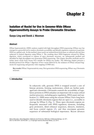
Genomic DNA sequence
HS HS HSHS
DNase-Seq analysis
DNase I
hypersensitive sites
Mapped seq tags
Fragments
released by
DNase
cleavage
DHS peaks
Fig. 2. DNase-seq analysis. Sites of hypersensitivity (HS) are susceptible to cutting by
DNase I, which releases many fragments of variable length from each hypersensitive
region.The released fragments are then purified, sequenced on one end, and the resultant
sequence tags then mapped back to the genome. A peak detection algorithm is used to
identify DHS peaks, two of which are shown.
3. 152 Isolation of Nuclei for DNase-seq
disturbance of chromatin structure. In the protocol described here,
isolation of fresh, high quality nuclei from tissues is facilitated by
sucrose ultracentrifugation. In brief, fresh liver tissue is collected,
homogenized, and the nuclei are pelleted by ultracentrifugation
through a 2 M sucrose cushion, which helps maintain the integrity
of the nuclei and chromatin structure. Only intact nuclei are of
sufficiently high density to pass through the sucrose cushion (8).
Nuclei isolated and purified by this method give reproducible and
reliable DNase-seq results (7). The same protocol can also be used
to isolate nuclei from tissue culture cells, although in that case a
simpler, detergent-based method may work as well (5). We antici-
pate that the protocol described here can be applied to other tis-
sues that yield high quality nuclei without major modifications.
Prepare all solutions using ultrapure water and analytical grade
reagents. Store all buffers at 4 °C unless otherwise noted. Protease
inhibitors, spermine, spermidine, and DTT should be added fresh
to each solution just prior to their use.
1. Scissors, blades, and forceps for tissue dissection.
2. CO2
chamber for rodents.
3. Paper towels, Kimwipes, and petri dishes.
4. Ice-cold 1.15% KCl and 1× Phosphate-buffered saline (PBS).
5. 250 ml beaker, 50 ml conical tubes.
6. 1 ml syringes and 27G needles (optional).
1. Preparative ultracentrifuge and SW28 rotor, or equivalent.
Ultra-clear centrifugation tubes (Beckman Coulter, Brea, CA;
cat. # 344058).
2. Potter-Elvehjem Tissue Grinder with Teflon Pestle, 10, 15, 30 or
55 ml size (Wheaton Science Products, Millville, NJ; see Note 1),
and a power drill.
3. Nuclear homogenization buffer (NEHB): 10 mM HEPES-
KOH, pH 7.9, 25 mM KCl, 1 mM EDTA, 2 M sucrose, 10%
glycerol, 0.15 mM spermine, 0.5 mM spermidine, 10 mM
NaF, 1 mM orthovanadate, 1 mM PMSF, 0.5 mM DTT, and
1× protease inhibitor cocktail (Sigma, St Louis, MO; cat. #
P8340) (see Note 2).
4. Nuclear storage buffer: 20 mM Tris–HCl, pH 8.0, 75 mM
NaCl, 0.5 mM EDTA, 50% (v/v) glycerol, 1 mM DTT, and
0.1 mM PMSF.
2. Materials
2.1. Materials
for Dissection of Liver
Tissue
2.2. Tissue
Homogenization
and Isolation of Nuclei
4. 16 G. Ling and D.J. Waxman
5. 1 M HEPES-KOH, pH 7.9. Dissolve 238.3 g HEPES in
700 ml of ultrapure water. Use potassium hydroxide pellets
and 1 M KOH solution to adjust pH to 7.9. Bring up to
1,000 ml with ultrapure water.
6. 1 M Tris–HCl, pH 8.0. Dissolve 121.1 g Tris base in 800 ml
of ultrapure water. Add concentrated HCl to bring to pH 8.0.
Bring up to 1,000 ml with ultrapure water.
7. 0.5 M EDTA, pH 8.0 (Sigma, St. Louis, MO).
8. Dounce homogenizer with B pestle, chilled on ice (Wheaton
Science Products, Millville, NJ).
9. Inverted tissue culture microscope, hemocytometer, and try-
pan blue solution (Sigma, cat. #T8154 or equivalent) for
counting nuclei.
10. 1.5-ml microcentrifuge tubes, chilled on ice.
11. Liquid nitrogen for freezing nuclei.
All steps described here, except animal-related procedures, should
be carried out in a 4 °C cold room. Samples should be kept at 4 °C
to minimize nonspecific DNA degradation.
1. Prepare complete NEHB on ice (see Note 2). Precool the
ultracentrifuge, rotor, tubes, and the tissue grinder before
starting the experiments.
2. Euthanize the mice using approval protocols, e.g., CO2
asphy-
xiation followed by cervical dislocation. Subsequent steps
should be carried out promptly to ensure that the minced liver
tissue is submerged in ice-cold NEHB (step 6) as quickly as
possible.
3. If required, remove blood sample from the heart using a 1-ml
syringe with 50 μl of heparin and a 27G needle (optional).
4. Remove each liver and rinse in ice-cold PBS. Snap-freeze a
small piece of tissue and store at −80 °C, e.g., for RNA isola-
tion at a later time.
5. Place the liver in a 100×15 mm petri dish on ice containing
ice-cold 1.15% KCl. Cut the liver into small pieces and wash
twice with 1.15% KCl.
6. Carefully dry off the excess KCl solution with a Kimwipe.
Weigh the liver and submerge the minced liver in ice-cold
NEHB buffer.
3. Methods
3.1. Dissection of Liver
Tissue
5. 172 Isolation of Nuclei for DNase-seq
1. Homogenize the liver in a 30 ml Potter-Elvehjem Tissue
Grinder with three to four strokes. Use a buffer-to-tissue ratio
of ~6 (v/w) (see Note 3).
2. Carefully overlay 25 ml of homogenate on top of 10 ml NEHB
in a SW28 tube.
3. Centrifuge at 25,000 rpm (90,000´g) for 45 min at 2 °C. The
nuclei should be found in a clear pellet at the bottom of the
tube (see Note 4).
4. Remove tissue debris floating on the top and decant the super-
natant. Turn the tube upside down in a cold room and wipe
the sides of each tube with a Kimwipe soaked with PBS.
5. Resuspend the pellet in a minimal volume of nuclear storage
buffer by applying a few strokes of a Dounce homogenizer (see
Note 5).
6. Dilute a sample of the resuspended nuclei 100-fold with PBS
and count the nuclei using a hemocytometer. Trypan Blue can
be added to a final concentration of 0.04% to help visualize the
nuclei. Nuclei should be counted at least three times to ensure
that a reproducible count is obtained.
7. Snap-freeze the suspended nuclei in small aliquots using liquid
nitrogen, for example 300 μl aliquots in 1.5 ml micro-centrif-
ugation tubes, and store at −80 °C (see Note 6).
1. Nuclei may be isolated from a single adult mouse liver if
DNase-seq data are to be collected for individual animals, oth-
erwise it may be more convenient to pool livers from three to
four mice and prepare a single preparation of nuclei, using a
30 ml or 55 ml tissue grinder for liver homogenization. If the
tissue weight is ~2 g or less (e.g., when isolating nuclei from a
single mouse liver), a 10 or 15 ml tissue grinder can be used
and the buffer volumes and sizes of the ultracentrifuge tubes
and rotor should be scaled down accordingly.
2. NEHB is prepared and stored at 4 °C as 10 mM HEPES pH
7.9, 25 mM KCl, 1 mM EDTA, 2 M sucrose, and 10% glycerol.
The other buffer components: spermine, spermidine, NaF,
orthovanadate, PMSF, DTT, and protease inhibitor cocktail are
prepared separately as concentrated stock solutions (100× for
NaF, orthovanadate (both in water), PMSF (in isopropanol),
and protease inhibitor cocktail (in DMSO); 2,000× for sper-
mine, spermidine, and DTT (all in water)), and stored in ali-
quots at −20 °C. The concentrated components are then added
to NEHB just before tissue dissection and homogenization.
3.2. Tissue
Homogenization
and Isolation of Nuclei
(Scale: Pool of Three
to Four Mouse Livers)
(See Note 1)
4. Notes
6. 18 G. Ling and D.J. Waxman
3. A buffer-to-tissue ratio of 6:1 (v/w) is recommended for
mammalian tissues. Insufficient buffer during tissue homoge-
nization can lead to impure nuclear preparations. Note that
the presence of 2 M sucrose makes it difficult to carry out the
homogenization step. As such, gloves and goggles should
be worn during homogenization for personal protection in
the event of glassware failure. Three to four strokes of the
homogenizer are required to disrupt >90% of the liver tissue.
Other tissues may require more vigorous conditions for effec-
tive homogenization.
4. A nuclear pellet with a red tinge indicates that the nuclei are
contaminated by red blood cells and should be discarded if
replalcement material is readily available. Increasing the vol-
ume of buffer used for homogenization may help avoid this
problem. It may be possible to wash the nuclei by resuspend-
ing the nuclear pellet in NEHB and then repeating the cen-
trifugation step.
5. The nuclei should be resuspended in a minimal volume of
nuclear storage buffer prior to counting. For example, nuclei
from three to four livers should initially be resuspended in
approx. 0.5 ml storage buffer. Additional buffer can be added
as required once the count is known to adjust the final prepara-
tion to 50–100 million nuclei per ml.
6. Although fresh nuclei may in some cases be preferrable for
DHS assays, we have not noticed any difference in results when
frozen mouse liver nuclei are used after storage at −80°C for
several months.
Acknowledgments
Supported in part by NIH grant DK33765 (to DJW).
References
1. Bell O, Tiwari VK, Thomä NH, Schübeler D
(2011) Determinants and dynamics of genome
accessibility. Nat Rev Genet 12:554–564
2. Gross DS, Garrard WT (1988) Nuclease
hypersensitive sites in chromatin. Annu Rev
Biochem 57:159–197
3. Boyle AP, Davis S, Shulha HP, Meltzer P,
Margulies EH, Weng Z, Furey TS, Crawford
GE (2008) High-resolution mapping and
characterization of open chromatin across the
genome. Cell 132:311–322
4. Crawford GE, Davis S, Scacheri PC, Renaud
G, Halawi MJ, Erdos MR, Green R, Meltzer
PS, Wolfsberg TG, Collins FS (2006) DNase-
chip: a high-resolution method to identify
DNase I hypersensitive sites using tiled
microarrays. Nat Methods 3:503–509
5. Sabo PJ, Kuehn MS, Thurman R, Johnson BE,
Johnson EM, Cao H, Yu M, Rosenzweig E,
Goldy J, Haydock A, Weaver M, Shafer A, Lee K,
Neri F, Humbert R, Singer MA, Richmond TA,
Dorschner MO, McArthur M, Hawrylycz M,
Green RD, Navas PA, Noble WS,
Stamatoyannopoulos JA (2006) Genome-scale
mapping of DNase I sensitivity in vivo using til-
ing DNA microarrays. Nat Methods 3:511–518
7. 192 Isolation of Nuclei for DNase-seq
6. Song L, Crawford GE (2010) DNase-seq: a
high-resolution technique for mapping active
gene regulatory elements across the genome
from mammalian cells. Cold Spring Harb
Protoc 2010:pdb.prot5384
7. Ling G, Sugathan A, Mazor T, Fraenkel E,
Waxman DJ (2010) Unbiased, genome-wide
in vivo mapping of transcriptional regulatory
elements reveals sex differences in chromatin
structure associated with sex-specific liver
gene expression. Mol Cell Biol 30(23):
5531–5544
8. Lichtsteiner S, Wuarin J, Schibler U (1987)
The interplay of DNA-binding proteins on the
promoter of the mouse albumin gene. Cell
51:963–973