Accuracy of two ventral approaches to block the femoral nerve in the Dog
•
1 like•501 views

Comparative study between two ventral ultrasound-guided approaches to block the femoral nerve in the dog
Recommended

Recommended
More Related Content
Recently uploaded
Recently uploaded (20)
Featured
Featured (20)
Accuracy of two ventral approaches to block the femoral nerve in the Dog
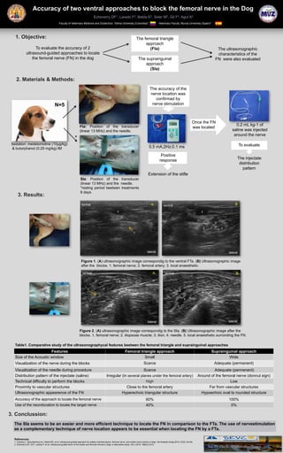
- 1. Accuracy of two ventral approaches to block the femoral nerve in the Dog Echeverry DF1, Laredo F2, Belda E2, Soler M2, Gil F2, Agut A2 Faculty of Veterinary Medicine and Zootechnic. Tolima University (Colombia)1 Veterinary Faculty. Murcia University (Spain)2 1. Objective: The femoral triangle approach To evaluate the accuracy of 2 (Fta) The ultrasonographic ultrasound-guided approaches to locate characteristics of the the femoral nerve (FN) in the dog The suprainguinal FN were also evaluated approach (SIa) 2. Materials & Methods: The accuracy of the nerve location was confirmad by N=5 nerve stimulation Once the FN Fta: Position of the transducer 0.2 mL kg-1 of was located (linear 13 MHz) and the needle. saline was injected around the nerve * Sedation: medetomidine (10μg/kg) To evaluate & butorphanol (0.25 mg/kg) IM 0.5 mA;2Hz;0.1 ms Positive The injectate response distribution pattern Extension of the stifle SIa: Position of the transducer (linear 13 MHz) and the needle. *resting period beetwen treatments 8 days 3. Results: ventral A ventral B 2 2 3 3 3 1 lateral lateral Figure 1. (A) ultrasonographic image correspondig to the ventral FTa. (B) Ultrasonographic image after the blocks. 1. femoral nerve; 2. femoral artery; 3. local anaesthetic. ventral A ventral B 2 2 4 1 4 5 3 1 5 5 2 lateral lateral Figure 2. (A) ultrasonographic image correspondig to the SIa. (B) Ultrasonographic image after the blocks. 1. femoral nerve; 2. iliopsoas muscle; 3. ilion; 4. needle. 5. local anaesthetic surronding the FN. Table1. Comparative study of the ultrasonographycal features beetwen the femoral triangle and suprainguinal approaches Features Femoral triangle approach Suprainguinal approach Size of the Acoustic window Small Wide Visualization of the nerve during the blocks Scarce Adequate (permanent) Visualization of the needle during procedure Scarce Adequate (permenent) Distribution pattern of the injectate (saline) Irregular (in several planes under the femoral artery) Around of the femoral nerve (donnut sign) Technical difficulty to perform the blocks High Low Proximity to vascular structures Close to the femoral artery Far from vascular structures Ultrasonographic appearence of the FN Hyperechoic triangular structure Hypoechoic oval to rounded structure Accuracy of the approach to locate the femoral nerve 60% 100% Use of the neurolocation to locate the target nerve 40% 0% 3. Conclussion: The SIa seems to be an easier and more efficient technique to locate the FN in comparison to the FTa. The use of nervestimulation as a complementary technique of nerve location appears to be essential when locating the FN by a FTa. References: 1. Campoy L, Bezuidenhout AJ, Gleed RD, et al: Ultrasound-guided approach for axillary brachial plexus, femoral nerve, and sciatic nerve blocks in dogs. Vet Anaesth Analg 2010; 37(2):144-53. 2. Echeverry DF, Gil F, Laredo F, et al: Ultrasound-guided block of the sciatic and femoral nerves in dogs: a descriptive study. Vet J 2010; 186(2):210-5.