Short case...Intramedullary cystic spinal cord metastasis
•
3 likes•186 views

Short case...Intramedullary cystic spinal cord metastasis. http://yassermetwally.com http://yassermetwally.net

Recommended
Recommended
More Related Content
Viewers also liked
Viewers also liked (15)
Similar to Short case...Intramedullary cystic spinal cord metastasis
Similar to Short case...Intramedullary cystic spinal cord metastasis (20)
More from Professor Yasser Metwally
More from Professor Yasser Metwally (20)
Recently uploaded
Recently uploaded (20)
Short case...Intramedullary cystic spinal cord metastasis
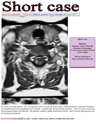
- 1. Short case Edited by Professor Yasser Metwally Professor of neurology Ain Shams university school of medicine Cairo, Egypt Visit my web site at: http://yassermetwally.com 48 Years old female patient- who was operated upon for grade III breast cancer (Adenocarcinoma)- presented clinically a with rapidly progressive quadriplegia over 4 months- 6 month after the mastectomy operation- with a C3 sensory level and brown Sequard spinal cord affection. The patient's condition rapidly deteriorated and she died 6 months following the start of neurological symptoms. DIAGNOSIS:INTRAMEDULLARY CYSTIC CERVICAL SPINAL CORD METASTASIS Short case publication... Version 4.5 | Edited by professor Yasser Metwally | December 2010
- 2. Figure 2. Postcontrast MRI T1 images showing densely enhanced well defined intramedullary oval nodules. Intramedullary T1 hypointensities probably represent cystic changes or cord edema. Notice the linear peripheral enhancement (A) which probably represents a concomitant meningeal carcinomatosis. Figure 1. Precontrast MRI T1 images showing mild dilatation of the spinal cord in cross section with intramedullary cystic changes. The spinal cord appears irregular in cross section with linear precontrast peripheral hyperintensity (D) probably representing blood products.
- 3. Figure 3. Precontrast MRI T1 image (A) and postcontrast MRI T1 images (B,C) showing irregular intramedullary patchy enhancement with central MRI T1 hypointensities which probably represent cord edema or cystic changes. The spinal cord is moderately enlarged from C2-D2. Notice the linear anterior peripheral meningeal enhancement which is -most probably- due to concomitant meningeal carcinomatosis. References 1. Metwally, MYM: Textbook of neurimaging, A CD-ROM publication, (Metwally, MYM editor) WEB-CD agency for electronic publishing, version 11.4a October 2010 Addendum A new version of short case is uploaded in my web site every week (every Saturday and remains available till Friday.) To download the current version follow the link "http://pdf.yassermetwally.com/short.pdf". You can download the long case version of this short case during the same week from: http://pdf.yassermetwally.com/case.pdf or visit web site: http://pdf.yassermetwally.com To download the software version of the publication (crow.exe) follow the link: http://neurology.yassermetwally.com/crow.zip At the end of each year, all the publications are compiled on a single CD-ROM, please contact the author to know more details. Also to view a list of the previously published case records follow the following link: (http://wordpress.com/tag/case-record/) or click on it if it appears as a link in your PDF reader To inspect the patient's full radiological study, click on the attachment icon (the paper clip icon in the left pane) of the acrobat reader then double click on the attached file Click here to download the long case version of this short case in PDF format