Short case...brain stem glioma
•
2 likes•560 views

Short case...brain stem glioma http://yassermetwally.com http://yassermetwally.net

Recommended
More Related Content
What's hot
What's hot (20)
Similar to Short case...brain stem glioma
Similar to Short case...brain stem glioma (20)
More from Professor Yasser Metwally
More from Professor Yasser Metwally (20)
Recently uploaded
Recently uploaded (20)
Short case...brain stem glioma
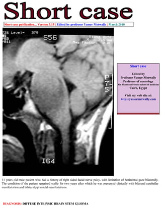
- 1. Short case publication... Version 3.15 | Edited by professor Yasser Metwally | March 2010 Short case Edited by Professor Yasser Metwally Professor of neurology Ain Shams university school of medicine Cairo, Egypt Visit my web site at: http://yassermetwally.com 11 years old male patient who had a history of right sided facial nerve palsy, with limitation of horizontal gaze bilaterally. The condition of the patient remained stable for two years after which he was presented clinically with bilateral cerebellar manifestation and bilateral pyramidal manifestations. DIAGNOSIS: DIFFUSE INTRINSIC BRAIN STEM GLIOMA
- 2. Figure 1. Precontrast MRI T1 (A) and MRI T2 images (B,C) showing a diffuse intrinsic brain stem glioma causing diffuse brain stem enlargement. The tumor is diffusely hypointense on the precontrast MRI T1 image and diffusely hyperintense on the MRI T2 images. The basilar artery is encased by the tumor (C). The tumor is mainly located at the pontine level. Notice the hydrocephalic changes. (B) The 4th ventricle is pushed posteriorly and compressed by the tumor. Figure 2. MRI T2 images, notice the hydrocephalic changes. Cystic tumor masses could be seen at the midbrain level indicating that the tumor has extended to the midbrain level. Two points must be addressed in this case report 1) the cause of the MRI signal intensity of pontine gliomas and 2) the pattern of spread of this tumor.
- 3. Radiologically low grade gliomas are usually identified by diffuse enlargement of the brain stem, abnormal signal intensity on MR or abnormal attenuation on CT. The lesions typically have precontrast CT attenuation and MRI signal changes suggesting increased water content and lower than normal specific gravity (lower CT scan densities with MRI T1 hypointensities and diffuse MRI T2 hyperintensities). It is tempting to consider that these changes represent edema. The question then arises: Is this vasogenic edema or cytotoxic edema? Because the blood-brain barrier is intact in these tumors, vasogenic edema is unlikely. The cells are not dead or dying, so that cytotoxic edema is also unlikely. Perhaps the edema results from the increased number of astrocytic cells that spread apart the normal myelinated axons of the white matter. The presence of significant amount of normal appearing astrocytes results in total increase in the water content of the brain stem. These cells may merely have different physical and chemical properties than the normal tightly packed bundles of axons that traverse through the brain stem. As the blood brain barrier is intact in low grade brain stem astrocytomas (grade II astrocytomas according to the WHO), no significant enhancement occurs, either on MRI or CT scan. Enhancement is characteristic of the more aggressive anaplastic astrocytomas (grade III) or glioblastoma multiforme. Diffuse intrinsic brain stem gliomas are actually diffuse astrocytomas Grade II,III,IV which have the following characteristics: Diffuse astrocytomas are tumors predominantly composed of astrocytes. Unless otherwise indicated, the term usually applies to diffusely infiltrating neoplasms (WHO grades II through IV). Diffuse astrocytoma is unusual in the first decade of life and most commonly presents in older children or young adults up to the age of 40 to 45. All diffuse astrocytomas, particularly the diffusely infiltrating variety, have a tendency toward progression to more malignant forms. Diffuse astrocytomas have a peculiar tendency to change its grade over time into the next higher grade of malignancy and the condition is age dependant. A change in the grade of diffuse astrocytoma is more likely to occur in the older age group. Diffuse astrocytomas commonly start as grade II at a younger age group then gradually change its grade over time into the next higher grade until they ultimately dedifferentiate into glioblastomas (secondary glioblastoma multiforme), on the other hand, glioblastoma multiforme in older patients are usually primary-that is, they occur as glioblastoma multiforme from their inception, without progression from a lower- grade tumor. Diffuse astrocytomas appear to form a continuum of both biological and histological aggression. They vary from lesions with almost normal cytology (grade I and grade II astrocytomas) through intermediate stages (grade III, anaplastic astrocytomas) and up to the most aggressive of all human brain tumours (grade IV astrocytomas or glioblastoma multiforme). Diffuse astrocytoma often spreads widely through the brain but without destruction and also without interruption of normal function. Microscopically, tumor cells infiltrate between myelinated fibers in a nondestructive manner (perineuronal satellitosis). The local spread of diffuse astrocytomas (forming gliomatosis cerebri and butterfly gliomas) does not mean that the tumour grade is grade IV (glioblastoma multiforme), local spread can occur in grade II and grade III and in the author experience gliomatosis cerebri and butterfly gliomas are much more commonly seen in grade II astrocytomas and has not been encountered in grade III (anaplastic astrocytomas) and grade IV (glioblastoma multiforme). It takes a long time for a diffuse astrocytoma to cross the corpus callosum to the opposite hemisphere to form a butterfly glioma. Patients harbouring glioblastomas have a much shorter life span for their tumours to form butterfly gliomas, however cases were reported for glioblastomas forming butterfly tumours. These glioma cells migrate through the normal parenchyma, collect just below the pial margin (subpial spread), surround neurons and vessels (perineuronal and perivascular satellitosis), and migrate through the white matter tracks (intrafacicular spread). This invasive behavior of the individual cells may correspond to the neoplastic cell's reacquisition of primitive migratory behavior during central nervous system development. The ultimate result of this behavior is the spread of individual tumor cells diffusely over long distances and into regions of brain essential for survival of the patient. The extreme example of this behavior is a condition referred to as gliomatosis cerebri, in which the entire brain is diffusely infiltrated by neoplastic cells with minimal or no central focal area of tumor per se. Furthermore, 25% of patients with GBM have multiple or multicentric GBMs at autopsy. Although GBMs can be visualized on MRI scans as mass lesions that enhance with contrast, the neoplastic cells extend far beyond the area of enhancement. In practice considerable histological heterogeneity in astrocytic tumours is found ( i.e., low grade areas with Rosenthal fibers and calcification can be intermixed with with frankly malignant ones). The differences in histologic features, potential for invasiveness, and extent of progression likely reflect genetic differences acquired during astrocytoma growth. Grade IV astrocytomas (glioblastoma multiforme) differ from diffuse astrocytoma grade II and grade III (anaplastic astrocytomas) in the presence of gross necrosis, and microscopically in the presence of vascular endothelial hyperplasia and tumour hemorrhage.
- 4. References 1. Metwally, MYM: Textbook of neurimaging, A CD-ROM publication, (Metwally, MYM editor) WEB-CD agency for electronic publishing, version 11.1a December 2010 Addendum A new version of short case is uploaded in my web site every week (every Saturday and remains available till Friday.) To download the current version follow the link "http://pdf.yassermetwally.com/short.pdf". You can download the long case version of this short case during the same week from: http://pdf.yassermetwally.com/case.pdf or visit web site: http://pdf.yassermetwally.com To download the software version of the publication (crow.exe) follow the link: http://neurology.yassermetwally.com/crow.zip At the end of each year, all the publications are compiled on a single CD-ROM, please contact the author to know more details. Also to view a list of the previously published case records follow the following link (http://wordpress.com/tag/case- record/) or click on it if it appears as a link in your PDF reader To inspect the patient's full radiological study, click on the attachment icon (the paper clip icon in the left pane) of the acrobat reader then double click on the attached file Click here to download the long case version of this short case in PDF format