SlideShare a Scribd company logo
1 of 15
Download to read offline
CASE OF THE WEEK
                    PROFESSOR YASSER METWALLY
CLINICAL PICTURE

CLINICAL PICTURE:

A 40 years old female patient presented clinically with manifestations of increased intracranial pressure, long tract
manifestations, ponto-medullary cranial nerve palsies. and cerebellar manifestations. Hearing was intact.

RADIOLOGICAL FINDINGS

RADIOLOGICAL FINDINGS:


Figure 1. Postcontrast MRI T1 images showing a densely enhanced cerebellopontine angle meningioma, Notice the
wide base attachment, the enhanced meningeal tail, and the moderate compression of the brain stem. A CSF cleft is
also evident.
Figure 2. Postcontrast MRI T1 images showing a densely enhanced cerebellopontine angle meningioma, Notice the
wide base attachment, the enhanced meningeal tail, and the moderate compression of the brain stem. A CSF cleft is
also evident.


Figure 3. Postcontrast MRI T1 images showing a densely enhanced cerebellopontine angle meningioma, Notice the
wide base attachment, the enhanced meningeal tail, and the moderate compression of the brain stem. A CSF cleft is
also evident.
Figure 4. The meningioma (syncytial subtype) is hyperintense on the
                                                MRI T2 images


Table 1. MRI appearance of the various types of meningiomas

       Type                                                    Comment
 Fibroblastic      Fibroblastic meningiomas are composed of large, narrow spindle cells. The distinct feature is the
 meningiomas       presence of abundant reticulum and collagen fibers between individual cells. On MR imaging,
                   fibroblastic meningiomas with cells embedded in a dense collagenous matrix appear as low signal
                   intensity in Tl-weighted and T2-weighted pulse sequences.
 Transitional      Transitional meningiomas are characterized by whorl formations in which the cells are wrapped
 meningiomas       together resembling onion skins. The whorls may degenerate and calcify, becoming psammoma
                   bodies. Marked calcifications can be seen in this histologic type. MR imaging of transitional
                   meningiomas thus also demonstrates low signal intensity on Tl- weighted and T2-weighted images,
                   with the calcifications contributing to the low signal intensity.
 Syncytial         Syncytial (meningothelial, endotheliomatous) meningiomas contain polygonal cells, poorly defined
 meningiomas       and arranged in lobules. Syncytial meningiomas composed of sheets of contiguous cells with
                   sparse interstitium might account for higher signal intensity in T2-weighted images. Microcystic
                   changes and nuclear vesicles can also contribute to increased signal intensity.
 Angioblastic      Angioblastic meningiomas are highly cellular and vascular tumors with a spongy appearance.
 meningiomas       Increased signal in T2-weighted pulse sequence of these tumors is due to high cellularity with
                   increase in water content of tumor.

Thus based on the correlation between histology and MR imaging appearance of meningiomas, it has been concluded
that meningiomas significantly hyperintense to cortex tend to be primarily of syncytial or angioblastic type, whereas
meningiomas hypointense to cortex tend to be primarily of fibrous or transitional type. Heterogeneous appearance of
meningiomas in T2-weighted pulse sequence can be due to tumor vascularity, calcifications, and cystic foci.

Table 2. MRI characteristics of meningiomas

  Pathological                                          T2 MRI appearance
      type
Fibroblastic     Hypointense on the T2 images because of the existence of dense collagen and fibrous tissue
    Transitional     Hypointense on the T2 images because of the existence of densely calcified psammoma bodies
    Syncytial        Hyperintense on the T2 images because of the existence of high cell count, microcysts or significant
                     tissue oedema
    Angioblastic     Same as the syncytial type. Blood vessels appear as signal void convoluted structures

Table 3. MRI characteristics of meningiomas

      MRI feature                                                 Description
    Vascular rim        The peripheries of meningiomas are supplied by branches from the anterior or middle cerebral
                        arteries that encircle the tumour and form the characteristic vascular rim
    Meningeal tail      The tail extends to a variable degree away from the meningioma site and probably represents a
                        meningeal reaction to the tumour
    Hypointense cleft Hypointense cleft between the tumour and the brain that probably represents blood vessels or a
                      CSF interface

          The dural tail or quot;dural flairquot;
      


The dural tail is a curvilinear region of dural enhancement adjacent to the bulky hemispheric tumor. The finding was
originally thought to represent dural infiltration by tumor, and resection of all enhancing dura mater was thought to be
appropriate. However, later studies helped confirm that most of the linear dural enhancement, especially when it was
more than a centimeter away from the tumor bulk, was probably caused by a reactive process. This reactive process
includes both vasocongestion and accumulation of interstitial edema, both of which increase the thickness of the dura
mater. Because the dural capillaries are quot;nonneural,quot; they do not form a blood-brain barrier, and, with accumulation
of water within the dura mater, contrast material enhancement occurs.


Figure 5. Dural tail enhancement with meningioma. (a) Diagram illustrates the thin, relatively curvilinear enhancement
that extends from the edge of a meningioma. Most of this enhancement is caused by vasocongestion and edema, rather
than neoplastic infiltration. The bulk of the neoplastic tissue is in the hemispheric extraaxial mass; nonetheless, the
dural tail must be carefully evaluated at surgery to avoid leaving neoplastic tissue behind. (b) Photograph of a resected
meningioma shows the dense, quot;meaty,quot; well-vascularized neoplastic tissue. At the margin of the lesion, there is a
quot;clawquot; of neoplastic tissue (arrowhead) overlying the dura mater (arrows) that is not directly involved with tumor.

DIAGNOSIS:

DIAGNOSIS: CEREBELLOPONTINE ANGLE MENINGIOMA

DISCUSSION

DISCUSSION:

Lesions that arise within or involve the internal auditory canal (IAC) can be considered together with lesions affecting
the cerebellopontine angle (CPA) cistern. This group of lesions consists of neoplastic and cystic masses arising from
normal or ectopic structures in the CPA region and also inflammatory and vascular processes that affect the CPA and
IAC cisterns and the cranial nerves located there. In addition to lesions specific to this region, any intraaxial or
extraaxial process that involves subarachnoid cisternal spaces in general can also be seen here.

       Vestibulocochlear schwannoma
   


Vestibulocochlear schwannoma (VCS) is a benign, slowly growing neoplasm that arises from Schwann cells in the
nerve sheath of the vestibulocochlear nerve. This tumor can affect other cranial nerves, but has a predilection for the
eighth nerve, and in particular the vestibular portion of the nerve, which has led to the preferred use of the term VCS
rather than the well-known synonym, acoustic neuroma [2]. The majority of intracranial schwannomas affect the
vestibular portion of the eighth nerve, and only a minority affect the cochlear portion.

VCS is a common tumor, accounting for approximately three fourths of all CPA masses and approximately one tenth
of all intracranial tumors [3,4]. Patients with VCS usually present in the fourth to sixth decades. The most common
symptom is progressive unilateral SNHL, often accompanied by tinnitus [5]. Vestibular symptoms of vertigo and
dizziness are less common, which is curious given that most of these tumors arise from the vestibular portion of the
nerve. This phenomenon may be because of a lesser susceptibility of the nerve to the effects of compression or because
of the central nervous system's better ability to compensate for unilateral vestibular denervation [6]. Very large tumors
in the CPA also may present with symptoms of cerebellar dysfunction or neuropathy of the lower cranial nerves.

VCS is associated with neurofibromatosis type 2 (NF-2) [7], and this condition should be suspected when VCS is
bilateral or is discovered in a child or young adult. The presence of NF-2 should also heighten the radiologist's
awareness of possible associated lesions, including meningiomas and ependymomas. Malignant degeneration of VCS is
rare and is associated with NF-1 [8]. Melanotic schwannoma is another malignant variant of VCS that may arise from
melanocytes that share their neural crest origin with Schwann cells [9].

MRI is the preferred imaging modality for detecting and describing VCS, unless MRI is contraindicated. [10]. On
unenhanced T1WI, schwannomas appear isointense to brain and can usually be detected as masses that displace the
normal CSF signal. After contrast administration, schwannomas typically show uniform enhancement, although
heterogeneity may be seen when areas of internal cystic or hemorrhagic change are present [11].

VCSs typically are centered near the porus acusticus and classically are described as having an elongated
intracanalicular component and bulbous CPA cisternal component that result in an “ice cream cone” configuration
(Fig. 1). Small lesions that are 15 mm or less may be contained entirely within the IAC and have a tubular shape [12].
Large lesions that are 30 mm or greater typically have a dominant CPA component. VCSs usually are solid but may
show cystic or hemorrhagic change, particularly in large or rapidly growing tumors [13] (Fig. 2).
Figure 6. Vestibulocochlear schwannoma. Axial T1-weighted MR imaging with
                                       contrast in patient with progressive SNHL shows an intensely enhancing
                                       schwannoma that nearly fills the IAC. The bulbous rounded portion of the
                                       tumor proximally projects into the CPA.


                                                                               Figure       7.     Vestibulocochlear
                                                                               schwannoma. (A) Axial T1-weighted
                                                                               MR imaging without contrast. Intrinsic
                                                                               high T1 signal in the posteromedial
                                                                               portion of the CPA tumor represents
                                                                               subacute blood products as a result of
                                                                               hemorrhage in this schwannoma. (B)
                                                                               Postcontrast    image   demonstrates
                                                                               typical intense enhancement in the
                                                                               remainder of the tumor.


Contrast-enhanced MRI is considered widely to be the first-line imaging technique for VCS evaluation, because of the
sensitivity afforded by the intense enhancement that these tumors typically manifest, even in small lesions [14]. High-
resolution T2 FSE imaging is, however, a supplemental noncontrast technique that can provide exquisite imaging of the
CSF and nerves in the CPA and IAC. On T2 FSE, VCSs appear as nodules or filling defects against the background of
bright CSF (Fig. 3). This technique allows for detection of tumors as small a few millimeters and can provide
adjunctive information that is useful in surgical planning, such as identifying from which nerve component the tumor
arises, defining the exact extent of the tumor boundaries, and whether there is a CSF cap at the fundus of the IAC. In
the proper clinical setting, T2 FSE can serve as an inexpensive screening technique for patients with uncomplicated
SNHL [15].


                                                                               Figure 8. Vestibular schwannoma. (A)
                                                                               Axial T1-weighted postcontrast image
                                                                               of the IAC. Enhanced image
                                                                               demonstrates a nodule of intense
                                                                               enhancement, corresponding to a small
                                                                               intracanalicular schwannoma. (B)
                                                                               Axial high-resolution T2 FSE MR
                                                                               image clearly demonstrates the tumor
                                                                               nodule, which originates from the
                                                                               inferior vestibular branch of the eighth
                                                                               cranial nerve.


       Meningioma
   


Meningiomas are neoplasms that arise from arachnoid cap cells, dural fibroblasts, or arachnoid membrane [16]. The
latter accounts for lesions in the CPA region. These tumors typically are benign and well circumscribed and are
epidemiologically and histologically similar to meningiomas that occur elsewhere in the cranial vault.

The incidence of meningioma in the CPA region is a distant second to VCS, accounting for approximately 5% to 10%
of CPA masses [17,18]. Depending on their location, CPA meningiomas may present with cranial neuropathy,
cerebellar dysfunction, or other symptoms resulting from local mass effect. Rarely, a meningioma that is limited to the
IAC may mimic clinically a VCS [19].

On noncontrast CT, meningiomas may be isointense or hyperintense, and unlike schwannomas, may show calcification
in up to one fourth of lesions [20]. The presence of hyperostosis on CT also suggests a diagnosis of meningioma. On
MRI, meningiomas show T1 signal that is roughly similar to brain and may be hyperintense or hypointense on T2WI.
The hallmark imaging features of meningioma include extra-axial dural-based location, intense enhancement,
hyperostosis, and calcification (Fig. 8). The dural origin of these tumors typically results in a hemispheric mass with a
broad dural margin that forms an obtuse angle against the adjacent bone compared with the spheroid or nodular mass
with acute angle typically seen in VCS [21,22]. The presence of a “dural tail” also favors the diagnosis of meningioma,
although this sign is not totally specific [23]. A CPA meningioma also may extend into the IAC and differentiation
between meningioma and VCS may be difficult (Fig. 9).


                                                                                Figure 9. Meningioma. (A) Axial CT at
                                                                                bone algorithm of a patient with a
                                                                                temporal bone meningioma (M)
                                                                                centered over the petrous bone at the
                                                                                posterior floor of the middle cranial
                                                                                fossa. The hyperostotic bony changes
                                                                                (arrows)     are   characteristic    for
                                                                                meningioma. (B) Postcontrast T1-
                                                                                weighted       MR     image       shows
                                                                                characteristic enhancement.


                                        Figure 10. CPA meningioma. Axial high-resolution T2 FSE scan shows a broad
                                        dural-based extra-axial mass in the prepontine and CPA cisterns (M), with
                                        extension into the IAC (arrow).


       Epidermoid and other cystic and congenital masses
   


              Epidermoid cyst
          


Epidermoid cyst (EC), also known as congenital cholesteatoma or epidermoid tumor, is caused by congenital rests of
ectoderm in the CPA cistern. EC also can occur in the ventricles, petrous bone, tympanic cavity, skull base, and
calvarium [24]. The squamous epithelium in these rests of ectoderm gives rise to accumulations of desquamated keratin
debris. This keratinaceous material grossly has a glistening, pearly appearance that has led to this lesion called “the
beautiful tumor” [25]. Patients with epidermoid typically present with symptoms in young or middle adulthood.

EC shows CT density that is similar to or slightly greater than that of CSF. On MR imaging, EC appears fairly
homogeneous, with signal characteristics that are similar to CSF. On T1WI, EC is hypointense but usually slightly
brighter than CSF, giving rise to the term “dirty CSF.” In contrast to other CPA region tumors, EC does not enhance.
EC is characteristically very hyperintense on T2WI and in some cases may be indistinguishable from CSF. When
conventional MR imaging sequences are indeterminate, diffusion-weighted imaging can be useful and greatly increases
the conspicuity of the lesion (Fig. 6).
Figure 11. Epidermoid. (A) Axial T1-
                                                                               weighted image of the CPA region in a
                                                                               35-year-old male with worsening
                                                                               vertigo. An epidermoid cyst is present
                                                                               (E), displacing the adjacent pons and
                                                                               cerebellum.      The     mass     appears
                                                                               hyperintense relative to CSF. (B) Axial
                                                                               high-resolution T2 FSE MR imaging
                                                                               allows      identification     of     the
                                                                               vestibulocochlear        nerves      and
                                                                               demonstrates anterior displacement of
                                                                               the cochlear branch (arrow) by the
                                                                               epidermoid cyst. The mass shows T2
                                                                               signal identical to CSF.

             Arachnoid cysts (AC)
         


Arachnoid cysts (AC) are simple loculated CSF collections that form as a result of a congenital focal defect or
duplication of the arachnoid membrane [26]. ACs can be seen in any location where the arachnoid membrane is found,
but the some of the more common locations include the middle cranial fossa, sylvian cisterns, and hemispheric
convexities. Less common locations include the CPA cisterns, suprasellar cistern, and cisterna magna. The imaging
features of AC are those of a large uniform CSF collection with smooth rounded margins. AC appears isodense to CSF
on CT and isointense to CSF on T1WI and T2WI, showing no contrast enhancement. AC may appear similar to EC.
Mass affect on adjacent brain or mild remodeling of adjacent bone often are present (Fig. 7). When the cyst wall
cannot be seen, differentiation between a simple enlarged CSF space and a small AC can be difficult.


                                        Figure 12. Arachnoid cyst. Axial T1-weighted MR images through the CPA
                                        cistern demonstrate a homogeneous CSF-signal collection () that causes mild
                                        mass effect on the adjacent brain structures. The cyst wall is incompletely
                                        perceptible.


             Dermoid cyst (DC)
         


Dermoid cyst (DC) is similar to EC in that it is the result of a congenital ectodermal inclusion, but differs in that it
includes tissues from all three ectodermal layers. DCs may contain fat, dermal appendages (including hair and teeth),
and calcification. DCs may rupture, resulting in spread of contents in the subarachnoid space and causing a chemical
meningitis.

             Lipomas
         


Lipomas are congenital hamartomatous lesions that are distinct from DCs. Lipomas are masses of ectopic fat that
probably form as a result of fatty maldevelopment of primitive meningeal mesodermal tissues [27]. Intracranial
lipomas most commonly are seen at the midline, often in association with callosal or other midline anomalies, but can
also occur in the CPA region [28]. Lipomas located within the IAC have been described [29]. Both DCs and lipomas on
MRI show characteristic hyperintense fat signal on T1WI. Lipomas tend to be smaller and more homogeneous (Fig. 8).
DCs often contain heterogeneous elements, including calcification, which is best appreciated CT. DC typically has
greater mass effect and may rupture, producing a characteristic appearance of scattered fat droplets in the CSF space.
Figure 13. Lipoma. (A) Axial T1-
                                                                                weighted MR image shows a CPA mass
                                                                                (arrow)       with      intrinsic    T1
                                                                                hyperintensity that is characteristic of
                                                                                lipoma. (B) Axial T1-weighted MR
                                                                                imaging with fat suppression confirms
                                                                                the fatty nature of the mass.


       Malignant and inflammatory meningitides
   


Any process that affects the meninges and subarachnoid spaces generally can affect the CPA and IAC cisterns. Spread
of malignancy to the subarachnoid space and CSF results in carcinomatosis and may be seen in patients with primary
CNS malignancy, adenocarcinoma (especially breast and lung), lymphoma, melanoma, leukemia, or other metastatic
malignancy [30]. Infectious meningitis, particularly tuberculous and fungal meningitis, results in focal meningeal
disease that is most prominent in the basilar cisterns. Noninfectious inflammatory conditions also cause localized
meningeal changes, including sarcoidosis, histiocytosis, idiopathic hypertrophic pachymeningitis, and postoperative
meningeal reaction [31–34].

Meningeal disease is evaluated best with contrast-enhanced MR imaging. Abnormal enhancement may appear as
multiple nodular deposits or as focal or diffuse leptomeningeal or pachymeningeal thickening [35,36] (Fig. 9). A solitary
meningeal deposit may mimic meningioma [37]. In general, when a focal meningeal process is discovered in the CPA or
IAC, the remainder of the intracranial contents and spinal axis should be examined to evaluate for possible
involvement.


                                                                                Figure 14. IAC metastasis. (A) Axial T2
                                                                                FSE image of the temporal bone in an
                                                                                elderly woman with acute onset
                                                                                hearing loss. Ill-defined thickening of
                                                                                the nerves in the IAC is seen. (B) Axial-
                                                                                enhanced T1-weighted image shows
                                                                                enhancement in the IAC as a result of
                                                                                carcinomatous implant. Bilateral IAC
                                                                                and additional cisternal enhancement
                                                                                were also present (not shown).


       Glomus tympanicum paraganglioma
   


Glomus tympanicum paraganglioma (GTP) is tumor of glomus cells, or paraganglia, that reside in the inferior
temporal bone. GTP is a subtype of paraganglioma that is localized to the glomus formations of the glossopharyngeal
(Jacobson's nerve) that are found near the cochlear promontory. Paragangliomas, which are located in the jugular
foramen, the proximal carotid sheath, and carotid bifurcation, are referred to as glomus jugulare, glomus vagale, and
carotid body tumor, respectively; the unique features of these lesions are not included in this discussion. When a large
glomus jugulare extends into the middle ear, the term “glomus jugulotympanicum” is used.

GTP is three times more common in women than men, usually presenting in middle age. Patients typically present with
pulsatile tinnitus, conductive hearing loss, or inner ear symptoms. Large jugulotympanicum tumors also may present
with multiple lower cranial neuropathies. On otologic examination, GTP appears as a vascular retrotympanic mass.

Small GTP tumors are seen readily on thin-section CT. The lesion appears a nodule of soft tissue at the medial wall of
the middle ear, classically located at the cochlear promontory (Fig. 10). Larger tumors fill the middle ear, but typically
do not cause bone erosion and tend to spare the ossicles. Glomus tumors show intense enhancement on MR imaging
because of their vascular nature. MR imaging plays a critical role in defining the extension of GTP intracranially and
into the skull base, inasmuch as the surgeon can appreciate only the tympanic part of the tumor on otoscopy.


                                                                                 Figure 15. Glomus tympanicum
                                                                                 paraganglioma. (A, B) Axial and
                                                                                 coronal CT in a 36-year-old woman
                                                                                 with     pulsatile     tinnitus     and
                                                                                 retrotympanic mass on otoscopic
                                                                                 examination. A small soft-tissue mass is
                                                                                 present at the medial wall of the middle
                                                                                 ear overlying the lower portion of the
                                                                                 cochlear promontory (arrows).


SUMMARY


SUMMARY

Meningiomas constitute between 13% and 19% of intracranial tumors. They are the most common tumors of nonglial
origin. These tumors most often occur in patients between the ages of 40 and 70 years. They are twice as common in
females. Meningiomas usually arise from the arachnoid, and there is a predilection for occurrence near arachnoid
granulations. Deep within the adult brain, the most frequent sites of occurrence are from the tuberculum sellae,
suprasellar and parasellar regions, sphenoid ridge, and tentorial incisura and the cerebellopontine angle.
Intraventricular tumors are also described. These arise from the tela choroidea or from meningeal cell rests in the
choroid plexus. They usually arise within the lateral ventricles and are more common on the left.

One of the keys to diagnosis of meningiomas is to recognize their extraaxial location. On CT, meningiomas usually
appear as homogeneous hyperdense masses. Approximately 20% of meningiomas show some calcification, which is
psammomatous in nature. About 10% of meningiomas are heterogeneous in density, which may reflect numerous
intratumoral blood vessels or intratumoral cysts or cysts adjacent to the tumor. These usually occur in the angioblastic
type. The vast majority of tumors show contrast enhancement, and peritumoral edema is common.

On MR imaging, meningiomas are isointense to gray matter in the majority of cases or mildly hypointense to gray
matter on Tl-weighted images. Heavily T2- weighted images show that these tumors are either isointense (50%) or
mildly hyperintense (40%) to gray matter. MR imaging is particularly helpful in delineating the extra-axial location of
these tumors. It may show a cerebrospinal fluid cleft between the tumor and adjacent brain or directly visualize the
displaced dural margins or pial vessels. A broad-based dural margin is frequently visible,the tentorial incisura is
frequently thickened.


       Addendum
   


             A new version of this PDF file (with a new case) is uploaded in my web site every week (every Saturday and
         
remains available till Friday.)

             To download the current version follow the link quot;http://pdf.yassermetwally.com/case.pdfquot;.
         
             You can also download the current version from my web site at quot;http://yassermetwally.comquot;.
         
             To download the software version of the publication (crow.exe) follow the link:
         
             http://neurology.yassermetwally.com/crow.zip
             The case is also presented as a short case in PDF format, to download the short case follow the link:
         
             http://pdf.yassermetwally.com/short.pdf
             At the end of each year, all the publications are compiled on a single CD-ROM, please contact the author to know
         
             more details.
             Screen resolution is better set at 1024*768 pixel screen area for optimum display.
         
             For an archive of the previously reported cases go to www.yassermetwally.net, then under pages in the right
         
             panel, scroll down and click on the text entry quot;downloadable case records in PDF formatquot;
             Also to view a list of the previously published case records follow the following link
         
             (http://wordpress.com/tag/case-record/) or click on it if it appears as a link in your PDF reader


REFERENCES

References

[1]. Curtin HD, Som PM, Bergeron RT. Temporal bone embryology and anatomy. In: Som PM, Curtin HD, eds. (3rd
edition) Head and neck imaging. volume 2:: Mosby-Year Book 1996:1330-1338

[2]. National Institutes of Health Consensus Development Conference Statement on Acoustic Neuroma. December 11–
13, 1991. The Consensus Development Panel. Arch Neurol. 1994;51:201-207

[3]. Lo WWM, Solti-Bohman LG. Tumors of the temporal bone and the cerebellopontine angle. In: Som PM, Curtin
HD, eds. (3rd edition) Head and neck imaging. volume 2:St. Louis: Mosby-Year Book 1996:1450

[4]. Swartz JD, Harnsberger HR. Imaging of the temporal bone. New York: Thieme Medical 1998p. 420

[5]. Kasantikul V, Netsky MG, Glassock Jr. ME, Hayes JW. Acoustic neurilemmoma. Clinicoanatomical study of 103
patients. J Neurosurg. 1980;52:28-35

[6]. Weissman JL. Hearing loss. Radiology. 1996;199:593-611

[7]. Lo WWM, Solti-Bohman LG. Tumors of the temporal bone and the cerebellopontine angle. In: Som PM, Curtin
HD, eds. (3rd edition) Head and neck imaging. volume 2:St.Louis: Mosby-Year Book 1996:1451

[8]. Gruskin P, Carberry JN. Pathology of acoustic tumors. In: House WF, Luetze CM, eds. Acoustic tumors. volume
1:Baltimore: University Park Press 1979:85-148

[9]. Earls JP, Robles HA, McAdams HP, Rao KC. General case of the day. Malignant melanotic schwannoma of the
eighth cranial nerve. Radiographics. 1994;14:1425-1427

[10]. Curtin HD. CT of acoustic neuroma and other tumors of the ear. Radiol Clin North Am. 1984;22:77-105

[11]. Tali ET, Yuh WT, Nguyen HD, et al. Cystic acoustic schwamnomas: MR characteristics. AJNR Am J
Neuroradiol. 1993;14:1241-1247

[12]. Harnsberger HR. The lower cranial nerves. Handbook of head and neck imaging. (2nd edition) Chicago: Mosby-
Year Book 1995:488-521

[13]. Charabi S, Mantoni M, Tos M, Thomson J. Cystic vestibular schwannomas: neuroimaging and growth rate. J
Laryngol Otol. 1994;108:375-379
[14]. Curati WL, Graif M, Kingsley DP, et al. Acoustic neuromas: Gd-DTPA enhancement in MR imaging. Radiology.
1986;158:447-451

[15]. Allen RW, Harnsberger HR, Shelton C, et al. Low-cost high-resolution fast spin-echo MR of acoustic
schwannoma: An alternative to enhanced conventional spin-echo MR?. AJNR Am J Neuroradiol. 1996;17:1205-1210

[16]. Black PM. Meningiomas. Neurosurgery. 1993;32:643-657

[17]. Hasso AN, Smith DS. The cerebellopontine angle. Semin Ultrasound CT MR. 1989;10:280-301

[18]. Lo WWM. Cerebellopontine angle tumors. San Diego (CA): Categorical Course on Neoplasms of the Central
Nervous System. American Society of Neuroradiology; 1990.

[19]. Langman AW, Jackler RK, Althaus SR. Meningioma of the internal auditory canal. Am J Otol. 1990;11:201-204

[20]. Valavanis A, Schubiger O, Hayek J, Pouliadis G. CT of meningiomas on the posterior surface of the petrous bone.
Neuroradiology. 1981;22:111-121

[21]. Lalwani AK, Jackler RK. Preoperative differentiation between meningioma of the cerebellopontine angel and
acoustic neruoma suing MR imaging. Otolaryngol Head Neck Surg. 1993;109:88-95

[22]. Mikhael MA, Ciric IS, Wolff AP. Differentiation of cerebellowpontine angle neuromas and meningiomas with MR
imaging. J Comput Assist Tomogr. 1985;9:852-856

[23]. Goldsher D, Litt AW, Pinto RS, et al. Dural “tail” associated with meningiomas on Gd-DTPA enhanced MR
images: characteristics, differential diagnostic value, and possible implicatins for treatment. Radiology. 1990;176:447-
450

[24]. Zimmerman RA, Bilaniuk LT, Dolinskas C. Cranial computed tomography of epidermoid and congenital fatty;
tumors of maldevelopmental origin. J Comput Tomogr. 1979;3:40-50

[25]. Osbom AG. Diagnostic neuroradiology. St. Louis: Mosby 1994p. 631

[26]. Rock IP, Zimmerman R, Bell WO, Fraser RA. Arachnoid cysts of the posterior fossa. Neurosurgery. 1986;18:176-
179

[27]. Smimiotopoulos JG, Yue NC, Rushing El. Cerebellopontine angle masses: radiologic-pathologic correlation.
Radiographics. 1993;13:1131-1147

[28]. Dalley RW, Robertson WD, Lapointe JS, Durity FA. Computed tomography of a cerebellopontine angle lipom. J
Comput Assist Tomogr. 1986;10:704-706

[29]. Cohen TI, Powers SK, Williams Jr. DW. MR appearance of intracanalicular eighth nerve lipoma. AJNR Am J
Neuroradiol. 1992;13:1188-1190

[30]. Russell DS, Rubinstein LJ. Pathology of tumors of the nervous system. Baltimore: Williams & Wilkins 1989

[31]. Song SK, Schwartz IS, Strauchen JA, et al. Meningeal nodules with features of extranodal sinus histiocytosis with
massive lymphadenoopathy. Am J Surg Pathol. 1989;13:406-412

[32]. Ranoux D, Devaux B, Lamy C, et al. Meningeal sarcoidosis, pseudo-meningioma, and pachymeningitis of the
convexity. J Neurol Neurosurg Psychiatry. 1992;55:300-303

[33]. Mayer SA, Yim GK, Onesti ST, et al. Biopsy-proven isolated sarcoid meningitis. Case report. J Neurosurg.
1993;78:994-996

[34]. Martin N, Masson C, Henin D, et al. Hypertrophic cranial pachymeningitis: assessment with CT and MR imaging.
AJNR Am J Neuroradiol. 1989;10:477-484
[35]. Sze G. Diseases of the intracranial meninges: MR imaging features. AJR Am J Roentgenol. 1993;160:727-733

[36]. Sze G, Soletsky S, Bronen R, Krol G. MR imaging of the cranial meninges with emphasis on contrast
enhancement and meningeal carcinomatosis. AJNR Am J Neuroradiol. 1989;10:965-975

[37]. Buff Jr. BL, Schick RM, Norrgaard T. Meningeal metastasis of leiomyosarcoma mimicking meningioma: CT and
MR findings. J Comput Assist Tomogr. 1991;15:166-167

[38]. Lefebvre PP, Leprince P, Weber T, et al. Neuronotrophic effect of developing otic vesicle on cochleo-vestibular
neurons: evidence for nerve growth facto involvement. Brain Res. 1990;507:254-260

[39]. Casselman JW, Offeciers FW, Govaerts PJ, et al. Aplasia and hypoplasia of the vestibulocochlear nerve: diagnosis
with MR imaging. Radiology. 1997;202:773-781

[40]. Glastonbury CM, Davidson HC, Harnsberger HR. Identification of deficiency of the cochlear nerve in patients
with sensorineural hearing loss using t2 fast-spin echo imaging. Presented at the American Society of Neuroradiology,
37th Annual Meeting, May 1999, San Diego, CA.

[41]. Kim HS, Kim DI, Chung IH, Lee WS, Kim KY. Topographical relationship of the facial and vestibulocochlear
nerves in the subarachnoid space and internal auditory canal. AJNR Am J Neuroradiol. 1998;19(6):1155-1161

[42]. Schuknecht HF. Pathology of the ear. (2nd edition) Philadelphia: Lea & Febiger 1993p. 332

[43]. Arnesen AR, Osen KK, Mugnaini E. Temporal and spatial sequence of anterograde degeneration in the cochlear
nerve fibers of the cat. A light microscopic study. J Comp Neurol. 1978;178(4):679-696

[44]. Hoeffding V, Feldman ML. Degeneration in the cochlear nerve of the rat following cochlear lesions. Brain Res.
1988;449(1–2):104-115

[45]. Ylikoski J, Collan Y, Palva T. Pathologic features of the cochlear nerve in profound deafness. Arch Otolaryngol.
1978;104(4):202-207

[46]. Valvassori GE, Clemis JD. The large vestibular aqueduct syndrome. Laryngoscope. 1978;88:723-728

[47]. Jackler RK, De La Cruz A. The large vestibular aqueduct syndrome. Laryngoscope. 1989;99:1238-1243

[48]. Mafee MF, Charletta D, Kumar A, Belmont H. Large vestibular aqueduct and congenital SNHL. AJNR Am J
Neuroradiol. 1992;13:805-819

[49]. Harnsberger HR, Dahlen RT, Shelton C. Advanced techniques in magnetic resonance imaging in the evaluation of
the large vestibular aqueduct syndrome. Laryngoscope. 1995;105:1037-1042

[50]. Jackler RK, Luxford WM, House WF. Congenital malformations of the inner ear: a classification based on
embryogenesis. Laryngoscope. 1987;97(pt 2, Suppl 40):2-14

[51]. Schuknecht HF. Pathology of the ear. Philadelphia: Lea & Febiger 1993p. 177

[52]. Schuknecht HF. Pathology of the ear. Philadelphia: Lea & Febiger 1993p. 115–189

[53]. Hasso AN, Casselman JW, Schmalbrock P. Temporal bone congenital anomalies. (3rd edition) In: Som PM,
Curtin HD, eds. Head and neck imaging. volume 2:St. Louis: Mosby-Year Book 1996:1361-1367

[54]. Michel EM. Memoire sur les anomalies congenitales de l'oreille interne, avec la premiere observation authentique
d'abscece complete d'oreille moyenne chez un sourd et muet de naissance, mort a l'age de onze ans. Gaz Med Strasb.
1863;23:55-58

[55]. Mondini C. Anatomia surdi nati sectio. De Bononiensi Scientiarum et Artium Instituto atzue Academia
Commentarii. Bononiae. 1791;7:28-29419–31
[56]. Lemmerling MM, Mancuso AA, Antonelli PJ, Kubilis PS. Normal modiolus: CT appearance in patients with a
large vestibular aqueduct. Radiology. 1997;204:213-219

[57]. Brogan M, Chakeres DW. Gd-DTPA-enhanced MR imaging of cochlear schwannoma. AJNR Am J Neuroradiol.
1990;11:407-408

[58]. Saeed SR, Birzgalis AR, Ramsden RT. Intralabyrinthine schwannoma shown by magnetic resonance imaging.
Neuroradiology. 1994;36:63-64

[59]. Mark AS. Contrast-enhanced magnetic resonance imaging of the temporal bone. Neuroimaging Clin North Am.
1994;4:117-131

[60]. Davidson HC, Krejei CS, Hamsberger HR. MR evaluation of labyrinthine schwannomas. Presented at the
American Society of Neuroradiology 36th Annual Meeting, Philadelphia, Apri1 1998.

[61]. Forton GE, Somers T, Hermans R, et al. Preoperatively diagnosed utricular neuroma treated by selective partial
labyrinthiectomy. Ann Otol Rhinol Laryngol. 1994;103:885-888

[62]. Swartz JD. Temporal bone inflammatory disease. (3rd edition) In: Som PM, Curtin HD, eds. Head and neck
imaging. Volume 2:St. Louis: Mosby-Year Book 1996:1415-1418

[63]. Seltzer S, Mark AS. Contrast enhancement of the labyrinth on MR scans in patients with sudden hearing loss and
vertigo: evidence of labyrinthine disease. AJNR Am J Neuroradiol. 1991;12:13-16

[64]. Mark AS, Seltzer S, Nelson-Drake J, et al. Labyrinthine enhancement on gadolinium-enhanced magnetic
resonance imaging in sudden deafness and vertigo: correlation with audiologic and electronystagmographic studies.
Ann Otol Rhinol Laryngol. 1992;101:459-464

[65]. Mark AS. Contrast-enhanced magnetic resonance imaging of the temporal bone. Neuroimaging Clin North Am.
1994;4:117-131

[66]. Downie AC, Howlett DC, Koefman RJ, et al. Case report: prolonged contrast enhancement of the inner ear on
magnetic resonance imaging in Ramsay Hunt syndrome. Br J Radiol. 1994;67:819-821

[67]. Davidson HC, Krejci CS, Harnsberger HR. MR Evaluation of labyrinthine schwannomas. Presented at the
American Society of Neuroradiology 36th Annual Meeting, Philadelphia, April 1998.

[68]. Swartz JD, Hamsberger HR. Imaging of the temporal bone. New York: Thieme Medical Publishers 1998p. 272–82

[69]. Weissman JL, Kamerer DB. Labyrinthitis ossificans. Am J Otolaryngol. 1993;14:363-365

[70]. Paparella MM. Labyrinthitis. In: Paparella MM, Shumrick DA, eds. Otolaryngology: the ear. Volume
2:Philadelphia: WB Saunders 1980:1735-1756

[71]. Swartz JD, Mandell DW, Wolfson RJ, et al. Fenestral and cochlear otosclerosis. Computed tomographic
evaluation. Am J Otolaryngol. 1985;6:476-481

[72]. Casselman JW, Kuhweide R, Ampe W, et al. Pathology of the membranous labyrinth: comparison of T1- and T2-
weighted and gadolinium-enhanced spin-echo and 3DFT-CISS imaging. AJNR Am J Neuroradiol. 1993;14:59-69

[73]. Heffner DK. Low-grade adenocarcinoma of probable endolymphatic sac origin: a clinicopathologic study of 20
cases. Cancer. 1989;64:2292-2302

[74]. Batsakis JG, el-Naggar AK. Papillary neoplasms (Heffner's tumors) of the endolymphatic sac. Ann Otol Rhinol
Laryngol. 1993;102(pt 1):648-651

[75]. MacDougal AD, Sangalang VE. A previously unrecognized papillary endolymphatic sac tumor presenting as a
cerebellopontine angle lesion. Can J Neurol Sci. 1985;189:203-204
[76]. Palmer JM, Coker NJ, Harper RL. Papillary adenoma of the temporal bone in von Hippel-Lindau disease.
Otolaryngol Head Neck Surg. 1989;100:64-68

[77]. Conley J, Janecka I. Schwan cell tumors of the facial nerve. Laryngoscope. 1974;84:958-962

[78]. Latack JT, Gabrielsen TO, Knake JE, et al. Facial nerve neruomas: radiologic evaluation. Radiology.
1983;149:731-739

[79]. Dort JC, Fisch U. Facial nerve schwanomma. Skull Base Surg. 1991;1:51-57

[80]. Pillsbury HC, Price HC, Gardiner LJ. Primary tumors of the facial nerve: diagnosis and management.
Laryngoscope. 1983;93:1045-1048

[81]. Jackson CG, Glasscock 3rd ME, Sismanis A. Facial paralysis of neoplastic origin: diagnosis and management.
Laryngoscope. 1980;90(pt 1):1581-1595

[82]. Nelson RA, House WF. Facial nerve neuroma in the posterior fossa: surgical considerations. In: Graham MD,
House WF, eds. Proceedings of the Fourth International Symposium on Facial Nerve Surgery. New York: Raven
1982:403-406

[83]. Lidov M, Som PM, Stacy C, Catalano P. Eccentric cystic facial schwannoma: CT and MR features. J Comp Assist
Tomogr. 1991;15:1065-1067

[84]. McMenomey SO, Glasscock 3rd ME, Nlinor LB, et al. Facial nerve neuromas presenting as acoustic tumors. Am J
Otol. 1994;15:307-312

[85]. Davidson HC. Head and neck digital teaching file. Salt Lake City (UT): Electronic Medical Education Resource
Group; 1999. Retrieved September 24, 2001, from DTF database.

[86] Metwally, MYM: Textbook of neuroimaging, A CD-ROM publication, (Metwally, MYM editor) WEB-CD agency
for electronic publication, version 10.1a January 2009

More Related Content

What's hot

Ocular tilt reaction and skew deviation
Ocular tilt reaction and skew deviationOcular tilt reaction and skew deviation
Ocular tilt reaction and skew deviationDr. Arghya Deb
 
Presentation1.pptx, radiological imaging of cerebello pontine angle mass lesi...
Presentation1.pptx, radiological imaging of cerebello pontine angle mass lesi...Presentation1.pptx, radiological imaging of cerebello pontine angle mass lesi...
Presentation1.pptx, radiological imaging of cerebello pontine angle mass lesi...Abdellah Nazeer
 
Orbital Apex Syndrome
Orbital Apex SyndromeOrbital Apex Syndrome
Orbital Apex SyndromeSahil Thakur
 
Neuro-otological aspects of Cerebellopontine angle SOL
Neuro-otological aspects of Cerebellopontine angle SOLNeuro-otological aspects of Cerebellopontine angle SOL
Neuro-otological aspects of Cerebellopontine angle SOLDr Fakir Mohan Sahu
 
Extraconal orbital tumors
Extraconal orbital tumorsExtraconal orbital tumors
Extraconal orbital tumorsAakriti Hasija
 
Orbital apex syndrome
Orbital apex syndromeOrbital apex syndrome
Orbital apex syndromeReshma Peter
 
imaging of Orbital tumours
imaging of Orbital tumoursimaging of Orbital tumours
imaging of Orbital tumoursvinothmezoss
 
Orbital tumours
Orbital tumoursOrbital tumours
Orbital tumoursairwave12
 
Mri imaging finding in wilson disease
Mri imaging finding in wilson diseaseMri imaging finding in wilson disease
Mri imaging finding in wilson diseasegulabsoni
 
Topic review spinal cord tumor new
Topic review  spinal cord tumor newTopic review  spinal cord tumor new
Topic review spinal cord tumor newTeerapong
 
207&356 moya moya &adult moyamoya disease
207&356 moya moya &adult moyamoya disease207&356 moya moya &adult moyamoya disease
207&356 moya moya &adult moyamoya diseaseNeurosurgery Vajira
 
Cerebellopontine Angle Tumors
Cerebellopontine Angle Tumors Cerebellopontine Angle Tumors
Cerebellopontine Angle Tumors Ade Wijaya
 
Orbital tumor and surgical approaches
Orbital tumor and surgical approachesOrbital tumor and surgical approaches
Orbital tumor and surgical approachesdrajay02
 
Radiology of demyelinating diseases
Radiology of demyelinating diseases Radiology of demyelinating diseases
Radiology of demyelinating diseases NeurologyKota
 

What's hot (20)

Ocular tilt reaction and skew deviation
Ocular tilt reaction and skew deviationOcular tilt reaction and skew deviation
Ocular tilt reaction and skew deviation
 
Presentation1.pptx, radiological imaging of cerebello pontine angle mass lesi...
Presentation1.pptx, radiological imaging of cerebello pontine angle mass lesi...Presentation1.pptx, radiological imaging of cerebello pontine angle mass lesi...
Presentation1.pptx, radiological imaging of cerebello pontine angle mass lesi...
 
Orbital Apex Syndrome
Orbital Apex SyndromeOrbital Apex Syndrome
Orbital Apex Syndrome
 
Neuro-otological aspects of Cerebellopontine angle SOL
Neuro-otological aspects of Cerebellopontine angle SOLNeuro-otological aspects of Cerebellopontine angle SOL
Neuro-otological aspects of Cerebellopontine angle SOL
 
Extraconal orbital tumors
Extraconal orbital tumorsExtraconal orbital tumors
Extraconal orbital tumors
 
Imaging cns tb
Imaging   cns tbImaging   cns tb
Imaging cns tb
 
MRI Tractography IMAGING
MRI Tractography IMAGINGMRI Tractography IMAGING
MRI Tractography IMAGING
 
Orbital apex syndrome
Orbital apex syndromeOrbital apex syndrome
Orbital apex syndrome
 
10 triangles 360°
10 triangles 360°10 triangles 360°
10 triangles 360°
 
imaging of Orbital tumours
imaging of Orbital tumoursimaging of Orbital tumours
imaging of Orbital tumours
 
Mri anatomy of orbit & pns
Mri anatomy of orbit & pnsMri anatomy of orbit & pns
Mri anatomy of orbit & pns
 
Orbital tumours
Orbital tumoursOrbital tumours
Orbital tumours
 
6 th nerve palsy
6 th nerve palsy6 th nerve palsy
6 th nerve palsy
 
Mri imaging finding in wilson disease
Mri imaging finding in wilson diseaseMri imaging finding in wilson disease
Mri imaging finding in wilson disease
 
Topic review spinal cord tumor new
Topic review  spinal cord tumor newTopic review  spinal cord tumor new
Topic review spinal cord tumor new
 
207&356 moya moya &adult moyamoya disease
207&356 moya moya &adult moyamoya disease207&356 moya moya &adult moyamoya disease
207&356 moya moya &adult moyamoya disease
 
Cerebellopontine Angle Tumors
Cerebellopontine Angle Tumors Cerebellopontine Angle Tumors
Cerebellopontine Angle Tumors
 
Orbital tumor and surgical approaches
Orbital tumor and surgical approachesOrbital tumor and surgical approaches
Orbital tumor and surgical approaches
 
CP angle 360°
CP angle 360°CP angle 360°
CP angle 360°
 
Radiology of demyelinating diseases
Radiology of demyelinating diseases Radiology of demyelinating diseases
Radiology of demyelinating diseases
 

Similar to Case record...Cerebellopontine angle meningioma

Short case...Cerebellopontine angle meningioma
Short case...Cerebellopontine angle meningiomaShort case...Cerebellopontine angle meningioma
Short case...Cerebellopontine angle meningiomaProfessor Yasser Metwally
 
Presentation1, radiological imaging of cavernous sinus lesions.
Presentation1, radiological imaging of cavernous sinus lesions.Presentation1, radiological imaging of cavernous sinus lesions.
Presentation1, radiological imaging of cavernous sinus lesions.Abdellah Nazeer
 
Brain tumor neuroimaging 4 17th may 02
Brain tumor neuroimaging   4 17th may 02Brain tumor neuroimaging   4 17th may 02
Brain tumor neuroimaging 4 17th may 02PS Deb
 
Topic of the month: Radiological pathology of meningiomas
Topic of the month: Radiological pathology of meningiomasTopic of the month: Radiological pathology of meningiomas
Topic of the month: Radiological pathology of meningiomasProfessor Yasser Metwally
 
imaging of itraconal mass EOS 2015.pptx
imaging of itraconal mass EOS 2015.pptximaging of itraconal mass EOS 2015.pptx
imaging of itraconal mass EOS 2015.pptxayman972811
 
pre sacral lesion sept5.pptx RADIOLOGY
pre sacral lesion sept5.pptx RADIOLOGYpre sacral lesion sept5.pptx RADIOLOGY
pre sacral lesion sept5.pptx RADIOLOGYranjitharadhakrishna3
 
Imaging of Intracranial Meningioma
Imaging of Intracranial MeningiomaImaging of Intracranial Meningioma
Imaging of Intracranial Meningiomahazem youssef
 
U nguyên sống (Chordoma) nội sọ
U nguyên sống (Chordoma) nội sọU nguyên sống (Chordoma) nội sọ
U nguyên sống (Chordoma) nội sọTran Vo Duc Tuan
 
Cystic masses of neck
Cystic masses of neckCystic masses of neck
Cystic masses of neckPRAMODG11
 
Case record...Epidural secondary CNS lymphoma
Case record...Epidural secondary CNS lymphomaCase record...Epidural secondary CNS lymphoma
Case record...Epidural secondary CNS lymphomaProfessor Yasser Metwally
 
Presentation1.pptx, radiological imaging of soft tissue masses of the hand an...
Presentation1.pptx, radiological imaging of soft tissue masses of the hand an...Presentation1.pptx, radiological imaging of soft tissue masses of the hand an...
Presentation1.pptx, radiological imaging of soft tissue masses of the hand an...Abdellah Nazeer
 

Similar to Case record...Cerebellopontine angle meningioma (20)

Case record...Spinal meningioma
Case record...Spinal meningiomaCase record...Spinal meningioma
Case record...Spinal meningioma
 
Case record...Multiple meningiomas
Case record...Multiple meningiomasCase record...Multiple meningiomas
Case record...Multiple meningiomas
 
Short case...Cerebellopontine angle meningioma
Short case...Cerebellopontine angle meningiomaShort case...Cerebellopontine angle meningioma
Short case...Cerebellopontine angle meningioma
 
Short case...Multiple meningiomas
Short case...Multiple meningiomasShort case...Multiple meningiomas
Short case...Multiple meningiomas
 
Case record...Parasagittal meningioma
Case record...Parasagittal meningiomaCase record...Parasagittal meningioma
Case record...Parasagittal meningioma
 
Presentation1, radiological imaging of cavernous sinus lesions.
Presentation1, radiological imaging of cavernous sinus lesions.Presentation1, radiological imaging of cavernous sinus lesions.
Presentation1, radiological imaging of cavernous sinus lesions.
 
Brain tumor neuroimaging 4 17th may 02
Brain tumor neuroimaging   4 17th may 02Brain tumor neuroimaging   4 17th may 02
Brain tumor neuroimaging 4 17th may 02
 
Topic of the month: Radiological pathology of meningiomas
Topic of the month: Radiological pathology of meningiomasTopic of the month: Radiological pathology of meningiomas
Topic of the month: Radiological pathology of meningiomas
 
imaging of itraconal mass EOS 2015.pptx
imaging of itraconal mass EOS 2015.pptximaging of itraconal mass EOS 2015.pptx
imaging of itraconal mass EOS 2015.pptx
 
Meninges and related tumors
Meninges and related tumorsMeninges and related tumors
Meninges and related tumors
 
Short case...Spinal meningioma
Short case...Spinal meningiomaShort case...Spinal meningioma
Short case...Spinal meningioma
 
Case record...Neuromyelitis optica
Case record...Neuromyelitis opticaCase record...Neuromyelitis optica
Case record...Neuromyelitis optica
 
pre sacral lesion sept5.pptx RADIOLOGY
pre sacral lesion sept5.pptx RADIOLOGYpre sacral lesion sept5.pptx RADIOLOGY
pre sacral lesion sept5.pptx RADIOLOGY
 
Imaging of Intracranial Meningioma
Imaging of Intracranial MeningiomaImaging of Intracranial Meningioma
Imaging of Intracranial Meningioma
 
U nguyên sống (Chordoma) nội sọ
U nguyên sống (Chordoma) nội sọU nguyên sống (Chordoma) nội sọ
U nguyên sống (Chordoma) nội sọ
 
Harbor UCLA Neuro-Radiology Case 6
Harbor UCLA Neuro-Radiology Case 6Harbor UCLA Neuro-Radiology Case 6
Harbor UCLA Neuro-Radiology Case 6
 
Cystic masses of neck
Cystic masses of neckCystic masses of neck
Cystic masses of neck
 
Meningioma
MeningiomaMeningioma
Meningioma
 
Case record...Epidural secondary CNS lymphoma
Case record...Epidural secondary CNS lymphomaCase record...Epidural secondary CNS lymphoma
Case record...Epidural secondary CNS lymphoma
 
Presentation1.pptx, radiological imaging of soft tissue masses of the hand an...
Presentation1.pptx, radiological imaging of soft tissue masses of the hand an...Presentation1.pptx, radiological imaging of soft tissue masses of the hand an...
Presentation1.pptx, radiological imaging of soft tissue masses of the hand an...
 

More from Professor Yasser Metwally

The Snake, the Scorpion, the turtle in Egypt
The Snake, the Scorpion, the turtle in EgyptThe Snake, the Scorpion, the turtle in Egypt
The Snake, the Scorpion, the turtle in EgyptProfessor Yasser Metwally
 
Radiological pathology of epileptic disorders
Radiological pathology of epileptic disordersRadiological pathology of epileptic disorders
Radiological pathology of epileptic disordersProfessor Yasser Metwally
 
Radiological pathology of cerebrovascular disorders
Radiological pathology of cerebrovascular disordersRadiological pathology of cerebrovascular disorders
Radiological pathology of cerebrovascular disordersProfessor Yasser Metwally
 
Radiological pathology of spontaneous cerebral hemorrhage
Radiological pathology of spontaneous cerebral hemorrhageRadiological pathology of spontaneous cerebral hemorrhage
Radiological pathology of spontaneous cerebral hemorrhageProfessor Yasser Metwally
 
Radiological pathology of cerebral amyloid angiography
Radiological pathology of cerebral amyloid angiographyRadiological pathology of cerebral amyloid angiography
Radiological pathology of cerebral amyloid angiographyProfessor Yasser Metwally
 
Radiological pathology of cerebral microbleeds
Radiological pathology of cerebral microbleedsRadiological pathology of cerebral microbleeds
Radiological pathology of cerebral microbleedsProfessor Yasser Metwally
 
Issues in radiological pathology: Radiological pathology of watershed infarct...
Issues in radiological pathology: Radiological pathology of watershed infarct...Issues in radiological pathology: Radiological pathology of watershed infarct...
Issues in radiological pathology: Radiological pathology of watershed infarct...Professor Yasser Metwally
 
Radiological pathology of cortical laminar necrosis
Radiological pathology of cortical laminar necrosisRadiological pathology of cortical laminar necrosis
Radiological pathology of cortical laminar necrosisProfessor Yasser Metwally
 

More from Professor Yasser Metwally (20)

The Egyptian Zoo in Cairo 2015
The Egyptian Zoo in Cairo 2015The Egyptian Zoo in Cairo 2015
The Egyptian Zoo in Cairo 2015
 
End of the great nile river in Ras Elbar
End of the great nile river in Ras ElbarEnd of the great nile river in Ras Elbar
End of the great nile river in Ras Elbar
 
The Lion and The tiger in Egypt
The Lion and The tiger in EgyptThe Lion and The tiger in Egypt
The Lion and The tiger in Egypt
 
The monkeys in Egypt
The monkeys in EgyptThe monkeys in Egypt
The monkeys in Egypt
 
The Snake, the Scorpion, the turtle in Egypt
The Snake, the Scorpion, the turtle in EgyptThe Snake, the Scorpion, the turtle in Egypt
The Snake, the Scorpion, the turtle in Egypt
 
The Egyptian Parrot
The Egyptian ParrotThe Egyptian Parrot
The Egyptian Parrot
 
The Egyptian Deer
The Egyptian DeerThe Egyptian Deer
The Egyptian Deer
 
The Egyptian Pelican
The Egyptian PelicanThe Egyptian Pelican
The Egyptian Pelican
 
The Flamingo bird in Egypt
The Flamingo bird in EgyptThe Flamingo bird in Egypt
The Flamingo bird in Egypt
 
Egyptian Cats
Egyptian CatsEgyptian Cats
Egyptian Cats
 
Radiological pathology of epileptic disorders
Radiological pathology of epileptic disordersRadiological pathology of epileptic disorders
Radiological pathology of epileptic disorders
 
Radiological pathology of cerebrovascular disorders
Radiological pathology of cerebrovascular disordersRadiological pathology of cerebrovascular disorders
Radiological pathology of cerebrovascular disorders
 
Radiological pathology of spontaneous cerebral hemorrhage
Radiological pathology of spontaneous cerebral hemorrhageRadiological pathology of spontaneous cerebral hemorrhage
Radiological pathology of spontaneous cerebral hemorrhage
 
Radiological pathology of cerebral amyloid angiography
Radiological pathology of cerebral amyloid angiographyRadiological pathology of cerebral amyloid angiography
Radiological pathology of cerebral amyloid angiography
 
Radiological pathology of cerebral microbleeds
Radiological pathology of cerebral microbleedsRadiological pathology of cerebral microbleeds
Radiological pathology of cerebral microbleeds
 
The Egyptian Zoo in Cairo
The Egyptian Zoo in CairoThe Egyptian Zoo in Cairo
The Egyptian Zoo in Cairo
 
Progressive multifocal leukoencephalopathy
Progressive multifocal leukoencephalopathyProgressive multifocal leukoencephalopathy
Progressive multifocal leukoencephalopathy
 
Progressive multifocal leukoencephalopathy
Progressive multifocal leukoencephalopathyProgressive multifocal leukoencephalopathy
Progressive multifocal leukoencephalopathy
 
Issues in radiological pathology: Radiological pathology of watershed infarct...
Issues in radiological pathology: Radiological pathology of watershed infarct...Issues in radiological pathology: Radiological pathology of watershed infarct...
Issues in radiological pathology: Radiological pathology of watershed infarct...
 
Radiological pathology of cortical laminar necrosis
Radiological pathology of cortical laminar necrosisRadiological pathology of cortical laminar necrosis
Radiological pathology of cortical laminar necrosis
 

Recently uploaded

AUDIENCE THEORY -CULTIVATION THEORY - GERBNER.pptx
AUDIENCE THEORY -CULTIVATION THEORY -  GERBNER.pptxAUDIENCE THEORY -CULTIVATION THEORY -  GERBNER.pptx
AUDIENCE THEORY -CULTIVATION THEORY - GERBNER.pptxiammrhaywood
 
Q4-PPT-Music9_Lesson-1-Romantic-Opera.pptx
Q4-PPT-Music9_Lesson-1-Romantic-Opera.pptxQ4-PPT-Music9_Lesson-1-Romantic-Opera.pptx
Q4-PPT-Music9_Lesson-1-Romantic-Opera.pptxlancelewisportillo
 
Concurrency Control in Database Management system
Concurrency Control in Database Management systemConcurrency Control in Database Management system
Concurrency Control in Database Management systemChristalin Nelson
 
Music 9 - 4th quarter - Vocal Music of the Romantic Period.pptx
Music 9 - 4th quarter - Vocal Music of the Romantic Period.pptxMusic 9 - 4th quarter - Vocal Music of the Romantic Period.pptx
Music 9 - 4th quarter - Vocal Music of the Romantic Period.pptxleah joy valeriano
 
Grade 9 Quarter 4 Dll Grade 9 Quarter 4 DLL.pdf
Grade 9 Quarter 4 Dll Grade 9 Quarter 4 DLL.pdfGrade 9 Quarter 4 Dll Grade 9 Quarter 4 DLL.pdf
Grade 9 Quarter 4 Dll Grade 9 Quarter 4 DLL.pdfJemuel Francisco
 
Inclusivity Essentials_ Creating Accessible Websites for Nonprofits .pdf
Inclusivity Essentials_ Creating Accessible Websites for Nonprofits .pdfInclusivity Essentials_ Creating Accessible Websites for Nonprofits .pdf
Inclusivity Essentials_ Creating Accessible Websites for Nonprofits .pdfTechSoup
 
Food processing presentation for bsc agriculture hons
Food processing presentation for bsc agriculture honsFood processing presentation for bsc agriculture hons
Food processing presentation for bsc agriculture honsManeerUddin
 
What is Model Inheritance in Odoo 17 ERP
What is Model Inheritance in Odoo 17 ERPWhat is Model Inheritance in Odoo 17 ERP
What is Model Inheritance in Odoo 17 ERPCeline George
 
USPS® Forced Meter Migration - How to Know if Your Postage Meter Will Soon be...
USPS® Forced Meter Migration - How to Know if Your Postage Meter Will Soon be...USPS® Forced Meter Migration - How to Know if Your Postage Meter Will Soon be...
USPS® Forced Meter Migration - How to Know if Your Postage Meter Will Soon be...Postal Advocate Inc.
 
HỌC TỐT TIẾNG ANH 11 THEO CHƯƠNG TRÌNH GLOBAL SUCCESS ĐÁP ÁN CHI TIẾT - CẢ NĂ...
HỌC TỐT TIẾNG ANH 11 THEO CHƯƠNG TRÌNH GLOBAL SUCCESS ĐÁP ÁN CHI TIẾT - CẢ NĂ...HỌC TỐT TIẾNG ANH 11 THEO CHƯƠNG TRÌNH GLOBAL SUCCESS ĐÁP ÁN CHI TIẾT - CẢ NĂ...
HỌC TỐT TIẾNG ANH 11 THEO CHƯƠNG TRÌNH GLOBAL SUCCESS ĐÁP ÁN CHI TIẾT - CẢ NĂ...Nguyen Thanh Tu Collection
 
INTRODUCTION TO CATHOLIC CHRISTOLOGY.pptx
INTRODUCTION TO CATHOLIC CHRISTOLOGY.pptxINTRODUCTION TO CATHOLIC CHRISTOLOGY.pptx
INTRODUCTION TO CATHOLIC CHRISTOLOGY.pptxHumphrey A Beña
 
Transaction Management in Database Management System
Transaction Management in Database Management SystemTransaction Management in Database Management System
Transaction Management in Database Management SystemChristalin Nelson
 
Incoming and Outgoing Shipments in 3 STEPS Using Odoo 17
Incoming and Outgoing Shipments in 3 STEPS Using Odoo 17Incoming and Outgoing Shipments in 3 STEPS Using Odoo 17
Incoming and Outgoing Shipments in 3 STEPS Using Odoo 17Celine George
 
MULTIDISCIPLINRY NATURE OF THE ENVIRONMENTAL STUDIES.pptx
MULTIDISCIPLINRY NATURE OF THE ENVIRONMENTAL STUDIES.pptxMULTIDISCIPLINRY NATURE OF THE ENVIRONMENTAL STUDIES.pptx
MULTIDISCIPLINRY NATURE OF THE ENVIRONMENTAL STUDIES.pptxAnupkumar Sharma
 
Activity 2-unit 2-update 2024. English translation
Activity 2-unit 2-update 2024. English translationActivity 2-unit 2-update 2024. English translation
Activity 2-unit 2-update 2024. English translationRosabel UA
 
Earth Day Presentation wow hello nice great
Earth Day Presentation wow hello nice greatEarth Day Presentation wow hello nice great
Earth Day Presentation wow hello nice greatYousafMalik24
 
ANG SEKTOR NG agrikultura.pptx QUARTER 4
ANG SEKTOR NG agrikultura.pptx QUARTER 4ANG SEKTOR NG agrikultura.pptx QUARTER 4
ANG SEKTOR NG agrikultura.pptx QUARTER 4MiaBumagat1
 
4.18.24 Movement Legacies, Reflection, and Review.pptx
4.18.24 Movement Legacies, Reflection, and Review.pptx4.18.24 Movement Legacies, Reflection, and Review.pptx
4.18.24 Movement Legacies, Reflection, and Review.pptxmary850239
 

Recently uploaded (20)

AUDIENCE THEORY -CULTIVATION THEORY - GERBNER.pptx
AUDIENCE THEORY -CULTIVATION THEORY -  GERBNER.pptxAUDIENCE THEORY -CULTIVATION THEORY -  GERBNER.pptx
AUDIENCE THEORY -CULTIVATION THEORY - GERBNER.pptx
 
Q4-PPT-Music9_Lesson-1-Romantic-Opera.pptx
Q4-PPT-Music9_Lesson-1-Romantic-Opera.pptxQ4-PPT-Music9_Lesson-1-Romantic-Opera.pptx
Q4-PPT-Music9_Lesson-1-Romantic-Opera.pptx
 
Concurrency Control in Database Management system
Concurrency Control in Database Management systemConcurrency Control in Database Management system
Concurrency Control in Database Management system
 
Music 9 - 4th quarter - Vocal Music of the Romantic Period.pptx
Music 9 - 4th quarter - Vocal Music of the Romantic Period.pptxMusic 9 - 4th quarter - Vocal Music of the Romantic Period.pptx
Music 9 - 4th quarter - Vocal Music of the Romantic Period.pptx
 
YOUVE GOT EMAIL_FINALS_EL_DORADO_2024.pptx
YOUVE GOT EMAIL_FINALS_EL_DORADO_2024.pptxYOUVE GOT EMAIL_FINALS_EL_DORADO_2024.pptx
YOUVE GOT EMAIL_FINALS_EL_DORADO_2024.pptx
 
Grade 9 Quarter 4 Dll Grade 9 Quarter 4 DLL.pdf
Grade 9 Quarter 4 Dll Grade 9 Quarter 4 DLL.pdfGrade 9 Quarter 4 Dll Grade 9 Quarter 4 DLL.pdf
Grade 9 Quarter 4 Dll Grade 9 Quarter 4 DLL.pdf
 
Inclusivity Essentials_ Creating Accessible Websites for Nonprofits .pdf
Inclusivity Essentials_ Creating Accessible Websites for Nonprofits .pdfInclusivity Essentials_ Creating Accessible Websites for Nonprofits .pdf
Inclusivity Essentials_ Creating Accessible Websites for Nonprofits .pdf
 
Food processing presentation for bsc agriculture hons
Food processing presentation for bsc agriculture honsFood processing presentation for bsc agriculture hons
Food processing presentation for bsc agriculture hons
 
What is Model Inheritance in Odoo 17 ERP
What is Model Inheritance in Odoo 17 ERPWhat is Model Inheritance in Odoo 17 ERP
What is Model Inheritance in Odoo 17 ERP
 
USPS® Forced Meter Migration - How to Know if Your Postage Meter Will Soon be...
USPS® Forced Meter Migration - How to Know if Your Postage Meter Will Soon be...USPS® Forced Meter Migration - How to Know if Your Postage Meter Will Soon be...
USPS® Forced Meter Migration - How to Know if Your Postage Meter Will Soon be...
 
HỌC TỐT TIẾNG ANH 11 THEO CHƯƠNG TRÌNH GLOBAL SUCCESS ĐÁP ÁN CHI TIẾT - CẢ NĂ...
HỌC TỐT TIẾNG ANH 11 THEO CHƯƠNG TRÌNH GLOBAL SUCCESS ĐÁP ÁN CHI TIẾT - CẢ NĂ...HỌC TỐT TIẾNG ANH 11 THEO CHƯƠNG TRÌNH GLOBAL SUCCESS ĐÁP ÁN CHI TIẾT - CẢ NĂ...
HỌC TỐT TIẾNG ANH 11 THEO CHƯƠNG TRÌNH GLOBAL SUCCESS ĐÁP ÁN CHI TIẾT - CẢ NĂ...
 
INTRODUCTION TO CATHOLIC CHRISTOLOGY.pptx
INTRODUCTION TO CATHOLIC CHRISTOLOGY.pptxINTRODUCTION TO CATHOLIC CHRISTOLOGY.pptx
INTRODUCTION TO CATHOLIC CHRISTOLOGY.pptx
 
Transaction Management in Database Management System
Transaction Management in Database Management SystemTransaction Management in Database Management System
Transaction Management in Database Management System
 
Incoming and Outgoing Shipments in 3 STEPS Using Odoo 17
Incoming and Outgoing Shipments in 3 STEPS Using Odoo 17Incoming and Outgoing Shipments in 3 STEPS Using Odoo 17
Incoming and Outgoing Shipments in 3 STEPS Using Odoo 17
 
MULTIDISCIPLINRY NATURE OF THE ENVIRONMENTAL STUDIES.pptx
MULTIDISCIPLINRY NATURE OF THE ENVIRONMENTAL STUDIES.pptxMULTIDISCIPLINRY NATURE OF THE ENVIRONMENTAL STUDIES.pptx
MULTIDISCIPLINRY NATURE OF THE ENVIRONMENTAL STUDIES.pptx
 
Activity 2-unit 2-update 2024. English translation
Activity 2-unit 2-update 2024. English translationActivity 2-unit 2-update 2024. English translation
Activity 2-unit 2-update 2024. English translation
 
Earth Day Presentation wow hello nice great
Earth Day Presentation wow hello nice greatEarth Day Presentation wow hello nice great
Earth Day Presentation wow hello nice great
 
YOUVE_GOT_EMAIL_PRELIMS_EL_DORADO_2024.pptx
YOUVE_GOT_EMAIL_PRELIMS_EL_DORADO_2024.pptxYOUVE_GOT_EMAIL_PRELIMS_EL_DORADO_2024.pptx
YOUVE_GOT_EMAIL_PRELIMS_EL_DORADO_2024.pptx
 
ANG SEKTOR NG agrikultura.pptx QUARTER 4
ANG SEKTOR NG agrikultura.pptx QUARTER 4ANG SEKTOR NG agrikultura.pptx QUARTER 4
ANG SEKTOR NG agrikultura.pptx QUARTER 4
 
4.18.24 Movement Legacies, Reflection, and Review.pptx
4.18.24 Movement Legacies, Reflection, and Review.pptx4.18.24 Movement Legacies, Reflection, and Review.pptx
4.18.24 Movement Legacies, Reflection, and Review.pptx
 

Case record...Cerebellopontine angle meningioma

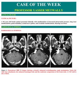
  • 1. CASE OF THE WEEK PROFESSOR YASSER METWALLY CLINICAL PICTURE CLINICAL PICTURE: A 40 years old female patient presented clinically with manifestations of increased intracranial pressure, long tract manifestations, ponto-medullary cranial nerve palsies. and cerebellar manifestations. Hearing was intact. RADIOLOGICAL FINDINGS RADIOLOGICAL FINDINGS: Figure 1. Postcontrast MRI T1 images showing a densely enhanced cerebellopontine angle meningioma, Notice the wide base attachment, the enhanced meningeal tail, and the moderate compression of the brain stem. A CSF cleft is also evident.
  • 2. Figure 2. Postcontrast MRI T1 images showing a densely enhanced cerebellopontine angle meningioma, Notice the wide base attachment, the enhanced meningeal tail, and the moderate compression of the brain stem. A CSF cleft is also evident. Figure 3. Postcontrast MRI T1 images showing a densely enhanced cerebellopontine angle meningioma, Notice the wide base attachment, the enhanced meningeal tail, and the moderate compression of the brain stem. A CSF cleft is also evident.
  • 3. Figure 4. The meningioma (syncytial subtype) is hyperintense on the MRI T2 images Table 1. MRI appearance of the various types of meningiomas Type Comment Fibroblastic Fibroblastic meningiomas are composed of large, narrow spindle cells. The distinct feature is the meningiomas presence of abundant reticulum and collagen fibers between individual cells. On MR imaging, fibroblastic meningiomas with cells embedded in a dense collagenous matrix appear as low signal intensity in Tl-weighted and T2-weighted pulse sequences. Transitional Transitional meningiomas are characterized by whorl formations in which the cells are wrapped meningiomas together resembling onion skins. The whorls may degenerate and calcify, becoming psammoma bodies. Marked calcifications can be seen in this histologic type. MR imaging of transitional meningiomas thus also demonstrates low signal intensity on Tl- weighted and T2-weighted images, with the calcifications contributing to the low signal intensity. Syncytial Syncytial (meningothelial, endotheliomatous) meningiomas contain polygonal cells, poorly defined meningiomas and arranged in lobules. Syncytial meningiomas composed of sheets of contiguous cells with sparse interstitium might account for higher signal intensity in T2-weighted images. Microcystic changes and nuclear vesicles can also contribute to increased signal intensity. Angioblastic Angioblastic meningiomas are highly cellular and vascular tumors with a spongy appearance. meningiomas Increased signal in T2-weighted pulse sequence of these tumors is due to high cellularity with increase in water content of tumor. Thus based on the correlation between histology and MR imaging appearance of meningiomas, it has been concluded that meningiomas significantly hyperintense to cortex tend to be primarily of syncytial or angioblastic type, whereas meningiomas hypointense to cortex tend to be primarily of fibrous or transitional type. Heterogeneous appearance of meningiomas in T2-weighted pulse sequence can be due to tumor vascularity, calcifications, and cystic foci. Table 2. MRI characteristics of meningiomas Pathological T2 MRI appearance type
  • 4. Fibroblastic Hypointense on the T2 images because of the existence of dense collagen and fibrous tissue Transitional Hypointense on the T2 images because of the existence of densely calcified psammoma bodies Syncytial Hyperintense on the T2 images because of the existence of high cell count, microcysts or significant tissue oedema Angioblastic Same as the syncytial type. Blood vessels appear as signal void convoluted structures Table 3. MRI characteristics of meningiomas MRI feature Description Vascular rim The peripheries of meningiomas are supplied by branches from the anterior or middle cerebral arteries that encircle the tumour and form the characteristic vascular rim Meningeal tail The tail extends to a variable degree away from the meningioma site and probably represents a meningeal reaction to the tumour Hypointense cleft Hypointense cleft between the tumour and the brain that probably represents blood vessels or a CSF interface The dural tail or quot;dural flairquot;  The dural tail is a curvilinear region of dural enhancement adjacent to the bulky hemispheric tumor. The finding was originally thought to represent dural infiltration by tumor, and resection of all enhancing dura mater was thought to be appropriate. However, later studies helped confirm that most of the linear dural enhancement, especially when it was more than a centimeter away from the tumor bulk, was probably caused by a reactive process. This reactive process includes both vasocongestion and accumulation of interstitial edema, both of which increase the thickness of the dura mater. Because the dural capillaries are quot;nonneural,quot; they do not form a blood-brain barrier, and, with accumulation of water within the dura mater, contrast material enhancement occurs.   Figure 5. Dural tail enhancement with meningioma. (a) Diagram illustrates the thin, relatively curvilinear enhancement
  • 5. that extends from the edge of a meningioma. Most of this enhancement is caused by vasocongestion and edema, rather than neoplastic infiltration. The bulk of the neoplastic tissue is in the hemispheric extraaxial mass; nonetheless, the dural tail must be carefully evaluated at surgery to avoid leaving neoplastic tissue behind. (b) Photograph of a resected meningioma shows the dense, quot;meaty,quot; well-vascularized neoplastic tissue. At the margin of the lesion, there is a quot;clawquot; of neoplastic tissue (arrowhead) overlying the dura mater (arrows) that is not directly involved with tumor. DIAGNOSIS: DIAGNOSIS: CEREBELLOPONTINE ANGLE MENINGIOMA DISCUSSION DISCUSSION: Lesions that arise within or involve the internal auditory canal (IAC) can be considered together with lesions affecting the cerebellopontine angle (CPA) cistern. This group of lesions consists of neoplastic and cystic masses arising from normal or ectopic structures in the CPA region and also inflammatory and vascular processes that affect the CPA and IAC cisterns and the cranial nerves located there. In addition to lesions specific to this region, any intraaxial or extraaxial process that involves subarachnoid cisternal spaces in general can also be seen here. Vestibulocochlear schwannoma  Vestibulocochlear schwannoma (VCS) is a benign, slowly growing neoplasm that arises from Schwann cells in the nerve sheath of the vestibulocochlear nerve. This tumor can affect other cranial nerves, but has a predilection for the eighth nerve, and in particular the vestibular portion of the nerve, which has led to the preferred use of the term VCS rather than the well-known synonym, acoustic neuroma [2]. The majority of intracranial schwannomas affect the vestibular portion of the eighth nerve, and only a minority affect the cochlear portion. VCS is a common tumor, accounting for approximately three fourths of all CPA masses and approximately one tenth of all intracranial tumors [3,4]. Patients with VCS usually present in the fourth to sixth decades. The most common symptom is progressive unilateral SNHL, often accompanied by tinnitus [5]. Vestibular symptoms of vertigo and dizziness are less common, which is curious given that most of these tumors arise from the vestibular portion of the nerve. This phenomenon may be because of a lesser susceptibility of the nerve to the effects of compression or because of the central nervous system's better ability to compensate for unilateral vestibular denervation [6]. Very large tumors in the CPA also may present with symptoms of cerebellar dysfunction or neuropathy of the lower cranial nerves. VCS is associated with neurofibromatosis type 2 (NF-2) [7], and this condition should be suspected when VCS is bilateral or is discovered in a child or young adult. The presence of NF-2 should also heighten the radiologist's awareness of possible associated lesions, including meningiomas and ependymomas. Malignant degeneration of VCS is rare and is associated with NF-1 [8]. Melanotic schwannoma is another malignant variant of VCS that may arise from melanocytes that share their neural crest origin with Schwann cells [9]. MRI is the preferred imaging modality for detecting and describing VCS, unless MRI is contraindicated. [10]. On unenhanced T1WI, schwannomas appear isointense to brain and can usually be detected as masses that displace the normal CSF signal. After contrast administration, schwannomas typically show uniform enhancement, although heterogeneity may be seen when areas of internal cystic or hemorrhagic change are present [11]. VCSs typically are centered near the porus acusticus and classically are described as having an elongated intracanalicular component and bulbous CPA cisternal component that result in an “ice cream cone” configuration (Fig. 1). Small lesions that are 15 mm or less may be contained entirely within the IAC and have a tubular shape [12]. Large lesions that are 30 mm or greater typically have a dominant CPA component. VCSs usually are solid but may show cystic or hemorrhagic change, particularly in large or rapidly growing tumors [13] (Fig. 2).
  • 6. Figure 6. Vestibulocochlear schwannoma. Axial T1-weighted MR imaging with contrast in patient with progressive SNHL shows an intensely enhancing schwannoma that nearly fills the IAC. The bulbous rounded portion of the tumor proximally projects into the CPA. Figure 7. Vestibulocochlear schwannoma. (A) Axial T1-weighted MR imaging without contrast. Intrinsic high T1 signal in the posteromedial portion of the CPA tumor represents subacute blood products as a result of hemorrhage in this schwannoma. (B) Postcontrast image demonstrates typical intense enhancement in the remainder of the tumor. Contrast-enhanced MRI is considered widely to be the first-line imaging technique for VCS evaluation, because of the sensitivity afforded by the intense enhancement that these tumors typically manifest, even in small lesions [14]. High- resolution T2 FSE imaging is, however, a supplemental noncontrast technique that can provide exquisite imaging of the CSF and nerves in the CPA and IAC. On T2 FSE, VCSs appear as nodules or filling defects against the background of bright CSF (Fig. 3). This technique allows for detection of tumors as small a few millimeters and can provide adjunctive information that is useful in surgical planning, such as identifying from which nerve component the tumor arises, defining the exact extent of the tumor boundaries, and whether there is a CSF cap at the fundus of the IAC. In the proper clinical setting, T2 FSE can serve as an inexpensive screening technique for patients with uncomplicated SNHL [15]. Figure 8. Vestibular schwannoma. (A) Axial T1-weighted postcontrast image of the IAC. Enhanced image demonstrates a nodule of intense enhancement, corresponding to a small intracanalicular schwannoma. (B) Axial high-resolution T2 FSE MR image clearly demonstrates the tumor nodule, which originates from the inferior vestibular branch of the eighth cranial nerve. Meningioma  Meningiomas are neoplasms that arise from arachnoid cap cells, dural fibroblasts, or arachnoid membrane [16]. The latter accounts for lesions in the CPA region. These tumors typically are benign and well circumscribed and are epidemiologically and histologically similar to meningiomas that occur elsewhere in the cranial vault. The incidence of meningioma in the CPA region is a distant second to VCS, accounting for approximately 5% to 10%
  • 7. of CPA masses [17,18]. Depending on their location, CPA meningiomas may present with cranial neuropathy, cerebellar dysfunction, or other symptoms resulting from local mass effect. Rarely, a meningioma that is limited to the IAC may mimic clinically a VCS [19]. On noncontrast CT, meningiomas may be isointense or hyperintense, and unlike schwannomas, may show calcification in up to one fourth of lesions [20]. The presence of hyperostosis on CT also suggests a diagnosis of meningioma. On MRI, meningiomas show T1 signal that is roughly similar to brain and may be hyperintense or hypointense on T2WI. The hallmark imaging features of meningioma include extra-axial dural-based location, intense enhancement, hyperostosis, and calcification (Fig. 8). The dural origin of these tumors typically results in a hemispheric mass with a broad dural margin that forms an obtuse angle against the adjacent bone compared with the spheroid or nodular mass with acute angle typically seen in VCS [21,22]. The presence of a “dural tail” also favors the diagnosis of meningioma, although this sign is not totally specific [23]. A CPA meningioma also may extend into the IAC and differentiation between meningioma and VCS may be difficult (Fig. 9). Figure 9. Meningioma. (A) Axial CT at bone algorithm of a patient with a temporal bone meningioma (M) centered over the petrous bone at the posterior floor of the middle cranial fossa. The hyperostotic bony changes (arrows) are characteristic for meningioma. (B) Postcontrast T1- weighted MR image shows characteristic enhancement. Figure 10. CPA meningioma. Axial high-resolution T2 FSE scan shows a broad dural-based extra-axial mass in the prepontine and CPA cisterns (M), with extension into the IAC (arrow). Epidermoid and other cystic and congenital masses  Epidermoid cyst  Epidermoid cyst (EC), also known as congenital cholesteatoma or epidermoid tumor, is caused by congenital rests of ectoderm in the CPA cistern. EC also can occur in the ventricles, petrous bone, tympanic cavity, skull base, and calvarium [24]. The squamous epithelium in these rests of ectoderm gives rise to accumulations of desquamated keratin debris. This keratinaceous material grossly has a glistening, pearly appearance that has led to this lesion called “the beautiful tumor” [25]. Patients with epidermoid typically present with symptoms in young or middle adulthood. EC shows CT density that is similar to or slightly greater than that of CSF. On MR imaging, EC appears fairly homogeneous, with signal characteristics that are similar to CSF. On T1WI, EC is hypointense but usually slightly brighter than CSF, giving rise to the term “dirty CSF.” In contrast to other CPA region tumors, EC does not enhance. EC is characteristically very hyperintense on T2WI and in some cases may be indistinguishable from CSF. When conventional MR imaging sequences are indeterminate, diffusion-weighted imaging can be useful and greatly increases the conspicuity of the lesion (Fig. 6).
  • 8. Figure 11. Epidermoid. (A) Axial T1- weighted image of the CPA region in a 35-year-old male with worsening vertigo. An epidermoid cyst is present (E), displacing the adjacent pons and cerebellum. The mass appears hyperintense relative to CSF. (B) Axial high-resolution T2 FSE MR imaging allows identification of the vestibulocochlear nerves and demonstrates anterior displacement of the cochlear branch (arrow) by the epidermoid cyst. The mass shows T2 signal identical to CSF. Arachnoid cysts (AC)  Arachnoid cysts (AC) are simple loculated CSF collections that form as a result of a congenital focal defect or duplication of the arachnoid membrane [26]. ACs can be seen in any location where the arachnoid membrane is found, but the some of the more common locations include the middle cranial fossa, sylvian cisterns, and hemispheric convexities. Less common locations include the CPA cisterns, suprasellar cistern, and cisterna magna. The imaging features of AC are those of a large uniform CSF collection with smooth rounded margins. AC appears isodense to CSF on CT and isointense to CSF on T1WI and T2WI, showing no contrast enhancement. AC may appear similar to EC. Mass affect on adjacent brain or mild remodeling of adjacent bone often are present (Fig. 7). When the cyst wall cannot be seen, differentiation between a simple enlarged CSF space and a small AC can be difficult. Figure 12. Arachnoid cyst. Axial T1-weighted MR images through the CPA cistern demonstrate a homogeneous CSF-signal collection () that causes mild mass effect on the adjacent brain structures. The cyst wall is incompletely perceptible. Dermoid cyst (DC)  Dermoid cyst (DC) is similar to EC in that it is the result of a congenital ectodermal inclusion, but differs in that it includes tissues from all three ectodermal layers. DCs may contain fat, dermal appendages (including hair and teeth), and calcification. DCs may rupture, resulting in spread of contents in the subarachnoid space and causing a chemical meningitis. Lipomas  Lipomas are congenital hamartomatous lesions that are distinct from DCs. Lipomas are masses of ectopic fat that probably form as a result of fatty maldevelopment of primitive meningeal mesodermal tissues [27]. Intracranial lipomas most commonly are seen at the midline, often in association with callosal or other midline anomalies, but can also occur in the CPA region [28]. Lipomas located within the IAC have been described [29]. Both DCs and lipomas on MRI show characteristic hyperintense fat signal on T1WI. Lipomas tend to be smaller and more homogeneous (Fig. 8). DCs often contain heterogeneous elements, including calcification, which is best appreciated CT. DC typically has greater mass effect and may rupture, producing a characteristic appearance of scattered fat droplets in the CSF space.
  • 9. Figure 13. Lipoma. (A) Axial T1- weighted MR image shows a CPA mass (arrow) with intrinsic T1 hyperintensity that is characteristic of lipoma. (B) Axial T1-weighted MR imaging with fat suppression confirms the fatty nature of the mass. Malignant and inflammatory meningitides  Any process that affects the meninges and subarachnoid spaces generally can affect the CPA and IAC cisterns. Spread of malignancy to the subarachnoid space and CSF results in carcinomatosis and may be seen in patients with primary CNS malignancy, adenocarcinoma (especially breast and lung), lymphoma, melanoma, leukemia, or other metastatic malignancy [30]. Infectious meningitis, particularly tuberculous and fungal meningitis, results in focal meningeal disease that is most prominent in the basilar cisterns. Noninfectious inflammatory conditions also cause localized meningeal changes, including sarcoidosis, histiocytosis, idiopathic hypertrophic pachymeningitis, and postoperative meningeal reaction [31–34]. Meningeal disease is evaluated best with contrast-enhanced MR imaging. Abnormal enhancement may appear as multiple nodular deposits or as focal or diffuse leptomeningeal or pachymeningeal thickening [35,36] (Fig. 9). A solitary meningeal deposit may mimic meningioma [37]. In general, when a focal meningeal process is discovered in the CPA or IAC, the remainder of the intracranial contents and spinal axis should be examined to evaluate for possible involvement. Figure 14. IAC metastasis. (A) Axial T2 FSE image of the temporal bone in an elderly woman with acute onset hearing loss. Ill-defined thickening of the nerves in the IAC is seen. (B) Axial- enhanced T1-weighted image shows enhancement in the IAC as a result of carcinomatous implant. Bilateral IAC and additional cisternal enhancement were also present (not shown). Glomus tympanicum paraganglioma  Glomus tympanicum paraganglioma (GTP) is tumor of glomus cells, or paraganglia, that reside in the inferior temporal bone. GTP is a subtype of paraganglioma that is localized to the glomus formations of the glossopharyngeal (Jacobson's nerve) that are found near the cochlear promontory. Paragangliomas, which are located in the jugular foramen, the proximal carotid sheath, and carotid bifurcation, are referred to as glomus jugulare, glomus vagale, and carotid body tumor, respectively; the unique features of these lesions are not included in this discussion. When a large glomus jugulare extends into the middle ear, the term “glomus jugulotympanicum” is used. GTP is three times more common in women than men, usually presenting in middle age. Patients typically present with pulsatile tinnitus, conductive hearing loss, or inner ear symptoms. Large jugulotympanicum tumors also may present with multiple lower cranial neuropathies. On otologic examination, GTP appears as a vascular retrotympanic mass. Small GTP tumors are seen readily on thin-section CT. The lesion appears a nodule of soft tissue at the medial wall of
  • 10. the middle ear, classically located at the cochlear promontory (Fig. 10). Larger tumors fill the middle ear, but typically do not cause bone erosion and tend to spare the ossicles. Glomus tumors show intense enhancement on MR imaging because of their vascular nature. MR imaging plays a critical role in defining the extension of GTP intracranially and into the skull base, inasmuch as the surgeon can appreciate only the tympanic part of the tumor on otoscopy. Figure 15. Glomus tympanicum paraganglioma. (A, B) Axial and coronal CT in a 36-year-old woman with pulsatile tinnitus and retrotympanic mass on otoscopic examination. A small soft-tissue mass is present at the medial wall of the middle ear overlying the lower portion of the cochlear promontory (arrows). SUMMARY SUMMARY Meningiomas constitute between 13% and 19% of intracranial tumors. They are the most common tumors of nonglial origin. These tumors most often occur in patients between the ages of 40 and 70 years. They are twice as common in females. Meningiomas usually arise from the arachnoid, and there is a predilection for occurrence near arachnoid granulations. Deep within the adult brain, the most frequent sites of occurrence are from the tuberculum sellae, suprasellar and parasellar regions, sphenoid ridge, and tentorial incisura and the cerebellopontine angle. Intraventricular tumors are also described. These arise from the tela choroidea or from meningeal cell rests in the choroid plexus. They usually arise within the lateral ventricles and are more common on the left. One of the keys to diagnosis of meningiomas is to recognize their extraaxial location. On CT, meningiomas usually appear as homogeneous hyperdense masses. Approximately 20% of meningiomas show some calcification, which is psammomatous in nature. About 10% of meningiomas are heterogeneous in density, which may reflect numerous intratumoral blood vessels or intratumoral cysts or cysts adjacent to the tumor. These usually occur in the angioblastic type. The vast majority of tumors show contrast enhancement, and peritumoral edema is common. On MR imaging, meningiomas are isointense to gray matter in the majority of cases or mildly hypointense to gray matter on Tl-weighted images. Heavily T2- weighted images show that these tumors are either isointense (50%) or mildly hyperintense (40%) to gray matter. MR imaging is particularly helpful in delineating the extra-axial location of these tumors. It may show a cerebrospinal fluid cleft between the tumor and adjacent brain or directly visualize the displaced dural margins or pial vessels. A broad-based dural margin is frequently visible,the tentorial incisura is frequently thickened. Addendum  A new version of this PDF file (with a new case) is uploaded in my web site every week (every Saturday and 
  • 11. remains available till Friday.) To download the current version follow the link quot;http://pdf.yassermetwally.com/case.pdfquot;.  You can also download the current version from my web site at quot;http://yassermetwally.comquot;.  To download the software version of the publication (crow.exe) follow the link:  http://neurology.yassermetwally.com/crow.zip The case is also presented as a short case in PDF format, to download the short case follow the link:  http://pdf.yassermetwally.com/short.pdf At the end of each year, all the publications are compiled on a single CD-ROM, please contact the author to know  more details. Screen resolution is better set at 1024*768 pixel screen area for optimum display.  For an archive of the previously reported cases go to www.yassermetwally.net, then under pages in the right  panel, scroll down and click on the text entry quot;downloadable case records in PDF formatquot; Also to view a list of the previously published case records follow the following link  (http://wordpress.com/tag/case-record/) or click on it if it appears as a link in your PDF reader REFERENCES References [1]. Curtin HD, Som PM, Bergeron RT. Temporal bone embryology and anatomy. In: Som PM, Curtin HD, eds. (3rd edition) Head and neck imaging. volume 2:: Mosby-Year Book 1996:1330-1338 [2]. National Institutes of Health Consensus Development Conference Statement on Acoustic Neuroma. December 11– 13, 1991. The Consensus Development Panel. Arch Neurol. 1994;51:201-207 [3]. Lo WWM, Solti-Bohman LG. Tumors of the temporal bone and the cerebellopontine angle. In: Som PM, Curtin HD, eds. (3rd edition) Head and neck imaging. volume 2:St. Louis: Mosby-Year Book 1996:1450 [4]. Swartz JD, Harnsberger HR. Imaging of the temporal bone. New York: Thieme Medical 1998p. 420 [5]. Kasantikul V, Netsky MG, Glassock Jr. ME, Hayes JW. Acoustic neurilemmoma. Clinicoanatomical study of 103 patients. J Neurosurg. 1980;52:28-35 [6]. Weissman JL. Hearing loss. Radiology. 1996;199:593-611 [7]. Lo WWM, Solti-Bohman LG. Tumors of the temporal bone and the cerebellopontine angle. In: Som PM, Curtin HD, eds. (3rd edition) Head and neck imaging. volume 2:St.Louis: Mosby-Year Book 1996:1451 [8]. Gruskin P, Carberry JN. Pathology of acoustic tumors. In: House WF, Luetze CM, eds. Acoustic tumors. volume 1:Baltimore: University Park Press 1979:85-148 [9]. Earls JP, Robles HA, McAdams HP, Rao KC. General case of the day. Malignant melanotic schwannoma of the eighth cranial nerve. Radiographics. 1994;14:1425-1427 [10]. Curtin HD. CT of acoustic neuroma and other tumors of the ear. Radiol Clin North Am. 1984;22:77-105 [11]. Tali ET, Yuh WT, Nguyen HD, et al. Cystic acoustic schwamnomas: MR characteristics. AJNR Am J Neuroradiol. 1993;14:1241-1247 [12]. Harnsberger HR. The lower cranial nerves. Handbook of head and neck imaging. (2nd edition) Chicago: Mosby- Year Book 1995:488-521 [13]. Charabi S, Mantoni M, Tos M, Thomson J. Cystic vestibular schwannomas: neuroimaging and growth rate. J Laryngol Otol. 1994;108:375-379
  • 12. [14]. Curati WL, Graif M, Kingsley DP, et al. Acoustic neuromas: Gd-DTPA enhancement in MR imaging. Radiology. 1986;158:447-451 [15]. Allen RW, Harnsberger HR, Shelton C, et al. Low-cost high-resolution fast spin-echo MR of acoustic schwannoma: An alternative to enhanced conventional spin-echo MR?. AJNR Am J Neuroradiol. 1996;17:1205-1210 [16]. Black PM. Meningiomas. Neurosurgery. 1993;32:643-657 [17]. Hasso AN, Smith DS. The cerebellopontine angle. Semin Ultrasound CT MR. 1989;10:280-301 [18]. Lo WWM. Cerebellopontine angle tumors. San Diego (CA): Categorical Course on Neoplasms of the Central Nervous System. American Society of Neuroradiology; 1990. [19]. Langman AW, Jackler RK, Althaus SR. Meningioma of the internal auditory canal. Am J Otol. 1990;11:201-204 [20]. Valavanis A, Schubiger O, Hayek J, Pouliadis G. CT of meningiomas on the posterior surface of the petrous bone. Neuroradiology. 1981;22:111-121 [21]. Lalwani AK, Jackler RK. Preoperative differentiation between meningioma of the cerebellopontine angel and acoustic neruoma suing MR imaging. Otolaryngol Head Neck Surg. 1993;109:88-95 [22]. Mikhael MA, Ciric IS, Wolff AP. Differentiation of cerebellowpontine angle neuromas and meningiomas with MR imaging. J Comput Assist Tomogr. 1985;9:852-856 [23]. Goldsher D, Litt AW, Pinto RS, et al. Dural “tail” associated with meningiomas on Gd-DTPA enhanced MR images: characteristics, differential diagnostic value, and possible implicatins for treatment. Radiology. 1990;176:447- 450 [24]. Zimmerman RA, Bilaniuk LT, Dolinskas C. Cranial computed tomography of epidermoid and congenital fatty; tumors of maldevelopmental origin. J Comput Tomogr. 1979;3:40-50 [25]. Osbom AG. Diagnostic neuroradiology. St. Louis: Mosby 1994p. 631 [26]. Rock IP, Zimmerman R, Bell WO, Fraser RA. Arachnoid cysts of the posterior fossa. Neurosurgery. 1986;18:176- 179 [27]. Smimiotopoulos JG, Yue NC, Rushing El. Cerebellopontine angle masses: radiologic-pathologic correlation. Radiographics. 1993;13:1131-1147 [28]. Dalley RW, Robertson WD, Lapointe JS, Durity FA. Computed tomography of a cerebellopontine angle lipom. J Comput Assist Tomogr. 1986;10:704-706 [29]. Cohen TI, Powers SK, Williams Jr. DW. MR appearance of intracanalicular eighth nerve lipoma. AJNR Am J Neuroradiol. 1992;13:1188-1190 [30]. Russell DS, Rubinstein LJ. Pathology of tumors of the nervous system. Baltimore: Williams & Wilkins 1989 [31]. Song SK, Schwartz IS, Strauchen JA, et al. Meningeal nodules with features of extranodal sinus histiocytosis with massive lymphadenoopathy. Am J Surg Pathol. 1989;13:406-412 [32]. Ranoux D, Devaux B, Lamy C, et al. Meningeal sarcoidosis, pseudo-meningioma, and pachymeningitis of the convexity. J Neurol Neurosurg Psychiatry. 1992;55:300-303 [33]. Mayer SA, Yim GK, Onesti ST, et al. Biopsy-proven isolated sarcoid meningitis. Case report. J Neurosurg. 1993;78:994-996 [34]. Martin N, Masson C, Henin D, et al. Hypertrophic cranial pachymeningitis: assessment with CT and MR imaging. AJNR Am J Neuroradiol. 1989;10:477-484
  • 13. [35]. Sze G. Diseases of the intracranial meninges: MR imaging features. AJR Am J Roentgenol. 1993;160:727-733 [36]. Sze G, Soletsky S, Bronen R, Krol G. MR imaging of the cranial meninges with emphasis on contrast enhancement and meningeal carcinomatosis. AJNR Am J Neuroradiol. 1989;10:965-975 [37]. Buff Jr. BL, Schick RM, Norrgaard T. Meningeal metastasis of leiomyosarcoma mimicking meningioma: CT and MR findings. J Comput Assist Tomogr. 1991;15:166-167 [38]. Lefebvre PP, Leprince P, Weber T, et al. Neuronotrophic effect of developing otic vesicle on cochleo-vestibular neurons: evidence for nerve growth facto involvement. Brain Res. 1990;507:254-260 [39]. Casselman JW, Offeciers FW, Govaerts PJ, et al. Aplasia and hypoplasia of the vestibulocochlear nerve: diagnosis with MR imaging. Radiology. 1997;202:773-781 [40]. Glastonbury CM, Davidson HC, Harnsberger HR. Identification of deficiency of the cochlear nerve in patients with sensorineural hearing loss using t2 fast-spin echo imaging. Presented at the American Society of Neuroradiology, 37th Annual Meeting, May 1999, San Diego, CA. [41]. Kim HS, Kim DI, Chung IH, Lee WS, Kim KY. Topographical relationship of the facial and vestibulocochlear nerves in the subarachnoid space and internal auditory canal. AJNR Am J Neuroradiol. 1998;19(6):1155-1161 [42]. Schuknecht HF. Pathology of the ear. (2nd edition) Philadelphia: Lea & Febiger 1993p. 332 [43]. Arnesen AR, Osen KK, Mugnaini E. Temporal and spatial sequence of anterograde degeneration in the cochlear nerve fibers of the cat. A light microscopic study. J Comp Neurol. 1978;178(4):679-696 [44]. Hoeffding V, Feldman ML. Degeneration in the cochlear nerve of the rat following cochlear lesions. Brain Res. 1988;449(1–2):104-115 [45]. Ylikoski J, Collan Y, Palva T. Pathologic features of the cochlear nerve in profound deafness. Arch Otolaryngol. 1978;104(4):202-207 [46]. Valvassori GE, Clemis JD. The large vestibular aqueduct syndrome. Laryngoscope. 1978;88:723-728 [47]. Jackler RK, De La Cruz A. The large vestibular aqueduct syndrome. Laryngoscope. 1989;99:1238-1243 [48]. Mafee MF, Charletta D, Kumar A, Belmont H. Large vestibular aqueduct and congenital SNHL. AJNR Am J Neuroradiol. 1992;13:805-819 [49]. Harnsberger HR, Dahlen RT, Shelton C. Advanced techniques in magnetic resonance imaging in the evaluation of the large vestibular aqueduct syndrome. Laryngoscope. 1995;105:1037-1042 [50]. Jackler RK, Luxford WM, House WF. Congenital malformations of the inner ear: a classification based on embryogenesis. Laryngoscope. 1987;97(pt 2, Suppl 40):2-14 [51]. Schuknecht HF. Pathology of the ear. Philadelphia: Lea & Febiger 1993p. 177 [52]. Schuknecht HF. Pathology of the ear. Philadelphia: Lea & Febiger 1993p. 115–189 [53]. Hasso AN, Casselman JW, Schmalbrock P. Temporal bone congenital anomalies. (3rd edition) In: Som PM, Curtin HD, eds. Head and neck imaging. volume 2:St. Louis: Mosby-Year Book 1996:1361-1367 [54]. Michel EM. Memoire sur les anomalies congenitales de l'oreille interne, avec la premiere observation authentique d'abscece complete d'oreille moyenne chez un sourd et muet de naissance, mort a l'age de onze ans. Gaz Med Strasb. 1863;23:55-58 [55]. Mondini C. Anatomia surdi nati sectio. De Bononiensi Scientiarum et Artium Instituto atzue Academia Commentarii. Bononiae. 1791;7:28-29419–31
  • 14. [56]. Lemmerling MM, Mancuso AA, Antonelli PJ, Kubilis PS. Normal modiolus: CT appearance in patients with a large vestibular aqueduct. Radiology. 1997;204:213-219 [57]. Brogan M, Chakeres DW. Gd-DTPA-enhanced MR imaging of cochlear schwannoma. AJNR Am J Neuroradiol. 1990;11:407-408 [58]. Saeed SR, Birzgalis AR, Ramsden RT. Intralabyrinthine schwannoma shown by magnetic resonance imaging. Neuroradiology. 1994;36:63-64 [59]. Mark AS. Contrast-enhanced magnetic resonance imaging of the temporal bone. Neuroimaging Clin North Am. 1994;4:117-131 [60]. Davidson HC, Krejei CS, Hamsberger HR. MR evaluation of labyrinthine schwannomas. Presented at the American Society of Neuroradiology 36th Annual Meeting, Philadelphia, Apri1 1998. [61]. Forton GE, Somers T, Hermans R, et al. Preoperatively diagnosed utricular neuroma treated by selective partial labyrinthiectomy. Ann Otol Rhinol Laryngol. 1994;103:885-888 [62]. Swartz JD. Temporal bone inflammatory disease. (3rd edition) In: Som PM, Curtin HD, eds. Head and neck imaging. Volume 2:St. Louis: Mosby-Year Book 1996:1415-1418 [63]. Seltzer S, Mark AS. Contrast enhancement of the labyrinth on MR scans in patients with sudden hearing loss and vertigo: evidence of labyrinthine disease. AJNR Am J Neuroradiol. 1991;12:13-16 [64]. Mark AS, Seltzer S, Nelson-Drake J, et al. Labyrinthine enhancement on gadolinium-enhanced magnetic resonance imaging in sudden deafness and vertigo: correlation with audiologic and electronystagmographic studies. Ann Otol Rhinol Laryngol. 1992;101:459-464 [65]. Mark AS. Contrast-enhanced magnetic resonance imaging of the temporal bone. Neuroimaging Clin North Am. 1994;4:117-131 [66]. Downie AC, Howlett DC, Koefman RJ, et al. Case report: prolonged contrast enhancement of the inner ear on magnetic resonance imaging in Ramsay Hunt syndrome. Br J Radiol. 1994;67:819-821 [67]. Davidson HC, Krejci CS, Harnsberger HR. MR Evaluation of labyrinthine schwannomas. Presented at the American Society of Neuroradiology 36th Annual Meeting, Philadelphia, April 1998. [68]. Swartz JD, Hamsberger HR. Imaging of the temporal bone. New York: Thieme Medical Publishers 1998p. 272–82 [69]. Weissman JL, Kamerer DB. Labyrinthitis ossificans. Am J Otolaryngol. 1993;14:363-365 [70]. Paparella MM. Labyrinthitis. In: Paparella MM, Shumrick DA, eds. Otolaryngology: the ear. Volume 2:Philadelphia: WB Saunders 1980:1735-1756 [71]. Swartz JD, Mandell DW, Wolfson RJ, et al. Fenestral and cochlear otosclerosis. Computed tomographic evaluation. Am J Otolaryngol. 1985;6:476-481 [72]. Casselman JW, Kuhweide R, Ampe W, et al. Pathology of the membranous labyrinth: comparison of T1- and T2- weighted and gadolinium-enhanced spin-echo and 3DFT-CISS imaging. AJNR Am J Neuroradiol. 1993;14:59-69 [73]. Heffner DK. Low-grade adenocarcinoma of probable endolymphatic sac origin: a clinicopathologic study of 20 cases. Cancer. 1989;64:2292-2302 [74]. Batsakis JG, el-Naggar AK. Papillary neoplasms (Heffner's tumors) of the endolymphatic sac. Ann Otol Rhinol Laryngol. 1993;102(pt 1):648-651 [75]. MacDougal AD, Sangalang VE. A previously unrecognized papillary endolymphatic sac tumor presenting as a cerebellopontine angle lesion. Can J Neurol Sci. 1985;189:203-204
  • 15. [76]. Palmer JM, Coker NJ, Harper RL. Papillary adenoma of the temporal bone in von Hippel-Lindau disease. Otolaryngol Head Neck Surg. 1989;100:64-68 [77]. Conley J, Janecka I. Schwan cell tumors of the facial nerve. Laryngoscope. 1974;84:958-962 [78]. Latack JT, Gabrielsen TO, Knake JE, et al. Facial nerve neruomas: radiologic evaluation. Radiology. 1983;149:731-739 [79]. Dort JC, Fisch U. Facial nerve schwanomma. Skull Base Surg. 1991;1:51-57 [80]. Pillsbury HC, Price HC, Gardiner LJ. Primary tumors of the facial nerve: diagnosis and management. Laryngoscope. 1983;93:1045-1048 [81]. Jackson CG, Glasscock 3rd ME, Sismanis A. Facial paralysis of neoplastic origin: diagnosis and management. Laryngoscope. 1980;90(pt 1):1581-1595 [82]. Nelson RA, House WF. Facial nerve neuroma in the posterior fossa: surgical considerations. In: Graham MD, House WF, eds. Proceedings of the Fourth International Symposium on Facial Nerve Surgery. New York: Raven 1982:403-406 [83]. Lidov M, Som PM, Stacy C, Catalano P. Eccentric cystic facial schwannoma: CT and MR features. J Comp Assist Tomogr. 1991;15:1065-1067 [84]. McMenomey SO, Glasscock 3rd ME, Nlinor LB, et al. Facial nerve neuromas presenting as acoustic tumors. Am J Otol. 1994;15:307-312 [85]. Davidson HC. Head and neck digital teaching file. Salt Lake City (UT): Electronic Medical Education Resource Group; 1999. Retrieved September 24, 2001, from DTF database. [86] Metwally, MYM: Textbook of neuroimaging, A CD-ROM publication, (Metwally, MYM editor) WEB-CD agency for electronic publication, version 10.1a January 2009