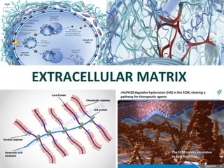
2. ECM
Many types of animal cells are surrounded by an extra cellular matrix
(ECM)- An Organized network of extra cellular materials.
It often plays a key regulatory role in determining the shape and
activities of the cell.
One of the best defined ECM is the basement membrane (basal lamina )
a continuousSheet 50 to 200 nm thickness.
It surrounds nerve fibers, muscles, and fat cells, underlies the basal
surfaces of epithelialTissues such as epidermis of the skin, lining of
digestive and respiratory tracts.
Basement membrane provides mechanical support for attached cells,
generate signals, That serve as a substratum for cell migration-it act as a
barrier for the passage of
macromolecules
12. Structure of ECM
• collagen
– the main ECM component, forms the main fibres
• elastin
• proteoglycans
- heteropolysacharides
• structural glycoproteins
- fibronectin, laminin
13. Functions of ECM
• Provides support anchorage and for cells.
• Regulates and determine cells dynamic behaviour :
- polarity of cells
- cell differentiation
- adhesion
- migration
• Provides mechanical support for tissues and organ architecture.
- growth
- regenerative and healing processes
- determination and maintenance of the
structure
• Place for active exchange of different metabolites, ions, water.
14. Cells Need Receptors to Recognize and Respond
to ECM
• Integrins
• Dystroglycan
• Syndecans
• Muscle-Specific kinase (MuSK)
• Others
15. Types of ECMs
• Basement membrane (basal lamina)
– Epithelia, endothelia, muscle, fat, nerves
• Elastic fibers
– Skin, lung, large blood vessels
• Stromal or interstitial matrix
• Bone, tooth, and cartilage
• Tendon and ligament
16. Why do all multicellular animals have ECM?
• Act as structural support to maintain cell organization and
integrity (epithelial tubes; mucosal lining of gut; skeletal
muscle fiber integrity)
• Compartmentalize tissues (pancreas: islets vs. exocrine
component; skin: epidermis vs. dermis)
• Provide hardness to bone and teeth (collagen fibrils
become mineralized)
• Present information to adjacent cells:
– Inherent signals (e.g., RGD motif in fibronectin)
– Bound signals (BMP7, TGFb, FGF, SHH)
• Serve as a highway for cell migration during development
(neural crest migration), in normal tissue maintenance
(intestinal mucosa), and in injury or disease (wound
healing; cancer)
17. Types of ECM Components
• Collagens
• Proteoglycans
– Perlecan, aggrecan, agrin, collagen XVIII
• Hyaluronan (no protein core)
• Large Glycoproteins
– Laminins, nidogens, fibronectin, vitronectin
• Fibrillins, elastin, LTBPs, MAGPs, fibulins
• “Matricellular” Proteins
– SPARC, Thrombospondins, Osteopontin, tenascins
18. Basement Membranes
• Specialized layers of extracellular matrix surrounding
or adjacent to all epithelia, endothelia, peripheral
nerves, muscle cells, and fat cells
• Originally defined by electron microscopy as ribbon-like
extracellular structures beneath epithelial cells
19. Generic Tissue Structure
Stratified,
pseudostratified,
or monolayer
(aka stroma)
“Tube within a Tube” concept
20. Basement Membrane
M. Loots, Univ. of Pretoria, S.A. J. Schwarzbauer, Curr. Biol. 1999
21. Basement Membranes
• In general, basement membranes appear very
similar to each other by EM.
• But all are not alike!
• There is a wealth of molecular and functional
heterogeneity among basement membranes,
due primarily to isoform variations of
basement membrane components.
22. Basement Membranes are Involved in a
Multitude of Biological Processes
• Influence cell proliferation, differentiation, and migration
• Maintain cell polarization and organization, as well as
tissue structure
• Act as a filtration barrier in the kidney between the
vasculature and the urinary space
• Separate epithelia from the underlying
stroma/mesenchyme/interstitium, which contains a non-basement
membrane matrix
25. Primary Components of
All Basement Membranes
• Collagen IV 6 chains form α chain heterotrimers
• Laminin 12 chains form several α-β-γ heterotrimers
• Entactin/Nidogen 2 isoforms
• Sulfated proteoglycans Perlecan and Agrin are the major
ones; Collagen XVIII is another
26.
27.
28. Collagen
• The most abundant protein in the body, making 25%-35% of all the
whole-body proteins.
• Collagen contributes to the stability of tissues and organs.
• It maintains their structural integrity.
• It has great tensile strenght.
• The main component of fascia, cartilage, ligaments, tendons, bone and
skin.
• Plays an important role in cell differentiation, polarity, movement
• Plays an important role in tissue and organ development
29. Collagen structure
Collagen is insoluble glycoprotein (protein + carbohydrate)
Collagen polypeptide structure:
- G – X – A – G – A – A – G – Y – A – G – A – A – G – X – A – G – A –
– A – G – X – A – G – A – A – G – Y – A – G – A – A – G – X – A – G –
– A – A – G – X – A – G – A – A – G – Y – A – G – A – A – G – X – A –
G - glycine, X - proline or hydroxyproline, Y – lysin or hydroxylysine, A – amino acid
• Proline and hydroxyproline constitute about 1/6 of the total
sequence, provide the stifness of the polypeptide chain.
• Carbohydrates : glucose, galactose
31. Collagen provides structural support to
tissues
• The principal function of collagens is to
provide structural support to tissues.
• Collagens are a family of over 20 different
extracellular matrix proteins.
– Together they are the most abundant proteins
in the animal kingdom.
32. 15.3 Collagen provides structural support to tissues
• All collagens are organized into triple
helical, coiled-coil “collagen subunits.”
– They are composed of three separate
collagen polypeptides.
• Collagen subunits are:
– secreted from cells
– then assembled into larger fibrils and fibers
in the extracellular space
33.
34.
35. Cells Need Receptors to Recognize and Respond
to ECM
• Integrins
• Dystroglycan
• Syndecans
• Muscle-Specific kinase (MuSK)
• Others
36.
37.
38. Three helical polypeptide units twist to form a triple-helical
collagen molecule: a molecular "rope" which has some
bending stiffness and does not undergo rotation.
39. Diversity of Collagens
Type I fibrils Skin, tendon, bone, ligaments, dentin,
interstitium
Type II Fibrils Cartilage, vitreous humor
Type III Fibrils Skin, muscle, bv
Type IV 2D sheets All basement membranes
Type V Fibrils with
globular end
Cornea, teeth, bone, placenta, skin,
smooth muscle
Type VI Fibril-assoc. (I) Most interstitial tissues
Type VII Long anchoring
fibril
Skin--connects epidermal basement
membrane/hemidesmosome to dermis
Type IX Fibril-assoc. (II) Cartilage, vitreous humor
Type XIII Transmembrane Hemidesmosomes in skin
Type XV HSPG Widespread; near basement
membranes in muscle
Type XVII Transmembrane Hemidesmosomes in skin (aka BPAG2
or BP180)
40. Collagen IV: Network or Sheet Forming
• Six genetically distinct a chains: α1- α6, ~180 kDa each
• Chains form three types of heterotrimers:
– (a1)2(α2), α3α4α5, (α5)2(α6)
• Like all Collagens, comprised mainly of Gly-x-y repeats, y is frequently
proline
• Gly-x-y pattern has multiple interruptions
– Provides flexibility to the collagen network and to the basement
membrane
Hudson et al., NEJM 2003
41. Collagen IV Trimer
• 7S domain at N-terminus
• Interrupted Gly-x-y triple
helical domain
• C-terminal non-collagenous
domain--NC1
42. Collagen IV Network
Trimers (aka protomers)
associate with each other,
four at the N-terminus and
two at the C-terminus
(hexamer), to form a chicken
wire-like network that
provides strength and
flexibility to the basement
membrane.
46. Fibrillar Collagens (I, II, III, V)
• Connective tissue proteins that
provide tensile strength
• Triple helix, composed of three a
chains
• Glycine at every third position
(Gly-X-Y)
• High proline content
– Hydroxylation required for proper
folding and secretion
• Found in bone, skin, tendons,
cartilage, arteries
47. CCoollllaaggeenn CCrroosssslliinnkkiinngg
• Once formed, collagen fibrils are greatly strengthened by covalent crosslinks that form
between the constituent collagen molecules.
• The first step in crosslink formation is the deamination by the enzyme lysyl oxidase of
specific lysine and hydroxylysine side chains to form reactive aldehyde groups.
• The aldehydes then form covalent bonds with each other or with other lysine or
hydroxylysine residues.
48. CCoollllaaggeenn CCrroosssslliinnkkiinngg
• If crosslinking is inhibited (Lysyl
hydroxylase mutations; vitamin C
deficiency), collagenous tissues
become fragile, and structures such
as skin, tendons, and blood vessels
tend to tear. There are also many
bone manifestations of under-crosslinked
collagen.
• Hydroxylation of specific lysines
governs the nature of the cross-link
formed, which affects the
biomechanical properties of the
tissue. Collagen is especially highly
crosslinked in the Achilles tendon,
where tensile strength is crucial.
49. Bone is Composed of Mineralized
Type I Collagen Fibrils
Bone is 70%
mineral and 30%
protein, mostly
collagen
Mineral is Dahllite,
similar to
hydroxyapatite
(contains calcium,
phosphate,
carbonate)
50. 15.3 Collagen provides structural support to tissues
• Mutations of collagen genes can lead to
a wide range of diseases, from mild
wrinkling to brittle bones to fatal
blistering of the skin.
51. Type IV Collagen Mutations and Human
Disease
• COL4A1 mutations
– Small vessel disease/retinal vascular
tortuosity
– Hemorrhagic stroke
– Porencephaly
– HANAC syndrome
• COL4A3/A4/A5 mutations
– Alport syndrome/hereditary
glomerulonephritis
Kidney Glomerular BM
52. Scurvy
• Liver spots on skin, spongy gums,
bleeding from mucous membranes,
depression, immobility
• Vitamin C deficiency
• Ascorbate is required for prolyl
hydroxylase and lysyl hydroxylase
activities
• Acquired disease of fibrillar collagen
Illustration from Man-of-War by Stephen Biesty (Dorling-Kindersley, NY, 1993)
53. Some Genetic Diseases of Collagen
• Collagen I
– Osteogenesis imperfecta
– Ehlers-Danlos syndrome type VII
• Collagen II
– Multiple diseases of cartilage
• Collagen III
– Ehlers-Danlos syndrome type IV
• Collagen IV
– Alport syndrome, stroke, hemorrhage, porencephaly
• Collagen VII
– Dystrophic epidermolysis bullosa (skin blistering)
54. Different Types of Mutations in Collagen I a Chain Genes
Cause Different Disease Severities
Gene location mutation Syndrome
COL1A1 17q22 Null alleles OI type I
Partial deletions; C-terminal
substitutions
OI type II
N-terminal substitutions OI types I, III or IV
Deletion of exon 6 EDS type VII
COL1A2 7q22.1 Splice mutations; exon deletions OI type I
C-terminal mutations OI type II, IV
N-terminal substitutions OI type III
Deletion of exon 6 EDS type VII
55. Osteogenesis Imperfecta
(brittle bone disease)
Clinical:
Ranges in severity from mild to perinatal lethal
bone fragility, short stature, bone deformities, teeth abnormalities,
gray-blue sclerae, hearing loss
Biochemical:
reduced and/or abnormal type I collagen
Molecular:
mutations in either type I collagen gene, COL1A1 or COL1A2, resulting
in haploinsufficiency or disruption of the triple helical domain
(dominant negative: glycine substitutions most common)
56. Generalizations
• Most ECM proteins are large, modular,
multidomain glycosylated or glycanated proteins
• Some domains recur in
different ECM proteins
– Fibronectin type III repeats
– Immunoglobulin repeats
– EGF-like repeats
– Laminin Globular (G) domain
– von Willebrand factor
Perlecan
58. Type IV Collagen NC1 Domains
• Exhibit anti-angiogenic activity
• Target tumor vasculature
59. Osteogenesis Imperfecta
(brittle bone disease)
Clinical:
Ranges in severity from mild to perinatal lethal
bone fragility, short stature, bone deformities, teeth abnormalities,
gray-blue sclerae, hearing loss
Biochemical:
reduced and/or abnormal type I collagen
Molecular:
mutations in either type I collagen gene, COL1A1 or COL1A2, resulting
in haploinsufficiency or disruption of the triple helical domain
(dominant negative: glycine substitutions most common)
60. Dominant Negative COL1 Mutations
* Gly subst. in COL4A2
*
Gly subst. in COL4A1
Byers P. Connective Tissue and Its Inheritable Disorders 1993, pp317-50.
½ of the
trimers are
abnormal
¾ of the
trimers are
abnormal
61. Elastin aanndd EEllaassttiicc FFiibbeerrss EExxhhiibbiitt
RRuubbbbeerr--LLiikkee PPrrooppeerrttiieess
• Physiological importance lies in the unique
elastomeric properties of elastin. Found in tissues
in which reversible extensibility or deformability
are crucial, such as the major arterial vessels (esp.
aorta), the lung and the skin.
• Elastin is characterized by a high index of
hydrophobicity (90% of all the amino acid residues
are nonpolar). One-third of the amino acid
residues are glycine with a preponderance of the
nonpolar amino acids Ala, Val, Leu, and Ile. As in
collagen, one-ninth of the residues are proline
(but with very little hydroxylation).
• Early in development, the elastic fibers consists of
microfibrils, which define fiber location and
morphology. Over time, tropoelastin accumulates
within the bed of microfibrils.
62. Elastic Fiber Biogenesis
• Elastic fibers are very complex, difficult to
repair structures
• There are two morphologically
distinguishable components
– Microfibrils
– Elastin
• Assembly follows a well-defined sequence
of events:
1. Assembly of microfibrils
2. Association of tropoelastin aggregates with
microfibrils
3. Crosslinking of tropoelastins with each other
by lysyl oxidase to form polymers Shifren and Mecham, 2006
64. Microfibril Components: ~30
• Fibrillin--three forms
• Microfibril-associated glycoproteins
(MAGPs)--two forms
• Latent TGFb Binding Proteins (LTBPs)--
four forms
• Proteoglycans, MFAPs, Fibulins,
Emilins, Collagens, Decorin, et al.
65. Marfan Syndrome
• Caused by dominant Fibrillin-1
(FBN1) mutations
– Haploinsufficiency is the culprit
• Skeletal, ocular, and
cardiovascular defects
• Deficiency of elastin-associated
microfibrils
• Syndrome may result from
alterations in TGFb signaling,
rather than purely structural
changes in microfibrils
66. Evidence for FBN/BMP7 Interactions
Fbn2+/-; Bmp7+/- trans-heterozygous
animals
show limb patterning
defects.
Artaga-Solis et al., J. Cell Biol. 2001
Specific
fragments of
Fibrillin 1, but
not LTBP1, bind
to BMP7
Gregory et al., JBC 2005
67. Elastic Fiber Biogenesis
• Elastic fibers are very complex, difficult to
repair structures
• There are two morphologically
distinguishable components
– Microfibrils
– Elastin
• Assembly follows a well-defined sequence
of events:
1. Assembly of microfibrils
2. Association of tropoelastin aggregates with
microfibrils
3. Crosslinking of tropoelastins with each other
by lysyl oxidase to form polymers Shifren and Mecham, 2006
70. Sulfated Proteoglycans
• Have protein cores with large
glycosaminoglycan (GAG) side chains (from
1 to >100) attached to serines
• Some PGs contain heparan sulfate
– Perlecan, Agrin, Collagen XVIII
(endostatin)
• Others contain chondroitin, keratan or
dermatan sulfate
• GAG chains are responsible for most of the
biological properties of proteoglycans and
provide charge to basement membranes
Heparan sulfate:
Composed of D-glucuronate-2-sulfate +
N-sulfo-D-glucosamine-6-sulfate
71. Some Major Proteoglycan Family Members
From: Iozzo, R.V. (1998) Ann. Rev. Biochem. 67:609 From: Iozzo, R.V. (2001) J. Clinic. Invest. 108:165
72. Perlecan
• Found widely in basement membranes and in cartilage.
• Contains domains similar to LDL receptor, laminin, and N-CAM
• Binds to Collagen IV and to Entactin/Nidogen
73. Endorepellin: Domain V of Perlecan
• Exhibits anti-angiogenic activity
• Targets tumor vasculature
74. Glycosaminoglycan Classification
Proteoglycans can be categorised depending upon the
nature of their glycosaminoglycan chains.
• Hyaluronic acid (does not contain any sulfate)
- non-covalent link complex with proteoglycans
• Chondroitin sulfate cartilage, bone
• Dermatan sulfate skin, blood vessels
• Heparan sulfate basement membrane,
component of cells surface
• Keratan sulfate cornea, bone, cartilage
often aggregated with chondroitin
sulfate
75. Function of Proteoglycans
• organize water molecules
- resistant to compression
- return to original shape
- repel negative molecules
• occupy space between cells and collagen
• high viscosity
- lubricating fluid in the joints
• specific binding to other macromolecules
• link to collagen fibers
- form network
- in bone combine with calcium
salts (calcium carbonate,
hydroxyapatite)
• cell migration and adhesion
- passageways between cells
• anchoring cells to matrix fibers
76. Structural Glycoproteins
• Direct linkage to collagen or proteoglycans
- anchoring collagen fibers to cell membrane
- covalent attachment to membrane lipid
• Major adhesive structural glycoproteins
- fibronectin
- laminin
77. Fibronectin Structure
• Dimer connected at C-terminal by S-S linkage
• Rigid and flexible domains
• Cell binding domain RGDS
(arg, gly, asp, ser)
- binding receptor in cell membranes
• Domain is binding to
- collagen type I, II and III
- heparin sulfate
- hyaluronic acid
- fibrin
78.
79. Fibronectin Function
• cell adhesion
• cell differentiation
• anchoring basal laminae to other ECM
• blood clothing
- clothing process, link to fibrin
80. Laminin
Heterotrimers are composed of
one a, one b, and one g chain.
• 400 to 800 kDa cruciform, Y, or rod-shaped
macromolecules.
• Major glycoprotein of basement membranes
—it’s required!
• Chains are evolutionarily related.
• 5 alpha, 4 beta, and 3 gamma chains are
known. They assemble with each other non-randomly.
• 15 heterotrimers described to date.
LM-521
81. Laminin Structure
and Function
• cross-shaped glycoprotein
• 3 polypeptide chains
• domain bind
- collagen type IV
- heparin
- heparin sulfate
• cell surface receptor
• cell adhesion
• cell differentiation
• anchoring the glycoprotein to
basal laminae
83. Laminin
• All laminin chains share structural
homology
• Contain globular, rod (EGF-like
repeats), and coiled-coil domains
• Alpha chains are unique, contain a
C-terminal laminin globular “LG”
domain, ~100 kDa
(New nomenclature)
84. Laminin Trimers Polymerize
• Laminin chains assemble into
trimers in the ER and are
secreted as trimers into the
extracellular space.
• Full-sized laminin trimers can
self-polymerize into a
macromolecular network
through short arm-short arm
interactions.
• The a chain LG domain is left
free for interactions with
cellular receptors.
88. Proteases degrade extracellular
matrix components
MMPs
– A large family of zinc containing enzymes
called MMP that are either secreted into the
– Extra cellular space or anchored to plasma
membrane
• Cells must routinely degrade and replace
their extracellular matrix as a normal part
of
– development (embryonic cell migration)
– wound healing
– .
89. MMPS
MMPs have been implicated in a number of pathological conditions, including
Arthritis, hepatitis,atherosclerosis,tooth and gum diseases and tumor progression
Inherited skeletal disorders have been found mutations in MMP genes.
90.
91. • Extracellular matrix proteins are
degraded by specific proteases, which
cells secrete in an inactive form.
• These proteases are only activated in
the tissues where they are needed.
• Activation usually occurs by proteolytic
cleavage of a propeptide on the
protease.
92. Proteases degrade extracellular matrix components
• The matrix metalloproteinase (MMP)
family is one of the most abundant
classes of these proteases.
– It can degrade all of the major classes of
extracellular matrix proteins.
• MMPs can activate one another by
cleaving off their propeptides.
– This results in a cascade-like effect of
protease activation that can lead to rapid
degradation of extracellular matrix proteins.
93. Proteases degrade extracellular matrix components
• Cells secrete inhibitors of these
proteases to protect themselves from
unnecessary degradation.
• Mutations in the matrix
metalloproteinase-2 gene give rise to
numerous skeletal abnormalities in
humans.
– This reflects the importance of extracellular
matrix remodeling during development.
94. Interaction of cells with with extra cellular materials
The components of ECM such as Fibronectin, laminin, proteoglycan, and collagen
Are capable of binding to receptors situated on the cell surface.
The most important family of receptors that attach cells to their microenviroinment
Is the Integrins
Integrins which are found only in animals are a family of membrane proteins that play
a key role in integrating the extracellular and intracellular enviroinments. On the
One side of the plasma membrane – binds to array molecules (ligands) that are
Present in the extra cellular enviroinment.
On the intracellular side of the membrane, integrin binds to interact with directly or
Indirectly with different proteins.
Integrins composed of two membrane spanning polypeptides α ,ß that are
Non covalently linked
95. Integrins can be activated rapidly by events within the cell that alter the
conformation of Cytoplasmic domains of the integrin subunits.
Integrins have been implicated in two main types of activities
Adhesion of cells to their substratum and transmisssion of signals from the
external enviroinment to the cell interior.
The binding of the extra cellular domain of an integrin to a ligand such as
fibronectin, Collagen can induce confirmational changes at the opposite
cytoplasmic end of integrins.
97. Most integrins are receptors for
extracellular matrix proteins
• Virtually all animal cells express integrins.
– They are the most abundant and widely
expressed class of extracellular matrix protein
receptors.
• Some integrins associate with other
transmembrane proteins.
98. 15.13 Most integrins are receptors for extracellular matrix proteins
• Integrins are composed of two distinct
subunits, known as α and β chains.
• The extracellular portions of both chains
bind to extracellular matrix proteins
• The cytoplasmic portions bind to
cytoskeletal and signaling proteins.
99. Most integrins are receptors for extracellular matrix proteins
• In vertebrates, there are many α and
β integrin subunits.
– These combine to form at least 24 different
αβ heterodimeric receptors.
• Most cells express more than one type
of integrin receptor.
– The types of receptor expressed by a cell
can change:
• over time or
• in response to different environmental
conditions
100. Most integrins are receptors for extracellular matrix proteins
• Integrin receptors bind to specific amino
acid sequences in a variety of
extracellular matrix proteins.
• All of the known sequences contain at
least one acidic amino acid.
101. Integrin receptors participate in cell
signaling
• Integrins are signaling receptors that
control both:
– cell binding to extracellular matrix proteins
– intracellular responses following adhesion
• Integrins have no enzymatic activity of
their own.
– Instead, they interact with adaptor proteins
that link them to signaling proteins.
102. Integrin receptors participate in cell signaling
• Two processes regulate the strength of
integrin binding to extracellular matrix
proteins:
– affinity modulation
• varying the binding strength of individual
receptors
– avidity modulation
• varying the clustering of receptors
103. Integrin receptors participate in cell signaling
• Changes in integrin receptor
conformation are central to both types
of modulation.
• They can result from changes:
– at the cytoplasmic tails of the receptor
subunits or
– in the concentration of extracellular cations
104. Integrin receptors participate in cell signaling
• In inside-out signaling, changes in
receptor conformation result from
intracellular signals that originate
elsewhere in the cell.
– For example, at another receptor
• In outside-in signaling, signals initiated
at a receptor are propagated to other
parts of the cell.
– For example, upon ligand binding
105. Integrin receptors participate in cell signaling
• The cytoplasmic proteins associated with
integrin clusters vary greatly depending on:
– the types of integrins and extracellular matrix
proteins engaged.
• The resulting cellular responses to integrin
outside-in signaling vary accordingly.
• Many of the integrin signaling pathways
overlap with growth factor receptor pathways.
106. Integrins and extracellular matrix
molecules play key roles in
development
• Gene knockout by homologous
recombination has been applied in mice
to;
– over 40 different extracellular matrix proteins
– 21 integrin genes
• Some genetic knockouts are lethal, while
others have mild phenotypes.
107. Integrins and extracellular matrix molecules play key roles in
development
• Targeted disruption of the β1 integrin
gene has revealed that it plays a critical
role in:
– the organization of the skin
– red blood cell development
110. Selectins
Comprise a family of integral membrane glycoproteins that recognize and bind
To a particular arrangement of sugars in the oligo saccharides.
Selectins possess a small cytoplasmic domain, a single membrane spaning
Domain.
3 known selectins – E selectins- present in the endothelial cells.
L- Selectins LEUKOCYTE
P- Selectins- PLATELETS
All three selectins recognize a particular group of sugars that is found at the
Ends of the carbohydrate chains of certain complex glycoproteins.
The importance of integrins in the inflammatory response is demonstrated by
A rare disease called leukocyte adhesion deficiency (LAD), Leukocyte
Of these individuals lack the ability to adher to the endothelial layer of
Venules. These patients suffer from repeated bacterial infections.
111. Selectins control adhesion of
circulating immune cells
• Selectins are cell-cell adhesion receptors
expressed exclusively on cells in the vascular
system.
• Three forms of selectin have been identified:
– L-selectin
– P-selectin
– E-selectin
112.
113. Selectins control adhesion of circulating immune cells
• Selectins function to arrest circulating
leukocytes in blood vessels so that they
can crawl out into the surrounding
tissue.
• In a process called discontinuous cell-cell
adhesion, selectins on leukocytes
bind weakly and transiently to
glycoproteins on the endothelial cells.
– The leukocytes come to a “rolling stop”
along the blood vessel wall.
114. Tight junctions form selectively permeable barriers between
cells
• Tight junctions also preserve epithelial
cell polarity by serving as a “fence.”
– It prevents diffusion of plasma membrane
proteins between the apical and basal
regions.
115. Tight junctions : sealing the extra cellular space
Tight junctions are located at the very epical end of the junctional complex between
Adjacent epithelial cells.
116.
117. Septate junctions in invertebrates
are similar to tight junctions
• The septate junction:
– is found only in invertebrates
– is similar to the vertebrate tight junction
• Septate junctions appear as a series of
either straight or folded walls (septa)
between the plasma membranes of
adjacent epithelial cells.
118. Septate junctions in invertebrates are similar to tight junctions
• Septate junctions function principally as
barriers to paracellular diffusion.
• Septate junctions perform two functions
not associated with tight junctions:
– they control cell growth and cell shape
during development.
• A special set of proteins unique to septate
junctions performs these functions.
119. ADHEREN JUNCTIONS
Adherens junctions : are found in a variety of sites within the body.They are
particularly Common in epithelia such as the lining of the intestine
where they occur as belts Binding cell to its surrounding neighbours.
The cadherin clusters of adherin junction – connect the external enviroinment to
theActin cytoskeleton and provide a pathway for signals to be transmitted from the
Cell exterior to the cytoplasm.
Adherin junctions situated between endothelial cells that line walls of blood vessels
transmit signals that ensure the survival of the cells.
120. Adherens junctions link adjacent
cells
• Adherens junctions are a family of related
cell surface domains.
– They link neighboring cells together.
• Adherens junctions contain
transmembrane cadherin receptors.
121. Adherens junctions link adjacent cells
• The best-known adherens junction is
the zonula adherens.
– It is located within the junctional complex
that forms between neighboring epithelial
cells in some tissues.
• Within the zonula adherens, adaptor
proteins called catenins link cadherins
to actin filaments.
122. Desmosomes : are disk-shaped adhesive junctions approximately 1mm in diameter.
That are found in a variety of tissues.
Desmosomes are particularly numerous in tissues that are subjected to mechanical
stress such as cardiac muscle and the epithelial layers of the skin and uterine
Cervix.
123. Desmosomes are intermediate
filamentbased cell adhesion
complexes
• The principal function of desmosomes is
to:
– provide structural integrity to sheets of
epithelial cells by linking the intermediate
filament networks of cells.
124. Desmosomes are intermediate filament-based cell adhesion
complexes
• Desmosomes are components of the
junctional complex.
• At least seven proteins have been
identified in desmosomes.
• The molecular composition of
desmosomes varies in different cell and
tissue types.
125. Desmosomes are intermediate filament-based cell adhesion
complexes
• Desmosomes function as both:
– adhesive structures
– signal transducing complexes
• Mutations in desmosomal components
result in fragile epithelial structures.
– These mutations can be lethal, especially if
they affect the organization of the skin.
126. Hemidesmosomes attach epithelial
cells to the basal lamina
• Hemidesmosomes, like desmosomes,
provide structural stability to epithelial
sheets.
• Hemidesmosomes are found on the
basal surface of epithelial cells.
– There, they link the extracellular matrix to
the intermediate filament network via
transmembrane receptors.
127. Hemidesmosomes attach epithelial cells to the basal lamina
• Hemidesmosomes are structurally
distinct from desmosomes.
• They contain at least six unique
proteins.
128. Hemidesmosomes attach epithelial cells to the basal lamina
• Mutations in hemidesmosome genes
give rise to diseases similar to those
associated with desmosomal gene
mutations.
• The signaling pathways responsible for
regulating hemidesmosome assembly
are not well understood.
129. Gap junctions : are sites between animal cells that are specilized for
Intercellular communication.
Gap junctions have a simple molecular compostion they are composed entirely
Of an integral membrane protein called connexin. Connexins are organized
Into multi subunit complexes called connexons that are completely span the
Membrane.
Each connexin is composed of six connexin subunits arranged in a ring around
A central opening.
130.
131. Gap junctions allow direct transfer
of molecules between adjacent cells
• Gap junctions are protein structures that
facilitate direct transfer of small molecules
between adjacent cells.
• They are found in most animal cells.
132. Gap junctions allow direct transfer of molecules between adjacent
cells
• Gap junctions consist of clusters of
cylindrical gap junction channels, which:
– project outward from the plasma
membrane
– span a 2-3 nm gap between adjacent cells
• The gap junction channels consist of
two halves, called connexons or
hemichannels.
– Each consists of six protein subunits called
connexins.
133. Gap junctions allow direct transfer of molecules between adjacent
cells
• Over 20 different connexin genes are
found in humans.
– These combine to form a variety of
connexon types.
• Gap junctions:
– allow for free diffusion of molecules 1200
daltons in size
– exclude passage of molecules 2000
daltons
134. Gap junctions allow direct transfer of molecules between adjacent
cells
• Gap junction permeability is regulated
by opening and closing of the gap
junction channels, a process called
“gating.”
• Gating is controlled by changes in
– intracellular pH
– calcium ion flux
– direct phosphorylation of connexin subunits
135. Plasmodesmata : are cytoplasmic channels that pass through the cell walls of
Adjacent cells .
Plasmodesmata are lined by plasma membrane and usually contain a dense
Central structure called desmotubule, derived from the smooth endoplasmic
Reticulum of the two cells
136. Gap junctions allow direct transfer of molecules between adjacent
cells
• Two additional families of nonconnexin
gap junction proteins have been
discovered.
– This suggests that gap junctions evolved
more than once in the animal kingdom.
137. CADHERINS
The Cadherins are a large family of glycoproteins that mediate Ca2+ dependent
Cell- cell adhesion and transmit signals from the ECM to cytoplasm.
Cadherins typically join cells of similar type to one another and do so predominantly
By binding to the same cadherin present on the surface of neighbouring cell.
The best studied cadherins are E-Cadherins (epithelial ), N-cadherins (neural)
And P-Cadherin (Placental).
138. Calcium-dependent cadherins
mediate adhesion between cells
• Cadherins constitute a family of cell
surface transmembrane receptor
proteins that are organized into eight
groups.
• The best-known group of cadherins is
called the “classical cadherins.”
– It plays a role in establishing and
maintaining cell-cell adhesion complexes
such as the adherens junctions.
139. Calcium-dependent cadherins mediate adhesion between cells
• Classical cadherins function as clusters
of dimers.
• The strength of adhesion is regulated
by varying both:
– the number of dimers expressed on the cell
surface
– the degree of clustering
140. Calcium-dependent cadherins mediate adhesion between
cells
• Classical cadherins bind to cytoplasmic
adaptor proteins, called catenins.
– Catenins link cadherins to the actin
cytoskeleton.
• Cadherin clusters regulate intracellular
signaling by forming a cytoskeletal
scaffold.
– This organizes signaling proteins and their
substrates into a three-dimensional
complex.
141. Calcium-dependent cadherins mediate adhesion between
cells
• Classical cadherins are essential for
tissue morphogenesis, primarily by
controlling:
– specificity of cell-cell adhesion
– changes in cell shape and movement
142. Calcium-independent NCAMs
mediate adhesion between neural
cells
• Neural cell adhesion molecules (NCAMs)
are expressed only in neural cells.
• They function primarily as homotypic cell-cell
adhesion and signaling receptors.
143. Calcium-independent NCAMs mediate adhesion between neural
cells
• Nerve cells express three different
types of NCAM proteins.
– They arise from alternative splicing of a
single NCAM gene.
Editor's Notes
Of course, the answer relates to what ECM does.
Very diverse group; Matricellular proteins interact with cell-surface receptors, extracellular matrix (ECM), and/or growth factors and proteases, but do not in most cases serve as structural components. They modulate cell/matrix interactions.
What is an epithelial cell? Between tubes are blood vessels (more tube) musculoskeletal system (myotubes), other organs, lung and kidney, primarily tubes. It’s ECM that allows these tubes to maintain their integrity, compartmentalization, and separation from each other.
Cornea? Classically divided into lamina densa and lamina lucida. For many epithelial cells in the body, the BM is its only link to the rest of the body.
Could the LL be artifactual?
Source of matrigel; Steve Weiss’s talk suggested lots of laminin, very little collagen
Basement membrane associated. Fibrillar collagens don’t have interruptions and form stiff networks.
Hexamer formation is the best studied process. Hexamer formation is crucial for determining the identity of the network.
Site 1 is a 13 residue beta hairpin motif that is swapped into a docking site (15 residues) on its swapping partner.
Dimerization of protomers to form hexamers occurs via the equatorial faces of the NC trimers; hexamers are stabilized by local and extensive hydrophobic and hydrophilic forces.
Lysyl oxidase oxidatively deaminates a lysine to generate an α-aminoadipic-δ-semialdehyde (allysine),
which spontaneously reacts with corresponding aldehydes to form various di-, tri-, or tetrafunctional cross-links (Kagan 1986).
Col4 alpha1/2 is widely distributed throughout the vasculature and other basement membranes.
HANAC: Hematuria, cystic kidney disease, intracranial aneurysms, muscle cramps
Trivia: What does ascorbate mean? Without scurvy.
Why are British navy men called limeys?
The first clinical trial.
Exon VI encodes the N-proteinase recognition site, so the collagen chain can not be cleaved without it.
Anyone know what dominant negative means?
Most gene mutations are heterozygous normal, but not these.
Shuffling of domains during evolution.
Promoted by Judah Folkman, the founder of the concept of anti-angiogenesis therapy for cancer.
Will discuss in more detail after introducing collagens.
Anyone know what dominant negative means?
Most gene mutations are heterozygous normal, but not these.
At the top, half the collagen protomers are normal, but abnormal ones interfere with fibril assembly.
Bottom, only ¼ of the collagen protomers are normal.
Mesenteric muscular artery; high power is aorta (elastic artery)
Smooth muscle cells are interspersed between elastic laminae
There could be some crosslinking at the cell surface, before the tropos reach the growing elastin polymer on the microfibril
Elastic fibers are very stable. What you develop, you live with; no turnover because of complexity of the assembly process.
Emphysema.
Beads likely derive from folding of fibrillin monomers.
Aortic dissection, tall stature. Lincoln?
Even missense mutations that affect conserved cysteines in the cbEGF repeats likely have haploinsufficiency as mechanism
TGFbeta thought to be overactivated in marfan syndrome.
Microfibrils could regulate BMP7 mediated events during development
So between TGF beta and BMP7 regulation, it is clear that microfibrils can exert a dramatic effect on cell behavior and itssue morphogenesis,
Given the complexity of biogenesis, one would assume that if fibers are damaged, they can’t be repaired or replaced. There is very little turnover throughout life.
GAGs have an extended conformation that imparts high viscosity to the surroundings.
Along with the high viscosity of GAGs comes low compressibility, which makes these molecules ideal for a lubricating fluid in the joints (glucosamine).
Versican and perlecan involved in heart development. Aggrecan and perlecan are rich in cartilage; knockouts cause major skeletal defects. Brevican and neurocan are in brain; knockouts have mild learning deficits. Syndecans are involved in FGF signaling. Glypicans involved in a variety of developmental processes.
THE CAN IS A GIVEAWAY.
Perlecan mutant has both skeletal growth defects and focal basement membrane defects in heart and brain.
An ECM protein can have one function as a whole molecule and another as a fragment.
Although many laminins look alike, they can have very different functions imparted by the specific chains that are present.
LM-332 is unique. With certain domains missing, some laminins can be viewed as having partial function, may provide looseness to networks. A la decoys?
Similar to actins/myosins, globins; variation but common structure and similar but not identical functions.
Size of full length trimer is about 800 kDa
The ability of laminins to self-polymerize make them uniquely suited to initiate basement membrane formation.
The LN domain is crucial for polymer formation; only alpha beta gamma works.
Laminin functions revealed by diverse phenotypes of laminin chain mutant mice and humans.
Lamb2 mutant mouse phenotype facilitated identification of LAMB2 mutations in humans with similar syndrome.