
Journal of Molecular Structure 1311 (2024) 138400
Available online 18 April 2024
0022-2860/© 2024 Elsevier B.V. All rights reserved.
Unveiling structural features, chemical reactivity, and bioactivity of a
newly synthesized purine derivative through crystallography and
computational approaches
Nadeem Abad a,1
, Shafeek Buhlak a,1
, Melek Hajji b,**
, Sana Saffour a
, Jihane Akachar a
,
Yunus Kesgun a
, Hanan Al-Ghulikah c
, Essam Hanashalshahaby a
, Hasan Turkez d
,
Adil Mardinoglu e,f,*
a
Trustlife Labs, Drug Research & Development Center, 34774 Istanbul, Turkiye
b
Research Unit: Electrochemistry, Materials and Environment, University of Kairouan, 3100 Kairouan, Tunisia
c
Department of Chemistry, College of Science, Princess Nourah bint Abdulrahman University, P.O. Box 84428, Riyadh 11671, Saudi Arabia
d
Department of Medical Biology, Faculty of Medicine, Atatürk University, Erzurum, Turkiye
e
Science for Life Laboratory, KTH-Royal Institute of Technology, SE-17165 Stockholm, Sweden
f
Centre for Host-Microbiome Interactions, Faculty of Dentistry, Oral & Craniofacial Sciences, King’s College London, London, SE1 9RT, United Kingdom
A R T I C L E I N F O
Keywords:
Purines
Crystallographic analysis
Noncovalent interactions
Density functional theory
Admet
Cox-2 inhibition
A B S T R A C T
We introduce the synthesis and characterization of a novel purine derivative, 2-amino-6‑chloro-N,N-diphenyl-
7H-purine-7-carboxamide. X-ray crystallography was utilized to elucidate its molecular and crystal structure. A
comprehensive crystal packing analysis uncovered a network of diverse intermolecular interactions, including
classical and unconventional hydrogen bonding. Remarkably, a unique halogen⋅⋅⋅π (C—Cl⋅⋅⋅π(ring)) interaction
was identified and theoretically analyzed within a multi-approach quantum mechanics (QM) framework,
revealing its lone-pair⋯π (n→π*) nature. Furthermore, insights into the electronic and chemical reactivity
properties are provided by means of Conceptual Density Functional Theory (CDFT) at wB97X-D/aug-cc-pVTZ
level. The compound’s drug-likeness, pharmacokinetics, and toxicology profiles are assessed using ADMETlab
2.0. Finally, molecular docking simulations were conducted to evaluate its bioactivity as a potential
cyclooxygenase-2 (COX-2) inhibitor. This study significantly advances our understanding of purine structure and
reactivity, offering valuable insights for the development of targeted purine-based COX-2 inhibitors and anti­
cancer therapeutics.
1. Introduction
Purines, a double-ringed heterocycles resulting from the fusion of
six-membered pyrimidine ring and five-membered imidazole ring, play
essential roles in cellular processes such as DNA, RNA, and energy
metabolism. Recently, purines have emerged as intriguing candidates
for anticancer research [1–6]. The structures of some anticancer active
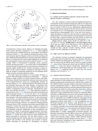
compounds containing-purines are shown in Fig. 1. A comprehensive
review identified diverse purine-heterocyclic hybrids as scaffolds for
anticancer drugs from the past decade [1]. Ma et al. have synthesized
6-mercapto-purine derivative (compound 4 in Fig. 1), exhibiting
antiproliferative effects against HepG2 and A2780 cancer cells [2].
Moreover, Attia et al. unveiled a novel class of purine derivatives with
potent antiproliferative activity, particularly compound 4b (identified
as 3 in Fig. 1) demonstrated effectiveness against colon, hepatic, and
breast cancer cells [3]. Nageswara Kode et al. disclosed the synthesis and
antitumor activity of a purine-based compound (compound 2 in Fig. 1),
exhibiting efficacy against various cancer cell lines [6]. Furthermore,
Salas et al. reported a highly effective 2,6,9-trisubstituted purine de­
rivative (7 h) inducing apoptosis and causing S-phase cell cycle arrest in
HL-60 cells, with notable efficacy in four out of seven tested cancer cell
lines [4]. In this context, motivated by cyclooxygenase-2 (COX-2)
* Corresponding author at: Trustlife Labs, Drug Research & Development Center, 34774 Istanbul, Turkiye.
** Corresponding author at: Research Unit, Electrochemistry, Materials and Environment, University of Kairouan, 3100 Kairouan, Tunisia.
E-mail addresses: melek.hajji@ipeik.u-kairouan.tn (M. Hajji), adilm@scilifelab.se (A. Mardinoglu).
1
These authors contributed equally.
Contents lists available at ScienceDirect
Journal of Molecular Structure
journal homepage: www.elsevier.com/locate/molstr
https://doi.org/10.1016/j.molstruc.2024.138400
Received 1 March 2024; Received in revised form 16 April 2024; Accepted 17 April 2024
Journal of Molecular Structure 1311 (2024) 138400
2
overexpression in various cancers, Hassan et al. designed and synthe­
sized a purine derivative, (compound 1 in Fig. 1), which exhibited
central heterocyclic scaffold bearing two vicinal phenyl and different
heteroaryl moieties [5]. The compound demonstrated potent antitu­
moral property against leukaemia, ovarian, and breast carcinoma cell
lines, exhibiting both antiproliferative and noteworthy COX-2 inhibitory
effects. It is noteworthy that various studies highlight the correlation of
the cyclooxygenase enzyme with numerous types of cancers [7–9]. For
example, COX-2 is overexpressed in pancreatic, breast, colorectal,
stomach, and lung carcinoma [9]. As a result, COX-2 is acknowledged as
a pivotal focus for the advancement of novel anticancer treatments. It is,
therefore, highly valuable for the scientific community to acquire in­
sights into how purine scaffolds, recognized as privileged structures,
might be utilized in forthcoming endeavours for the systematic devel­
opment of purine-containing anticancer agents.
This study reports the synthesis of a novel purine derivative, 2-
amino-6‑chloro-N,N-diphenyl-7H-purine-7-carboxamide, achieved
through a simple reaction of 6‑chloro-7H-purin-2-amine with diphe­
nylcarbamic chloride. We elucidated the molecular and crystal structure
of the prepared derivative using X-ray crystallography. Notably, the
structure exhibits a C—Cl⋅⋅⋅π interactions between the chlorine atom and
the pyrimidine heterocycle ring. These halogen⋅⋅⋅π interactions, have
frequently observed in diverse systems, contribute significantly to the
stability of halogenated organic compounds, supramolecular assem­
blies, and solid-state structures [10–14]. Their importance extends to
biomolecules as well [15,16], with documented roles in pyrimidine and
purine-containing systems [17]. In our case, this C—Cl⋅⋅⋅π interaction
offers a valuable opportunity to gain deeper insights into this stabilizing
force through a real-world solid-state structure. To delve beyond geo­
metric features, we employed multi-approach quantum mechanics (QM)
analysis to elucidate the nature (σ-Hole⋯π or lone pair⋯π) and ener­
getics of this interaction. Moreover, we employed Conceptual Density
Functional Theory (CDFT), molecular electrostatic potential (MEP), and
quantitative "atomic local molecular surface" analysis to enhance our
comprehension of the electronic and chemical reactivity properties of
the synthesized compound. Additionally, in silico investigations were
conducted to evaluate its drug-likeness, pharmacokinetic, and toxico­
logical profiles, utilizing the updated ADMETlab 2.0 integrated online
platform [18]. Finally, the in-silico bioactivity of the synthesized com­
pound concerning its interactions with the COX-2 enzyme is also
assessed through molecular docking approach. We anticipate that this
research will contribute to the structure and reactivity of purines,
thereby advancing our knowledge for development of selective
purine-based COX-2 inhibitors and anticancer therapeutics.
2. Material and methods
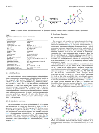
2.1. Synthesis of the investigated compound: 2-amino-6‑chloro-N,N-
diphenyl-7H-purine-7-carboxamide
The title compound 2-amino-6‑chloro-N,N-diphenyl-7H-purine-7-
carboxamide has been prepared starting from 200 mg of 6‑chloro-7H-
purin-2-amine which was stirred with K2CO3 (417.6 mg) and TBAB (36
mg, 0.11 mmol) in THF for 15 min. Subsequently, diphenylcarbamic
chloride (462 mg) was introduced into the reaction mixture and stirred
for a duration of 12 h. The progression of the reaction was monitored
using thin-layer chromatography (TLC). At the end of the reaction, a
simple decantation is carried out, then the solvent is evaporated under
reduced pressure, the residue acquired is chromatograph on a column of
gel silica using a mixture of hexane and ethyl acetate as eluent and
recrystallized by EtOH and obtained (80 %) as white powder. Scheme 1
Eluent (Hexane/ Ethyle acetate (50/50 %) Yield=80 %, (◦
F) =
120–122 [( ◦
C) = 48–50], 1
H NMR (DMSO‑d6 400 MHz) δ ppm:8.240 (s,
1H, N–CH–N); 7.388–7.47 (m, 10H, CHarom) 7.127 (s, 2H, NH2). 13
C
NMR (101 MHz, DMSO‑d6) δppm: 160.203 (C = O), 152.659 (C–Cl),
149.636 (C–NH2), 148.135, 141.676, 140.526, 129.333, 127.535,
126.874, 121.975. LCMS/MS: m/z 365.01 [M + H]+
.
2.2. Single crystal X-ray diffraction (SCXRD)
The molecular structure of prepared compound was determined
using single-crystal X-ray diffraction. Data were collected at 299.38 K on
a Bruker APEX-II CCD diffractometer. Using Olex2 1.3 [19], the struc­
ture was solved with the SHELXT 2014/5 structure solution program
[20] using Intrinsic Phasing and refined with the SHELXL refinement
package [21] using Least Squares minimization. Identification and
visualization of weak interactions was made utilizing Platon [22] and
Mercury 4.0 [23]. Additional details on crystal data and structure
refinement can be found in Table 1.
2.3. Quantum chemical simulations
All density functional theory (DFT) simulations were carried out
using GaussView 6.0 and Gaussian 09, Rev D.01 software [24,25]. The
geometry optimizations were performed at ωB97xD/aug-cc-pVDZ level.
The wB97X-D hybrid functional combines long-range corrections with
Grimme’s D2 dispersion model [26]. When used in conjunction with the
aug-cc-pVXZ Dunning correlation-consistent polarized valence basis set,
it yields accurate results for various noncovalent interactions, striking a
balance between accuracy and computational cost [27–32]. Subsequent
single-point energy computations were carried out considering disper­
sion effects, at ωB97xD/aug-cc-pVTZ level. All interaction energies were
obtained with supermolecule method and underwent correction for
basis set superposition errors (BSSE) through the counterpoise proced­
ure [33]. The quantum theory of atoms in molecules (QTAIM) [34] and
independent gradient model (IGM) [35] was accomplished, from the
DFT-based converged wave functions, using Multiwfn 3.8 [36] and
visualized by VMD 1.9.3 [37]. The electron localization function (ELF)
[38] and the 1D profiles of the electron density (ED) and electrostatic
potential (ESP) functions [39] were calculated in the Multiwfn program.
Natural bond orbital (NBO) [40] findings were obtained using the
Gaussian-NBO 3.1 module, implemented in Gaussian program. The
Molecular Electrostatic Potential (MEP) surfaces were performed with
GaussView 6.0 at 0.001 a.u. isosurface, combining with quantitative
"Atomic local molecular surface" analysis. Global chemical reactivity
indices were predicted through chemical reactivity theory [27]. Tech­
nical and theoretical background of these approaches is extensively
detailed in our previous works [27,29,41].
Fig. 1. Some purine-based compounds with potential activity as anticancer.
N. Abad et al.
Journal of Molecular Structure 1311 (2024) 138400
3
2.4. ADMET predictions
The drug-likeness and toxicity of the synthesized compound under­
went a comprehensive assessment using "ADMET Evaluation" module of
the integrated online platform ADMETlab 2.0 (https://admetmesh.
scbdd.com/) [18]. The compound’s SMILES string, “NC1NC(Cl)C2C(N1)
N(CN2)C(N(C1CCCCC1) C1CCCCC1)O”, was generated with ChemDraw
Professional 17.1, and served as input. A key feature of ADMETlab is its
extensive coverage, encompassing 75 endpoints across 17 physico­
chemical, 23 pharmacokinetic, 27 toxicologic, and 13 medicinal
chemistry parameters. Additionally, the user-friendly interface enables a
rapid and nuanced assessment of the ADMET profile, providing detailed
numerical predictions, supported with color-coded decision states
(green: excellent, yellow: medium, red: poor) and a radar plot consid­
ering 13 key properties.
2.5. In silico docking experiments
The crystallographic data for the cyclooxygenase-2 (COX-2) enzyme
was accessed from the Protein Data Bank (PDB ID: 3LN1) [42]. The li­
gands and one monomer of COX-2 were prepared for docking via stan­
dard methods using AutoDock Tools 1.5.6 [43]. The docking was carried
out using a flexible ligand docking approach with AutoDock Vina 1.2.0
[44]. A three-dimensional affinity grid (52 × 40 × 40) with a grid point
spacing of 0.375 Å was positioned around the COX-2 active site. The
best-ranked conformation complex was analyzed using PyMOL 2.5 [45]
assisted by ChatGPT-PyMOL Plugin [46], and through LigPlot+ V.2.2.8
program [47].
3. Results and discussion
3.1. Structural analysis
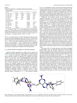
The asymmetric unit comprises two independent molecules charac­
terized by differences in the planarity of their central cores and the
orientations of substituents (Fig. 2). The purine moiety containing Cl2
exhibits slight non-planarity, evident in the dihedral angle of 1.55(16)
between the constituent rings, with a total puckering amplitude (Q) of
0.033(3) Å. In the second molecule, the corresponding angle and total
puckering amplitude are 0.94(15)◦
and 0.037(3) Å, respectively.
Furthermore, the C4-N5-C6-N6 torsion angle is 140.5(3)◦
, while the
C20-N11-C24-N12 angle is − 138.9(3)◦
. Additionally, the (C7/C8/C9/
C10/C11/C12) and (C13/C14/C15/C16/C17/C18) benzene rings are
inclined to each other by 83.55(18)◦
, whereas the corresponding angle
in the second molecule is 73.48(17)◦
. All bond lengths, however, remain
within expected ranges.
In the crystal, the two molecules within the asymmetric unit form
connections through C5—H5⋅⋅⋅O2 and N7—H7B⋅⋅⋅O1 hydrogen bonds
(Fig. S1). Crystal packing analysis reveals a network of diverse inter­
molecular interactions that stabilize the compound’s structure (Table 2),
which are categorized as: classical hydrogen bonding (N1—H1A⋅⋅⋅N2
and N1—H1B⋅⋅⋅O2), unconventional weak hydrogen bonds
(C10—H10⋅⋅⋅N8 and C23—H23⋅⋅⋅O1), C/N–H⋅⋅⋅π(ring) interactions
(N1—H1B⋅⋅⋅π, C32—H32⋅⋅⋅π and C34—H34⋅⋅⋅ π). Although aromatic
rings are present, no true π-π stacking interactions were observed due to
all inter-ring plane distances exceeding the typical limit of 3.8 Å [48,49].
The closest centroid separation was 4.188(2) Å, exceeding this
threshold, so these interactions were not included in Table 2. Interest­
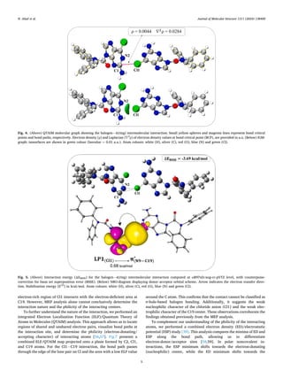
ingly, a halogen⋯π(ring) interaction, C2—Cl1⋯π(pyrimidine), was
observed, offering an additional stabilizing force (see Table 2, Fig. 3).
Scheme 1. Synthesis pathway and chemical structure of the investigated compound: 2-amino-6‑chloro-N,N-diphenyl-7H-purine-7-carboxamide.
Table 1
Crystal data and details of the structure determination.
CCDC No. 2,325,198
Empirical formula C18H13ClN6O
Formula weight 364.79
Temperature/K 299.38
Crystal system, Space group Monoclinic, P21/c
a, b, c /Å 9.279(2), 24.458(5), 17.659(4)
α, β, γ /◦
90, 93.883(4), 90
Volume/Å3
, Z 3998.3(15), 8
ρcalcg/cm3
, μ/mm-1
1.212, 0.209
F(000) 1504.0
Crystal size/mm3
0.133 × 0.122 × 0.107
Radiation MoKα (λ = 0.71073)
2θ range for data collection/◦
2.848 to 54.962
Index ranges − 11 ≤ h ≤ 12, -31 ≤ k ≤ 31, -22 ≤ l ≤ 22
Reflections collected 41,070
Independent reflections 9118 [Rint = 0.1039, Rsigma = 0.1146]
Data/restraints/parameters 9118/0/485
Goodness-of-fit on F2
0.918
Final R indexes [I>=2σ (I)] R1 = 0.0583, wR2 = 0.1392
Final R indexes [all data] R1 = 0.1550, wR2 = 0.1768
Largest diff. peak/hole / e Å− 3
0.27/− 0.23
Fig. 2. ORTEP-Illustration of the asymmetric unit of the crystal structure,
showing two independent molecules with their labelling scheme and 50 %
probability ellipsoids. Atom colours: white (H), grey (C), blue (N), red (O) and
green (Cl).
N. Abad et al.
Journal of Molecular Structure 1311 (2024) 138400
4
The Cl⋯π (centroid) distance is 3.556 Å. The distances between Cl and
the nearest atom from pyrimidine ring (3.237 Å for Cl1⋯C19) is shorter
than the sum of the van der Waals radii of the involved atoms (Cl = 1.75
Å, C = 1.70 Å). The angles surrounding chlorine (C2–Cl–centroid =
141.18◦
, C2–Cl–(π-plane) = 71.82 ◦
) deviate from the anticipated
directional characteristics of classical halogen bonds. The nature and
energetics of this halogen⋯π interaction are explored further in subse­
quent sections using quantum mechanics DFT-based approaches.
3.2. Quantum mechanical investigations on noncovalent interactions
Quantum chemical approaches are widely used to quantify and
visualize noncovalent interactions within different molecular systems
[50–55]. Herein, we conducted a comprehensive quantum mechanics
study, primarily focused on exploring the energetics and nature
(σ-Hole⋯π or lone pair⋯π) of the crystallographically observed halo­
gen⋯π interaction. This study utilized dispersion-corrected density
functional theory (DFT-D) at the ωB97xD/aug-cc-pVTZ level. To achieve
this, a dimeric sub-structure was retrieved from the crystallographic
structure and partially optimized. The optimization involved all
hydrogen atoms while keeping the positions of other atoms frozen. This
approach allowed the assessment of the interaction in its solid-state
configuration, avoiding geometric rearrangement and ensuring a
converged wavefunction. Initially, a Quantum Theory of ’Atom­
s-in-Molecules’ (QTAIM) analysis was performed, and the resulting
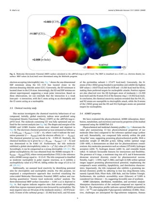
molecular graph is illustrated in Fig. 4. The interaction between
C2—Cl1⋯π(pyrimidine) is evident, with a bond critical point (BCP) and
bond path connecting Cl1 and the C19 atom of the pyrimidine ring. The
calculated electron density (ρ) and Laplacian (∇2
ρ) values of 0.0044 a.u.
and 0.0284 a.u., respectively, indicate a closed-shell type interaction.
Subsequently, Independent Gradient Model (IGM) analysis was con­
ducted, revealing an extended green IGM isosurface in the contact re­
gion, signifying the weak dispersive character of this interaction (Fig. 4).
Furthermore, the interaction energy for the homodimer was
computed, accounting for the basis set superposition error (BSSE)
through counterpoise correction. The resulting ΔEBSSE value was − 3.69
kcal/mol (Fig. 5). To further explore the characteristics of this inter­
molecular pattern, an examination was conducted using Natural Bond
Orbital (NBO) analysis, considering orbital interaction and charge-
transfer concepts. Fig. 5 displays an NBO diagram showing donor-
acceptor orbital interaction along with second-order perturbation en­
ergy (E(2)
). Notably, the C2—Cl1⋯π interaction is characterized by LP1
(Cl1)→ π*(N9–C19) electronic delocalization, with a stabilization E1
value of 0.69 kcal/mol. This orbital interaction corresponds to a lone-
pair⋯π interaction, with the lone pair bonding orbital on the Cl atom
acting as the donor orbital (Lewis base). Moreover, the analysis revealed
the presence of a π(N9-C19)→σ*(C2–Cl1) interaction with a very low
stabilization energy value (E(2)
) of 0.07 kcal/mol. In this case, the π(N9-
C19) bonding orbital in pyrimidine functions as the donor orbital (Lewis
base), suggesting a σ-hole interaction character. The E1 value being
larger than the E2 value, along with the absence of LP(N) → σ(Cl–C)
electronic delocalization, signifies that the investigated interaction is
likely a lone-pair⋯π (n → π*) interaction. This observation supports the
QTAIM results, which do not show evidence of a direct contact (bond
path and bond critical point) between the Cl atom and neighbouring N
atoms.
The nature of the Cl⋯π(ring) interaction was further investigated
electrostatically using the Molecular Electrostatic Potential (MEP)
analysis. MEP surface helps identify potential sites for noncovalent
bonding by highlighting regions of electron richness (negative potential)
and electron deficiency (positive potential) around the molecule. It can
also assess the size and strength of the σ-holes [56]. Fig. 6 shows the
MEP surface for the fully optimized monomer structure calculated at the
ωB97xD/aug-cc-pVTZ level. As observed, the chlorine atom (Cl1) ex­
hibits a low positive MEP value (+2.059 kcal/mol) opposite the C–Cl
bond, indicating an electron-deficient area corresponding to the Cl
σ-hole. This suggests a low propensity for Cl1 to act as a σ-hole donor in
noncovalent interactions. Conversely, the electron belt (σ-lump) around
Cl1 displays a negative MEP value (− 11.587 kcal/mol). On the other
hand, the C19 atom has a positive MEP value (+3.603 kcal/mol), albeit
lower in magnitude. This indicates its suitability to act as an electron
acceptor (electrophilic site). Therefore, the MEP analysis suggests an
electrostatically favorable, but likely weak, interaction where the
Table 2
Geometric parameters (A, ◦
) of potential noncovalent interactions.
Hydrogen Bonding:
D—H…
A a
D—H H…
A D…
A D—H…
A
N1—H1A⋅⋅⋅N2 (i)
0.78(4) 2.38(4) 3.153(4) 174(4)
N1—H1B⋅⋅⋅O2 (ii)
0.93(3) 2.48(3) 3.345(4) 155(3)
C10—H10⋅⋅⋅N8 (iii)
0.93 2.58 3.444(5) 154
N7—H7B⋅⋅⋅O1 1.00(5) 2.21(5) 3.165(4) 161(4)
C5—H5⋅⋅⋅O2 0.93 2.40 3.235(4) 149
C23—H23⋅⋅⋅O1(iv)
0.93 2.43 3.293(4) 155
π-(ring) interactions:
C/N—H…
Cg H…
Cg C/N…
Cg C—H…
Cg
N1—H1B⋅⋅⋅Cg1 3.0(3) 3.600(4) 124(2)
C32—H32⋅⋅⋅Cg2 2.77 3.620(3) 153
C34—H34⋅⋅⋅Cg3 2.96 3.730(4) 141
C—X…
Cg H…
Cg C—X, π b
C—X…
Cg
C2—Cl1⋅⋅⋅Cg4 3.556(2) 71.82 141.18(1)
a
A = acceptor, D = donor. b
C—X, π is the angle of the C–Cl bond with the
π-plane. Cg = centroid of the rings: Cg1, Cg2, Cg3 and Cg4 are the centroids of
the rings (C20-C21-N10-C23-N11), (C13-C14-C15-C16-C17-C18), (C1-N2-C2C3-
C4-N3) and (C19-N8-C20-C21-C22-N9), respectively. Symmetry codes: (i): 2-
x,1-y,1-z (ii): 1 + x,y,z (iii): x,1/2-y,1/2 + z (iv): − 1 + x,y,z.
Fig. 3. Illustration of intermolecular halogen⋯π(ring) interaction, C2—Cl1⋯π(pyrimidine) within the crystalline structure. Interaction between Cl1 atom and
centroid of pyrimidine ring is depicted by dashed magenta lines. Atom colours: white (H), grey (C), blue (N), red (O) and green (Cl).
N. Abad et al.
Journal of Molecular Structure 1311 (2024) 138400
5
electron-rich region of Cl1 interacts with the electron-deficient area at
C19. However, MEP analysis alone cannot conclusively determine the
interaction nature and the philicity of the interacting centers.
To further understand the nature of the interaction, we performed an
integrated Electron Localization Function (ELF)/Quantum Theory of
Atoms in Molecules (QTAIM) analysis. This approach allows us to locate
regions of shared and unshared electron pairs, visualize bond paths at
the interaction site, and determine the philicity (electron-donating/
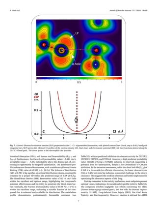
accepting character) of interacting atoms [56,57]. Fig.7 presents a
combined ELF/QTAIM map projected onto a plane formed by C2, Cl1,
and C19 atoms. For the Cl1⋅⋅⋅C19 interaction, the bond path passes
through the edge of the lone pair on Cl and the area with a low ELF value
around the C atom. This confirms that the contact cannot be classified as
σ-hole-based halogen bonding. Additionally, it suggests the weak
nucleophilic character of the chloride anion (Cl1) and the weak elec­
trophilic character of the C19 center. These observations corroborate the
findings obtained previously from the MEP analysis.
To complement our understanding of the philicity of the interacting
atoms, we performed a combined electron density (ED)/electrostatic
potential (ESP) study [39]. This analysis compares the minima of ED and
ESP along the bond path, allowing us to differentiate
electron-donor/acceptor sites [58,59]. In polar noncovalent in­
teractions, the ESP minimum shifts towards the electron-donating
(nucleophilic) center, while the ED minimum shifts towards the
Fig. 4. (Above) QTAIM molecular graph showing the halogen⋯π(ring) intermolecular interaction: Small yellow spheres and magenta lines represent bond critical
points and bond paths, respectively. Electron density (ρ) and Laplacian (∇2
ρ) of electron density values at bond critical point (BCP), are provided in a.u. (Below) IGM-
graph: isosurfaces are shown in green colour (Isovalue = 0.01 a.u.). Atom colours: white (H), silver (C), red (O), blue (N) and green (Cl).
Fig. 5. (Above) Interaction energy (ΔEBSSE) for the halogen⋯π(ring) intermolecular interaction computed at ωB97xD/aug-cc-pVTZ level, with counterpoise-
correction for basis set superposition error (BSSE). (Below) NBO-diagram displaying donor–acceptor orbital scheme. Arrow indicates the electron transfer direc­
tion. Stabilization energy (E(2)
) in kcal/mol. Atom colours: white (H), silver (C), red (O), blue (N) and green (Cl).
N. Abad et al.
Journal of Molecular Structure 1311 (2024) 138400
6
electron-accepting (electrophilic) site. Fig. 7 shows the one-dimensional
ESP minimum along the Cl1⋅⋅⋅C19 line located closer to the
electron-donating chloride anion (Cl1). Conversely, the ED minimum is
located closer to the C19 atom. Interestingly, the ED and ESP minima are
almost superimposed, suggesting a weak polar interaction. Based on
these observations, we can conclude that the interaction is a weak
lone-pair⋯π interaction, with the C atom acting as an electrophile and
the Cl centre acting as a nucleophile.
3.3. Chemical reactivity study
This section investigates the chemical reactivity behaviours of our
compound. Initially, global reactivity indices were predicted using
Conceptual Density Functional Theory (CDFT) at the ωB97xD/aug-cc-
pVTZ level. The molecule containing Cl1 was fully optimized and uti­
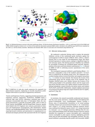
lized for the current analysis (see Fig. 8a). The shapes and energies of the
HOMO and LUMO frontier orbitals were obtained and depicted in
Fig. 8b. The electronic chemical potential (μ) was estimated as follows: μ
≈ 1/2(ELUMO + EHOMO) ≈ 1/2(I + A), where I and A indicate the ioni­
zation potential (I = − EHOMO) and the electron affinity (A = − ELUMO).
The chemical potential (μ) was found to be − 3.994 eV. Similarly, the
chemical hardness (η) is given by η ≈ (ELUMO − EHOMO) ≈ (I − A) and
was determined to be 4.661 eV. Furthermore, the title molecule
exhibited a global electrophilicity index (ω = μ2
/2η) value of 1.711 eV.
Accordingly, it can be categorized as a strong electrophile [29–31]. The
global nucleophilicity (N) was obtained from the HOMO energies: N =
EHOMO − EHOMO(TCE), where tetracyanoethylene (TCE) is the reference
with a HOMO energy equal to − 9.12 eV. The title compound is classified
as moderate nucleophile in polar organic reactions, as it exhibits a
nucleophilicity value of 2.795 eV, falling within the range of 2.0 to 3.0
eV [29–31].
Additionally, we assessed local reactivity by identifying potential
sites for electrophilic and nucleophilic attacks. For this purpose, we
employed a comprehensive approach that involved visualizing the
color-coded Molecular Electrostatic Potential (MEP) surface and con­
ducting quantitative "Atomic local molecular surface" analysis. The
three-dimensional MEP surface (isosurface = 0.001 a.u) is depicted in
Fig. 8c. Red regions indicate negative sites that attract electrophiles,
while blue regions represent positive sites favoured by nucleophiles. The
most negative sites are N4-atom of the imidazole moiety (− 23.972 kcal/
mol), O-atom of the carbonyl group (− 21.062 kcal/mol), and N2-atom
of the pyrimidine subunit (− 17.677 kcal/mol). Conversely, the H-
atoms of the (-NH2) group bonded to pyrimidine unit exhibit the highest
ESP values (+18.673 kcal/mol for H1B and +15.501 kcal/mol for H1A),
making them preferred targets for nucleophilic attacks. Positive regions
are also observed over the H5 hydrogen atom of imidazole (+14.359
kcal/mol) and the H-atom H12 of the benzene ring (+14.052 kcal/mol).
In conclusion, the chemical reactivity analysis revealed that the N4, O1,
and N2 atoms are susceptible to electrophilic attack, while the H-atoms
of the (-NH2) group and the H5 and H12 hydrogen atoms are potential
targets for nucleophiles.
3.4. ADMET properties
We have evaluated the physicochemical, ADME (absorption, distri­
bution, metabolism, and excretion) and toxicity properties of the studied
compound using the ADMETlab 2.0.
Physicochemical and medicinal chemistry profiles: Fig. 9 showcases the
radar plot summarizing 13 key physicochemical properties of our
molecule (blue line) compared to the reference optimal range (yellow
and red). Remarkably, our compound falls entirely within the pink
"drug-like" zone, suggesting promising physicochemical profile. With a
molecular weight of 386.260 g/mol, falling within optimal range
(100~600), it demonstrates an ideal size for pharmacokinetic consid­
erations. Key molecular parameters such as volume (375.640), hydrogen
acceptors (nHA: 7), hydrogen donors (nHD: 6), and rotatable bonds
(nRot: 4) indicate favourable features for interactions and flexibility.
The presence of 4 rings, a maximum ring size of 9, and 8 heteroatoms
showcase structural diversity crucial for pharmaceutical success.
Notably, LogS (− 1.559), logP (1.586), and logD (2.528) within optimal
ranges support promising pharmaceutical potential, emphasizing the
compound’s water solubility, lipophilicity, and distribution character­
istics. Importantly, the title compound also exhibits a promising me­
dicinal chemistry profile by adhering to four key drug-likeness rules,
namely Lipinski Rule, Pfizer Rule, GSK Rule, and the Golden Triangle.
Furthermore, the synthetic accessibility score (SAscore) of 5.009 is
considered low (SAscore < 6), suggesting favorable ease of synthesis.
ADME (absorption, distribution, metabolism, and excretion) analysis:
Absorption characteristics of the analysed compound are reported in
Table S1. The absorption profile indicates optimal MDCK permeability
(2.4 × 10− 06
) and negligeable P-glycoprotein inhibition (0.006). How­
ever, challenges are anticipated for Pgp-substrate endpoints, Human
Fig. 6. Molecular Electrostatic Potential (MEP) surface calculated at the ωB97xD/aug-cc-pVTZ level. The MEP is visualized on a 0.001 a.u. electron density iso­
surface. MEP values (in kcal/mol) were determined using the Multiwfn program.
N. Abad et al.
Journal of Molecular Structure 1311 (2024) 138400
7
Intestinal Absorption (HIA), and human oral bioavailability (F20 % and
F30 %). Furthermore, the Caco-2 cell permeability value (− 5.884 cm/s;
acceptable range > − 5.15) falls slightly above the desired cut-off, pre­
senting an opportunity for targeted optimization. The distribution pro­
file underscores favourable outcomes, with a satisfactory Plasma Protein
Binding (PPB) value of 65.932 % (< 90 %). The Volume of Distribution
(VD) at 0.781 L/kg signifies an optimal distribution volume, meeting the
criterion for a proper VD within the predicted range of 0.04–20 L/kg.
The Blood-Brain Barrier (BBB) Penetration value of 0.116 cm/s falls
within the excellent and optimal range, highlighting the compound’s
potential effectiveness and its ability to penetrate the blood-brain bar­
rier. Similarly, the Fraction Unbound (Fu) value of 50.98 % (≥ 5 %) is
within the excellent range, indicating a suitable fraction of the com­
pound that is unbound and available for distribution. The metabolism
profile demonstrates predominantly favourable outcomes (see
Table S2), with no predicted inhibition or substrate activity for CYP1A2,
CYP2C19, CYP2C9, and CYP3A4. However, a high predicted probability
value (0.852) of being a CYP2D6 substrate is observed, suggesting a
potential area for optimization, despite a low probability of CYP2D6
inhibition. In the excretion assessment, while the short half-life (T1/2) of
0.265 h is favourable for efficient elimination, the lower clearance rate
(CL) at 3.252 mL/min/kg indicates a potential challenge in the drug’s
clearance. This suggests the need for attention and further exploration in
optimizing the clearance aspects of the drug.
Toxicity evaluation: In the toxicity evaluation, most endpoints present
optimal values, indicating a favourable safety profile (refer to Table S3).
The compound exhibits negligible side effects concerning the hERG
(Human ether-a-go-go related gene), and low risks for Human Hepato­
toxicity (H–HT), Drug-Induced Liver Injury (DILI), Rat Oral Acute
Toxicity, and Carcinogenicity. However, caution is advised for AMES
Fig. 7. (Above) Electron localization function (ELF) projection for the C—Cl⋯π(pyrimidine) interaction, with plotted contour lines (black, step is 0.05), bond path
(magenta line), BCP (green dot). (Below) 1D profiles of the electron density (ED, black line) and electrostatic potential (ESP, red line) functions plotted along the
Cl1⋅⋅⋅C19 bond path.. The arrow points to the electrophilic site provider.
N. Abad et al.
Journal of Molecular Structure 1311 (2024) 138400
8
Toxicity and Respiratory Toxicity, suggesting potential genotoxicity and
respiratory concerns, respectively. Furthermore, FDAMDD shows a
relatively high value (0.975), indicating a potential issue with the
maximum recommended daily dose. In Toxicophore Rules, the com­
pound demonstrates a generally favourable profile, with no alerts in
Acute Toxicity, SureChEMBL, and FAF-Drugs4 Rules. However, specific
alerts linked to the Cl-C fragment raise concerns, particularly in Geno­
toxic and NonGenotoxic Carcinogenicity, Skin Sensitization, Aquatic
Toxicity, and NonBiodegradability Rules. These alerts underscore areas
requiring focused evaluation and optimization to comprehensively un­
derstand and mitigate potential risks associated with the Cl-C fragment.
3.5. Molecular docking analysis
We conducted a molecular docking study to explore the potential
inhibitory activity of the synthesized purine derivative against the
cyclooxygenase-2 (COX-2) enzyme (PDB ID: 3LN1). COX-2, a dimeric
isozyme that is a key target for anti-inflammatory drugs, also shows
elevated expression in various cancers, making it a significant target for
new anticancer agents. The docking protocol’s reliability was validated
by successfully re-docking the crystallized ligand celecoxib. Superim­
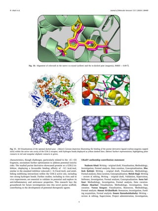
position of the native and newly docked conformations (Fig. 10) showed
an RMSD of 0.89 Å, confirming accurate reproduction of the native
conformation.
The investigated compound exhibited a good binding affinity (− 9.1
kcal/mol), comparable to that of the standard celecoxib (− 9.3 kcal/
mol), as evidenced by the docking energy score. The compound’s non­
covalent binding scheme involves both polar and nonpolar interactions
(Fig. 11). It forms two short H-bonds: One between the O-atom of the
carboxamide group and the nitrogen atom of the imidazole ring of
Histidine (His342) amino acid. Another between the H-atoms of the
(-NH2) group bonded to the pyrimidine subunit and the oxygen atom
from Serine (Ser339) amino acid. LigPlot analysis revealed several sta­
bilizing hydrophobic contacts between the purine moiety and residues
Thr79, His75, and Pro500. Additionally, nonpolar aliphatic interactions
occur between the phenyl rings and a region formed by Gln179, Asp501,
and Pro177 (Fig. S2).
4. Conclusion
In conclusion, this research successfully synthesized and character­
ized the novel purine derivative, 2-amino-6‑chloro-N,N-diphenyl-7H-
purine-7-carboxamide. X-ray crystallographic analysis revealed a
diverse array of intermolecular interactions in its crystal packing,
including classical and unconventional H-bonds, C—H⋅⋅⋅π interactions,
and a unique C—Cl⋅⋅⋅π(ring) interaction. Quantum chemical calcula­
tions confirmed the distinctive nature of the C—Cl⋅⋅⋅π(ring) interaction
as a lone-pair⋯π (n→π*) halogen interaction, differing from the com­
mon σ-hole-based halogen bond. DFT-based chemical reactivity analysis
identified the compound as a potent electrophile and a moderately
reactive nucleophile in polar organic reactions, highlighting potential
attack sites for both electrophilic and nucleophilic reactions. The com­
pound exhibited a promising drug-like profile with favourable ADMET
Fig. 8. (a) Optimized geometry structure with atom numbering scheme. (b) Electron density distribution (isosurface= 0.02 a.u.) and energy levels of the HOMO and
LUMO. Orbital energies are shown in eV. (c) Views of the molecular electrostatic potential (MEP) surface calculated at the ωB97xD/aug-cc-pVTZ level, visualized on
the 0.001 a.u. electron density isosurface. Maximum and minimum MEP values (in kcal/mol) were determined using Multiwfn 3.8.
Fig. 9. ADMETLab 2.0 radar plot visually summarizes the compound’s key
physicochemical properties, emphasizing its favorable drug-likeness profile.
Endpoint abbreviations are provided in Supplementary Information.
N. Abad et al.
Journal of Molecular Structure 1311 (2024) 138400
9
characteristics, though challenges, particularly related to the –(C—Cl)
fragment, necessitate further optimization to address potential toxicity
risks. The studied purine derivative showcased promise as a COX-2 in­
hibitor, displaying a favourable binding affinity of − 9.1 kcal/mol,
similar to the standard inhibitor celecoxib (− 9.3 kcal/mol), and estab­
lishing stabilizing interactions within the COX-2 active site, including
two strong hydrogen bonds. Further studies, including in vitro and in
vivo experiments, are essential to validate its potential and explore its
anti-inflammatory and anticancer properties. This research lays the
groundwork for future investigations into this novel purine scaffold,
contributing to the development of potential therapeutic agents.
CRediT authorship contribution statement
Nadeem Abad: Writing – original draft, Visualization, Methodology,
Investigation, Formal analysis, Data curation, Conceptualization. Sha­
feek Buhlak: Writing – original draft, Visualization, Methodology,
Formal analysis, Data curation, Conceptualization. Melek Hajji: Writing
– review & editing, Writing – original draft, Validation, Supervision,
Software, Investigation, Formal analysis, Conceptualization. Sana Saf­
four: Methodology, Investigation, Formal analysis, Data curation.
Jihane Akachar: Visualization, Methodology, Investigation, Data
curation. Yunus Kesgun: Visualization, Resources, Methodology,
Formal analysis. Hanan Al-Ghulikah: Resources, Investigation, Fund­
ing acquisition, Formal analysis. Essam Hanashalshahaby: Writing –
review & editing, Supervision, Project administration, Investigation,
Fig. 10. Alignment of celecoxib in the native co-crystal (yellow) and the re-docked pose (magenta), RMSD = 0.89 Å.
Fig. 11. 3D-Visualizations of the optimal docked pose - (Above) Cartoon depiction illustrating the binding of the purine derivative ligand (carbon-magenta capped
stick) within the active site cavity of the COX-2 receptor, with hydrogen bonds displayed as yellow dashed lines. (Below) Surface representation, highlighting polar
contacts in red and nonpolar aliphatic contacts in green.
N. Abad et al.
Journal of Molecular Structure 1311 (2024) 138400
10
Funding acquisition, Conceptualization. Hasan Turkez: Writing – re­
view & editing, Visualization, Validation, Software, Resources. Adil
Mardinoglu: Writing – review & editing, Validation, Supervision,
Project administration, Funding acquisition.
Declaration of competing interest
The authors declare that they have no known competing financial
interests or personal relationships that could have appeared to influence
the work reported in this paper.
AM and HT are the founder and shareholder of Molius Therapeutics.
The other authors declare no competing interests.
Data availability
Data will be made available on request.
Acknowledgments
The authors extend their appreciation to Princess Nourah bint
Abdulrahman University Researchers Supporting Project number
(PNURSP2024R95), Princess Nourah bint Abdulrahman University,
Riyadh, Saudi Arabia.
The authors gratefully acknowledge the financial support from
Molius Therapeutics and The Knut and Alice Wallenberg Foundation
(No.72254).
Supporting Information
CCDC 2325198 contains the supplementary crystallographic data
for this paper. These data can be obtained free of charge via http://
www.ccdc.cam.ac.uk/conts/retrieving.html (or from the Cambridge
Crystallographic Data Centre, 12, Union Road, Cambridge CB2 1EZ, UK;
fax: +44 1223 336033).
Supplementary materials
Supplementary material associated with this article can be found, in
the online version, at doi:10.1016/j.molstruc.2024.138400.
References
[1] A. Chaurasiya, C. Sahu, S.K. Wahan, P.A. Chawla, Targeting cancer through
recently developed purine clubbed heterocyclic scaffolds: an overview, J. Mol.
Struct. 1280 (2023) 134967, https://doi.org/10.1016/j.molstruc.2023.134967.
[2] Y. Ma, X. Yan, R. Du, X. Yang, Y. Li, Y. Gao, W. Li, Synthesis, anti-cancer activity
and mechanism study of 6-mercapto-purine derivatives, Lett. Drug Des. Discov. 13
(2016) 570–576, https://doi.org/10.2174/1570180812666150821003536.
[3] R.T. Attia, M.A. Ewida, E. Khaled, S.A. Fahmy, I.M. Fawzy, Newly synthesized
anticancer purine derivatives inhibiting p -EIF4E using surface-modified lipid
nanovesicles, ACS. Omega 8 (2023) 37864–37881, https://doi.org/10.1021/
acsomega.3c02991.
[4] C.O. Salas, A.M. Zarate, V. Kryštof, J. Mella, M. Faundez, J. Brea, M.I. Loza, I. Brito,
D. Hendrychová, R. Jorda, A.R. Cabrera, R.A. Tapia, C. Espinosa-Bustos, Promising
2,6,9-trisubstituted purine derivatives for anticancer compounds: synthesis, 3D-
QSAR, and preliminary biological assays, Int. J. Mol. Sci. 21 (2019) 161, https://
doi.org/10.3390/ijms21010161.
[5] A.Y. Hassan, E.S. Abou-Amra, S.A. El-Sebaey, Design and synthesis of new series of
chiral pyrimidine and purine analogs as COX-2 inhibitors: anticancer screening,
molecular modeling, and in silico studies, J. Mol. Struct. 1278 (2023) 134930,
https://doi.org/10.1016/j.molstruc.2023.134930.
[6] N. Kode, L. Chen, D. Murthy, D. Adewumi, S. Phadtare, New bis-N9-
(methylphenylmethyl)purine derivatives: synthesis and antitumor activity, Eur. J.
Med. Chem. 42 (2007) 327–333, https://doi.org/10.1016/j.ejmech.2006.10.017.
[7] N. ul A. Mohsin, S. Aslam, M. Ahmad, M. Irfan, S.A. Al-Hussain, M.E.A. Zaki,
Cyclooxygenase-2 (COX-2) as a target of anticancer agents: a review of novel
synthesized scaffolds having anticancer and COX-2 inhibitory potentialities,
Pharmaceuticals 15 (2022) 1471, https://doi.org/10.3390/ph15121471.
[8] W. Akhtar, L.M. Nainwal, M.F. Khan, G. Verma, G. Chashoo, A. Bakht, M. Iqbal,
M. Akhtar, M. Shaquiquzzaman, M.M. Alam, COX-2 inhibition and metabolic
stability studies of 6-(4-fluorophenyl)-pyrimidine-5-carbonitrile derivatives as
anticancer and anti-inflammatory agents, J. Fluor. Chem. 236 (2020) 109579,
https://doi.org/10.1016/j.jfluchem.2020.109579.
[9] C.R. Bell, V.S. Pelly, A. Moeini, S.C. Chiang, E. Flanagan, C.P. Bromley, C. Clark, C.
H. Earnshaw, M.A. Koufaki, E. Bonavita, S. Zelenay, Chemotherapy-induced COX-2
upregulation by cancer cells defines their inflammatory properties and limits the
efficacy of chemoimmunotherapy combinations, Nat. Commun. 13 (2022) 2063,
https://doi.org/10.1038/s41467-022-29606-9.
[10] G.A.M. Mersal, I.S. Yahia, H.S. El-Sheshtawy, Lone pair halogen (X2)…π
interactions stabilizes molecular halogens (X2=I2, Br2, Cl2, and F2) on reduced
graphene oxide surface: structural, solvent effect and optical properties, J. Mol.
Struct. 1244 (2021) 130963, https://doi.org/10.1016/j.molstruc.2021.130963.
[11] G. Berger, J. Soubhye, A. vanderLee, C. VandeVelde, R. Wintjens, P. Dubois,
S. Clément, F. Meyer, Interplay between halogen bonding and lone pair–π
interactions: a computational and crystal packing study, Chempluschem.
Chempluschem. 79 (2014) 552–558, https://doi.org/10.1002/cplu.201400005.
[12] K. Sharma, T.P. Mohan, U. Gangwar, D. Chopra, Role of lone pair- π interaction and
halogen bonding in the crystal packing of 1,2,4-oxadiazole derivatives, J. Mol.
Struct. 1197 (2019) 742–752, https://doi.org/10.1016/j.molstruc.2019.07.107.
[13] H. Zhuo, Q. Li, W. Li, J. Cheng, Is π halogen bonding or lone pair⋯π interaction
formed between borazine and some halogenated compounds? Phys. Chem. Chem.
Phys. 16 (2014) 159–165, https://doi.org/10.1039/C3CP54006E.
[14] H.R. Khavasi, N. Rahimi, Lone pair⋯π interaction versus σ-hole appearance in
metal-bonded halogens, CrystEngComm. 21 (2019) 2929–2939, https://doi.org/
10.1039/C8CE01795F.
[15] J. Kozelka, Lone pair–π interactions in biological systems: occurrence, function,
and physical origin, European Biophysics Journal 46 (2017) 729–737, https://doi.
org/10.1007/s00249-017-1210-1.
[16] S.K. Singh, P.R. Joshi, R.A. Shaw, J.G. Hill, A. Das, Interplay between hydrogen
bonding and n→π* interaction in an analgesic drug salicin, Physical Chemistry
Chemical Physics 20 (2018) 18361–18373, https://doi.org/10.1039/C8CP00655E.
[17] M. Chawla, E. Chermak, Q. Zhang, J.M. Bujnicki, R. Oliva, L. Cavallo, Occurrence
and stability of lone pair–π stacking interactions between ribose and nucleobases in
functional RNAs, Nucleic. Acids. Res. 45 (2017) 11019–11032, https://doi.org/
10.1093/nar/gkx757.
[18] G. Xiong, Z. Wu, J. Yi, L. Fu, Z. Yang, C. Hsieh, M. Yin, X. Zeng, C. Wu, A. Lu,
X. Chen, T. Hou, D. Cao, ADMETlab 2.0: an integrated online platform for accurate
and comprehensive predictions of ADMET properties, Nucl. Acids Res 49 (2021)
W5–W14, https://doi.org/10.1093/nar/gkab255.
[19] O.V. Dolomanov, L.J. Bourhis, R.J. Gildea, J.A.K. Howard, H. Puschmann, OLEX2 :
a complete structure solution, refinement and analysis program, J. Appl.
Crystallogr. 42 (2009) 339–341, https://doi.org/10.1107/S0021889808042726.
[20] G.M. Sheldrick, SHELXT – Integrated space-group and crystal-structure
determination, Acta Crystallogr. A Found Adv. 71 (2015) 3–8, https://doi.org/
10.1107/S2053273314026370.
[21] G.M. Sheldrick, Crystal structure refinement with SHELXL, Acta Crystallogr. C
Struct. Chem. 71 (2015) 3–8, https://doi.org/10.1107/S2053229614024218.
[22] A.L. Spek, Single-crystal structure validation with the program PLATON, J. Appl.
Crystallogr. 36 (2003) 7–13, https://doi.org/10.1107/S0021889802022112.
[23] C.F. Macrae, I. Sovago, S.J. Cottrell, P.T.A. Galek, P. McCabe, E. Pidcock,
M. Platings, G.P. Shields, J.S. Stevens, M. Towler, P.A. Wood, Mercury 4.0: from
visualization to analysis, design and prediction, J. Appl. Crystallogr. 53 (2020)
226–235, https://doi.org/10.1107/S1600576719014092.
[24] R. Dennington, T.A. Keith, J.M. Millam, GaussView Version 6, Semichem Inc,
Shawnee Mission KS (2016).
[25] M.J. Frisch, G.W. Trucks, H.B. Schlegel, G.E. Scuseria, M.A. Robb, J.R. Cheeseman,
G. Scalmani, V. Barone, B. Mennucci, G.A. Petersson, H. Nakatsuji, M. Caricato,
X. Li, H.P. Hratchian, A.F. Izmaylov, J. Bloino, G. Zheng, J.L. Sonnenberg, M. Hada,
M. Ehara, K. Toyota, R. Fukuda, J. Hasegawa, M. Ishida, T. Nakajima, Y. Honda,
O. Kitao, H. Nakai, T. Vreven, J.A. Montgomery Jr, J.E. Peralta, F. Ogliaro,
M. Bearpark, J.J. Heyd, E. Brothers, K.N. Kudin, V.N. Staroverov, R. Kobayashi,
J. Normand, K. Raghavachari, A. Rendell, J.C. Burant, S.S. Iyengar, J. Tomasi,
M. Cossi, N. Rega, J.M. Millam, M. Klene, J.E. Knox, J.B. Cross, V. Bakken,
C. Adamo, J. Jaramillo, R. Gomperts, R.E. Stratmann, O. Yazyev, A.J. Austin,
R. Cammi, C. Pomelli, J.W. Ochterski, R.L. Martin, K. Morokuma, V.G. Zakrzewski,
G.A. Voth, P. Salvador, J.J. Dannenberg, S. Dapprich, A.D. Daniels, Ö. Farkas, J.
B. Foresman, J.V. Ortiz, J. Cioslowski, D.J. Fox, Gaussian 09 Revision D.01,
Gaussian, Inc., Wallingford CT, 2013.
[26] S. Grimme, J. Antony, S. Ehrlich, H. Krieg, A consistent and accurate ab initio
parametrization of density functional dispersion correction (DFT-D) for the 94
elements H-Pu, J. Chem. Phys. 132 (2010) 154104, https://doi.org/10.1063/
1.3382344.
[27] M. Hajji, N. Abad, M.A. Habib, S.M.H. Elmgirhi, T. Guerfel, Computational
chemistry methods for modelling non-covalent interactions and chemical
reactivity— An overview, J. Indian Chem. Soc. 98 (2021) 100208, https://doi.org/
10.1016/j.jics.2021.100208.
[28] M. Hajji, A. Haouas, N. Abad, T. Guerfel, The Unconventional Noncovalent
Interactions Control: crystallographic and Theoretical Analyses of the Crystalline
Structure of 1,1′-(1-Chloro-4-methoxyphenyl)dibenzene as a Case Study,
ChemistrySelect. 8 (2023), https://doi.org/10.1002/slct.202302624.
[29] M. Hajji, H. Mtiraoui, N. Amiri, M. Msaddek, T. Guerfel, Crystallographic and first-
principles density functional theory study on the structure, noncovalent
interactions, and chemical reactivity of 1,5-benzodiazepin-2-ones derivatives, Int.
J. Quantum Chem. 119 (2019), https://doi.org/10.1002/qua.26000.
[30] N. Abad, L. Guelmami, A. Haouas, M. Hajji, M. El Hafi, J. Sebhaoui, T. Guerfel, J.
T. Mague, E.M. Essassi, Y. Ramli, Synthesis, non-covalent interactions and
chemical reactivity of 1-pentyl-3-phenylquinoxalin-2(1H)-one — Structural and
N. Abad et al.
Journal of Molecular Structure 1311 (2024) 138400
11
computational studies, J. Mol. Struct. 1286 (2023) 135622, https://doi.org/
10.1016/J.MOLSTRUC.2023.135622.
[31] M. Hajji, J.S. Al-Otaibi, M. Belkhiria, S. Dhifaoui, M.A. Habib, S.M.H. Elmgirhi,
H. Mtiraoui, R. Bel-Hadj-Tahar, M. Msaddek, T. Guerfel, Structural and
computational analyses of a 2-propanolammonium-chlorocadmate(II) assembly:
pivotal role of hydrogen bonding and H—H interactions, J. Mol. Struct. 1223
(2021) 128998, https://doi.org/10.1016/j.molstruc.2020.128998.
[32] S.F. Boys, F. Bernardi, The calculation of small molecular interactions by the
differences of separate total energies. Some procedures with reduced errors, Mol.
Phys. 19 (1970) 553–566, https://doi.org/10.1080/00268977000101561.
[33] R.F.W. Bader, A quantum theory of molecular structure and its applications, Chem.
Rev. 91 (1991) 893–928, https://doi.org/10.1021/cr00005a013.
[34] C. Lefebvre, H. Khartabil, J. Boisson, J. Contreras-García, J. Piquemal, E. Hénon,
The Independent Gradient Model: a New Approach for Probing Strong and Weak
Interactions in Molecules from Wave Function Calculations, Chemphyschem. 19
(2018) 724–735, https://doi.org/10.1002/cphc.201701325.
[35] T. Lu, F. Chen, Multiwfn: a multifunctional wavefunction analyzer, J. Comput.
Chem. 33 (2012) 580–592, https://doi.org/10.1002/jcc.22885.
[36] W. Humphrey, A. Dalke, K. Schulten, VMD: visual molecular dynamics, J. Mol.
Graph. 14 (1996) 33–38, https://doi.org/10.1016/0263-7855(96)00018-5.
[37] A. Savin, R. Nesper, S. Wengert, T.F. Fässler, ELF: the Electron Localization
Function, Angewandte Chemie International Edition in English 36 (1997)
1808–1832, https://doi.org/10.1002/anie.199718081.
[38] E. Bartashevich, S. Mukhitdinova, I. Yushina, V. Tsirelson, Electronic criterion for
categorizing the chalcogen and halogen bonds: sulfur–iodine interactions in
crystals, Acta Crystallogr. B Struct. Sci. Cryst. Eng. Mater. 75 (2019) 117–126,
https://doi.org/10.1107/S2052520618018280.
[39] F. Weinhold, C.R. Landis, E.D. Glendening, What is NBO analysis and how is it
useful? Int. Rev. Phys. Chem. 35 (2016) 399–440, https://doi.org/10.1080/
0144235X.2016.1192262.
[40] N. Abad, M. Hajji, Y. Ramli, M. Belkhiria, S. Moftah H. Elmgirhi, M.A. Habib,
T. Guerfel, J.T. Mague, E.M. Essassi, A newly synthesized nitrogen-rich derivative
of bicyclic quinoxaline—Structural and conceptual DFT reactivity study, J. Phys.
Org. Chem. 33 (2020), https://doi.org/10.1002/poc.4055.
[41] J.L. Wang, D. Limburg, M.J. Graneto, J. Springer, J.R.B. Hamper, S. Liao, J.
L. Pawlitz, R.G. Kurumbail, T. Maziasz, J.J. Talley, J.R. Kiefer, J. Carter, The novel
benzopyran class of selective cyclooxygenase-2 inhibitors. Part 2: the second
clinical candidate having a shorter and favorable human half-life, Bioorg. Med.
Chem. Lett. 20 (2010) 7159–7163, https://doi.org/10.1016/j.bmcl.2010.07.054.
[42] G.M. Morris, R. Huey, W. Lindstrom, M.F. Sanner, R.K. Belew, D.S. Goodsell, A.
J. Olson, AutoDock4 and AutoDockTools4: automated docking with selective
receptor flexibility, J. Comput. Chem. 30 (2009) 2785–2791, https://doi.org/
10.1002/jcc.21256.
[43] J. Eberhardt, D. Santos-Martins, A.F. Tillack, S. Forli, AutoDock Vina 1.2.0: new
docking methods, expanded force field, and python bindings, J. Chem. Inf. Model.
61 (2021) 3891–3898, https://doi.org/10.1021/acs.jcim.1c00203.
[44] Schrödinger L.L.C., The PyMOL molecular graphics system, Version 2.5, 2021.
[45] Jinyuan Sun, PyMOL ChatGPT Plugin (2023). https://github.com/JinyuanSun/Ch
atMol.git.
[46] R.A. Laskowski, M.B. Swindells, LigPlot+: multiple ligand–protein interaction
diagrams for drug discovery, J. Chem. Inf. Model. 51 (2011) 2778–2786, https://
doi.org/10.1021/ci200227u.
[47] C.R. Martinez, B.L. Iverson, Rethinking the term “pi-stacking, Chem. Sci. 3 (2012)
2191, https://doi.org/10.1039/c2sc20045g.
[48] C. Janiak, A critical account on π–π stacking in metal complexes with aromatic
nitrogen-containing ligands †, J. Chem. Soc. (2000) 3885–3896, https://doi.org/
10.1039/b003010o. Dalton Transactions.
[49] S. Nikoo, J.M. Rawson, Assessment of Computational Methods for Calculating
Accurate Non-covalent Interaction Energies in 1,2,3,5-Dithiadiazolyl Radicals,
Cryst. Growth Des. 21 (2021) 4878–4891, https://doi.org/10.1021/acs.
cgd.1c00244.
[50] S. Gatfaoui, N. Issaoui, T. Roisnel, H. Marouani, Synthesis, experimental and
computational study of a non-centrosymmetric material 3-methylbenzylammo­
nium trioxonitrate, J. Mol. Struct. 1225 (2021) 129132, https://doi.org/10.1016/j.
molstruc.2020.129132.
[51] F. Hammami, N. Issaoui, A DFT Study of the Hydrogen Bonded Structures of
Pyruvic Acid–Water Complexes, Front. Phys. 10 (2022), https://doi.org/10.3389/
fphy.2022.901736.
[52] C. Daghar, N. Issaoui, T. Roisnel, V. Dorcet, H. Marouani, Empirical and
computational studies on newly synthesis cyclohexylammonium perchlorate,
J. Mol. Struct. 1230 (2021) 129820, https://doi.org/10.1016/j.
molstruc.2020.129820.
[53] A.S. Kazachenko, M. Medimagh, N. Issaoui, O. Al-Dossary, M.J. Wojcik, A.
S. Kazachenko, A.V. Miroshnokova, Y.N. Malyar, Sulfamic acid/water complexes
(SAA-H2O(1-8)) intermolecular hydrogen bond interactions: fTIR,X-ray, DFT and
AIM analysis, J. Mol. Struct. 1265 (2022) 133394, https://doi.org/10.1016/j.
molstruc.2022.133394.
[54] R. Vargas, J. Garza, A. Martínez, I.A. Ibarra, Computational tools to study non-
covalent interactions and confinement effects in chemical systems, Chem.
Commun. 60 (2024) 3008–3018, https://doi.org/10.1039/D3CC06347J.
[55] D.F. Mertsalov, R.M. Gomila, V.P. Zaytsev, M.S. Grigoriev, E.V. Nikitina, F.
I. Zubkov, A. Frontera, On the importance of halogen bonding interactions in Two
X-ray structures containing all four (F, Cl, Br, I) halogen atoms, Crystals. (Basel) 11
(2021) 1406, https://doi.org/10.3390/cryst11111406.
[56] S.A. Katkova, E.V. Antonova, A.M. Cheranyova, D.M. Ivanov, M.A. Kinzhalov, Solid
state phosphorescence enhancement of PtII-based emitters via combination of
π-hole(Isocyano Group)⋅⋅⋅ dz2[PtII] and I⋅⋅⋅Cl halogen-bonding interactions,
Inorganics. (Basel) 11 (2023) 403, https://doi.org/10.3390/inorganics11100403.
[57] S. Burguera, A. Bauzá, A. Frontera, Hg(II)⋅d8[M] interactions: are they
metallophilic interactions or spodium bonds? Chemphyschem. 24 (2023) https://
doi.org/10.1002/cphc.202300585.
[58] M. Bulatova, D.M. Ivanov, J.M. Rautiainen, M.A. Kinzhalov, K.N. Truong,
M. Lahtinen, M. Haukka, Studies of nature of uncommon bifurcated I–I⋅⋅⋅(I–M)
Metal-involving noncovalent interaction in palladium(ii) and platinum(ii)
isocyanide cocrystals, Inorg. Chem. 60 (2021) 13200–13211, https://doi.org/
10.1021/acs.inorgchem.1c01591.
N. Abad et al.