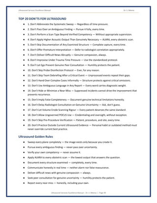
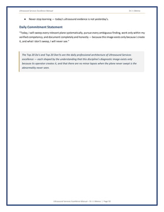
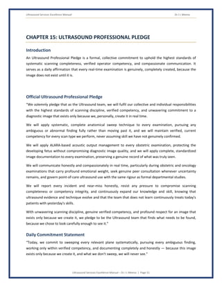
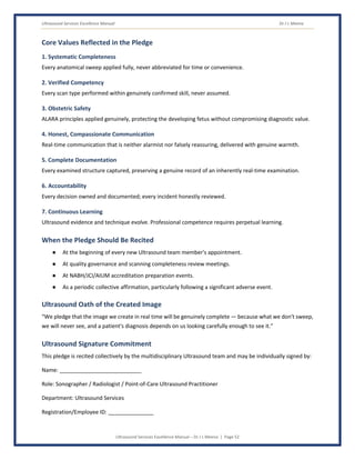
Ultrasound Services Excellence Manual Dr J L Meena
Ultrasound Services Excellence Manual – Dr J L Meena | Page 1
ULTRASOUND SERVICES
EXCELLENCE MANUAL
Global Standards for Ultrasound Excellence, Operator Competency, and Quality
Healthcare
A Comprehensive Reference for
THE MULTIDISCIPLINARY ULTRASOUND TEAM
Sonographers • Radiologists • Point-of-Care Ultrasound Practitioners • Obstetric & Allied Ultrasound
Professionals
“The Ultrasound Image Exists Only Because a Skilled Hand Creates It in Real Time — What We
Don't Sweep, We Will Never See.”
Dr J L Meena
Ultrasound Services Excellence Manual Dr J L Meena
Ultrasound Services Excellence Manual – Dr J L Meena | Page 2
DEDICATION
This book is dedicated to all those who are committed to advancing healthcare through a discipline unlike any other
in diagnostic imaging: the ultrasound image does not exist before the sonographer creates it, sweep by sweep, in
real time — unlike a CT or MRI scanner, which captures a fixed volume of data regardless of who presses the button,
an ultrasound examination's diagnostic value is created live, by a skilled hand and a trained eye, and whatever plane
is never swept, whatever structure is never interrogated, was simply never seen.
To Every Patient Whose Diagnosis Depended on a Sweep That Was Never Skipped
For whom a complete, systematic examination — every organ interrogated, every measurement taken correctly,
every abnormal finding pursued rather than passed over — represents a promise kept: that the image their diagnosis
depended on was genuinely, skillfully created, not merely acquired.
To Every Expecting Parent Whose Ultrasound Confirmed a Beating Heart, or Gently Delivered Difficult
News
For whom an obstetric scan performed with genuine technical skill and communicated with equal compassion
represents this discipline's highest calling — real-time images that carry some of medicine's most joyful and most
difficult moments, created by a hand that never rushed and never looked away.
To My Family
For their unwavering love, patience, and encouragement, which have been the foundation of this journey.
To My Mentors & Teachers
For inspiring my passion, guiding my path, and instilling in me the understanding that ultrasound, more than any
other imaging modality, places the entire diagnostic burden on the skill of the person holding the probe — there is
no fixed protocol that scans itself, no automated acquisition that guarantees completeness, only a trained hand's
real-time judgment about where to look next, and how carefully to look there.
To My Colleagues & Friends
For their support, collaboration, and shared dedication to a discipline where a systematic sweep technique, honestly
and completely performed, is the only thing standing between a diagnosis found and an abnormality quietly missed
because the probe simply never passed over it.
To Every Member of the Ultrasound Team
Sonographers, radiologists, point-of-care ultrasound practitioners, and every professional who has swept a probe
through one more plane, just to be certain, when a faster examination would have been just as easy to justify — the
guardians of a diagnostic image that exists only because they, personally, in that moment, chose to create it well.
Your skill, your systematic thoroughness, and your unwavering commitment to a discipline where the image
exists only because you created it are the silent pillars of a healthcare system that finds what needs to be
found, because someone chose to look carefully enough to see it. This book is for you, and because of you.
Thank you to everyone who has been a part of this journey.
Ultrasound Services Excellence Manual Dr J L Meena
Ultrasound Services Excellence Manual – Dr J L Meena | Page 3
FOREWORD
There is a discipline in diagnostic imaging unlike any other: the ultrasound image does not exist before the
sonographer creates it. A CT scanner captures a fixed volume of data in a single automated acquisition, largely
independent of who presses the button. An ultrasound examination is fundamentally different — it is created
live, in real time, by a skilled hand moving a probe and a trained eye interpreting what appears on the screen
in that instant. This makes ultrasound the most genuinely operator-dependent modality in all of medical
imaging: the diagnostic value of the examination is inseparable from the skill, the systematic thoroughness,
and the real-time judgment of the person performing it. Whatever anatomical plane is never swept, whatever
structure is never deliberately interrogated, was simply never seen — and there is frequently no second
automated pass to catch what the operator's hand and eye missed the first time.
The “Ultrasound Services Excellence Manual: Global Standards for Ultrasound Excellence, Operator
Competency, and Quality Healthcare” has been developed to provide a comprehensive professional framework
for the multidisciplinary Ultrasound team — sonographers, radiologists, point-of-care ultrasound practitioners,
and allied obstetric ultrasound professionals — working together to ensure that every real-time examination
is created with the systematic completeness and technical skill its operator-dependent nature demands.
This manual brings together internationally recognized principles of systematic scanning protocol and
complete anatomical sweep technique, operator competency and credentialing, obstetric ultrasound safety
across all trimesters, the ALARA principle applied to acoustic output, point-of-care ultrasound governance,
ultrasound-guided procedural safety, and image documentation supporting genuine quality review of an
inherently real-time examination. It is built around globally accepted accreditation frameworks — WHO
diagnostic ultrasound safety guidelines, NABH, JCI, and international standards from the American Institute of
Ultrasound in Medicine (AIUM) and the International Society of Ultrasound in Obstetrics and Gynecology
(ISUOG) — to provide a rigorous, regulation-compliant framework for Ultrasound Services excellence.
The chapters of this manual guide the Ultrasound team through every dimension of the role — from systematic
scanning protocols that ensure complete anatomical coverage, through operator competency and obstetric
safety, to ALARA-based acoustic output management, procedural guidance, and the professional conduct that
defines excellence across every category of ultrasound practice: the systematic abdominal sweep that finds
the mass a rushed examination would have missed, the obstetric scan whose careful, complete anatomical
survey catches an anomaly in time to matter, and the point-of-care examination performed at the bedside with
the same rigour as a formal departmental study.
Ultrasound Services is not simply an imaging service that produces pictures without radiation or a magnetic
field — it is a distinct discipline built on the understanding that its diagnostic reliability depends entirely on the
real-time skill of its operator, that a complete, systematic examination is the only defence against the
abnormality the probe never happened to sweep across, and that this team's technical competency is not a
supporting factor in diagnostic accuracy but its actual, direct source. Their work is defined by a discipline that
must never relax, because an image never created cannot later be recovered.
Ultrasound Services Excellence Manual Dr J L Meena
Ultrasound Services Excellence Manual – Dr J L Meena | Page 4
It is hoped that this manual will serve as an indispensable resource for Sonographers, Radiologists, Point-of-
Care Ultrasound Practitioners, hospital administrators, quality heads, and all those committed to building a
healthcare system where every real-time examination is created with the systematic, skilled completeness that
this uniquely operator-dependent technology demands.
“The Ultrasound Image Exists Only Because a Skilled Hand Creates It in Real Time — What We Don't
Sweep, We Will Never See.”
With Best Wishes, Dr J L Meena
Ultrasound Services Excellence Manual Dr J L Meena
Ultrasound Services Excellence Manual – Dr J L Meena | Page 5
TABLE OF CONTENTS
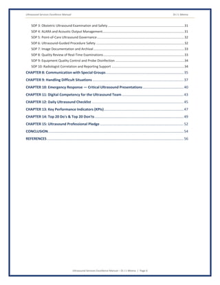
FOREWORD............................................................................................................................................3
LIST OF ABBREVIATIONS........................................................................................................................8
MANUAL MOTTO.................................................................................................................................10
CHAPTER 1: Introduction.....................................................................................................................11
Purpose...........................................................................................................................................................11
Objectives .......................................................................................................................................................12
Vision & Mission .............................................................................................................................................13
CHAPTER 2: Role of the Ultrasound Team in Quality Healthcare ......................................................14
Systematic Scanning Protocol & Complete Anatomical Sweep......................................................................14
Operator Competency & Credentialing ..........................................................................................................14
Obstetric Ultrasound Safety ...........................................................................................................................15
ALARA & Acoustic Output Management ........................................................................................................15
Point-of-Care Ultrasound (POCUS) Governance.............................................................................................16
Ultrasound-Guided Procedural Safety............................................................................................................16
Image Documentation & Quality Review........................................................................................................17
Radiologist Correlation & Reporting Support.................................................................................................17
CHAPTER 3: Professional Standards for the Ultrasound Team ..........................................................17
Professional Appearance & Behaviour ...........................................................................................................17
Communication Standards .............................................................................................................................18
Confidentiality & Information Security...........................................................................................................19
Ethical Standards ............................................................................................................................................19
Time Management, Teamwork & Continuous Learning.................................................................................20
CHAPTER 4: Patient-Centered Communication in Ultrasound...........................................................21
The A-T-C-R-C Communication Model............................................................................................................21
Communicating Findings & Delivering Difficult Obstetric News.....................................................................22
CHAPTER 5: Patient Safety and Examination Completeness Standards ............................................24
The R-T-A-C-A Framework ..............................................................................................................................24
Daily Quality Standards Checklist ...................................................................................................................26
CHAPTER 6: Confidentiality, Privacy, and Information Security ........................................................28
Privacy in Ultrasound Practice........................................................................................................................28
Cybersecurity of Ultrasound Information Systems.........................................................................................29
CHAPTER 7: Standard Operating Procedures (SOPs)..........................................................................30
SOP 1: Systematic Scanning Protocol and Anatomical Sweep........................................................................30
SOP 2: Operator Competency Verification and Credentialing........................................................................30
Ultrasound Services Excellence Manual Dr J L Meena
Ultrasound Services Excellence Manual – Dr J L Meena | Page 6
SOP 3: Obstetric Ultrasound Examination and Safety ....................................................................................31
SOP 4: ALARA and Acoustic Output Management..........................................................................................31
SOP 5: Point-of-Care Ultrasound Governance................................................................................................32
SOP 6: Ultrasound-Guided Procedure Safety .................................................................................................32
SOP 7: Image Documentation and Archival....................................................................................................33
SOP 8: Quality Review of Real-Time Examinations.........................................................................................33
SOP 9: Equipment Quality Control and Probe Disinfection ............................................................................34
SOP 10: Radiologist Correlation and Reporting Support ................................................................................34
CHAPTER 8: Communication with Special Groups..............................................................................35
CHAPTER 9: Handling Difficult Situations ...........................................................................................37
CHAPTER 10: Emergency Response — Critical Ultrasound Presentations.........................................40
CHAPTER 11: Digital Competency for the Ultrasound Team..............................................................43
CHAPTER 12: Daily Ultrasound Checklist ............................................................................................45
CHAPTER 13: Key Performance Indicators (KPIs)................................................................................47
CHAPTER 14: Top 20 Do's & Top 20 Don'ts.........................................................................................49
CHAPTER 15: Ultrasound Professional Pledge....................................................................................52
CONCLUSION........................................................................................................................................54
REFERENCES.........................................................................................................................................56
Ultrasound Services Excellence Manual Dr J L Meena
Ultrasound Services Excellence Manual – Dr J L Meena | Page 7
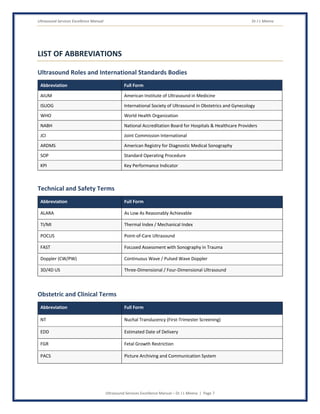
LIST OF ABBREVIATIONS
Ultrasound Roles and International Standards Bodies
Abbreviation Full Form
AIUM American Institute of Ultrasound in Medicine
ISUOG International Society of Ultrasound in Obstetrics and Gynecology
WHO World Health Organization
NABH National Accreditation Board for Hospitals & Healthcare Providers
JCI Joint Commission International
ARDMS American Registry for Diagnostic Medical Sonography
SOP Standard Operating Procedure
KPI Key Performance Indicator
Technical and Safety Terms
Abbreviation Full Form
ALARA As Low As Reasonably Achievable
TI/MI Thermal Index / Mechanical Index
POCUS Point-of-Care Ultrasound
FAST Focused Assessment with Sonography in Trauma
Doppler (CW/PW) Continuous Wave / Pulsed Wave Doppler
3D/4D US Three-Dimensional / Four-Dimensional Ultrasound
Obstetric and Clinical Terms
Abbreviation Full Form
NT Nuchal Translucency (First-Trimester Screening)
EDD Estimated Date of Delivery
FGR Fetal Growth Restriction
PACS Picture Archiving and Communication System
Ultrasound Services Excellence Manual Dr J L Meena
Ultrasound Services Excellence Manual – Dr J L Meena | Page 8
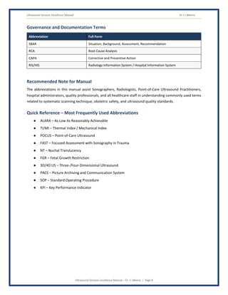
Governance and Documentation Terms
Abbreviation Full Form
SBAR Situation, Background, Assessment, Recommendation
RCA Root Cause Analysis
CAPA Corrective and Preventive Action
RIS/HIS Radiology Information System / Hospital Information System
Recommended Note for Manual
The abbreviations in this manual assist Sonographers, Radiologists, Point-of-Care Ultrasound Practitioners,
hospital administrators, quality professionals, and all healthcare staff in understanding commonly used terms
related to systematic scanning technique, obstetric safety, and ultrasound quality standards.
Quick Reference – Most Frequently Used Abbreviations
● ALARA – As Low As Reasonably Achievable
● TI/MI – Thermal Index / Mechanical Index
● POCUS – Point-of-Care Ultrasound
● FAST – Focused Assessment with Sonography in Trauma
● NT – Nuchal Translucency
● FGR – Fetal Growth Restriction
● 3D/4D US – Three-/Four-Dimensional Ultrasound
● PACS – Picture Archiving and Communication System
● SOP – Standard Operating Procedure
● KPI – Key Performance Indicator
Ultrasound Services Excellence Manual Dr J L Meena
Ultrasound Services Excellence Manual – Dr J L Meena | Page 9
MANUAL MOTTO
“The Ultrasound Image Exists Only Because”
“A Skilled Hand Creates It in Real Time —”
“What We Don't Sweep, We Will Never See.”
Ultrasound Services Excellence Manual Dr J L Meena
Ultrasound Services Excellence Manual – Dr J L Meena | Page 10
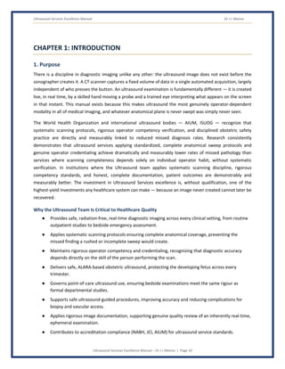
CHAPTER 1: INTRODUCTION
1. Purpose
There is a discipline in diagnostic imaging unlike any other: the ultrasound image does not exist before the
sonographer creates it. A CT scanner captures a fixed volume of data in a single automated acquisition, largely
independent of who presses the button. An ultrasound examination is fundamentally different — it is created
live, in real time, by a skilled hand moving a probe and a trained eye interpreting what appears on the screen
in that instant. This manual exists because this makes ultrasound the most genuinely operator-dependent
modality in all of medical imaging, and whatever anatomical plane is never swept was simply never seen.
The World Health Organization and international ultrasound bodies — AIUM, ISUOG — recognize that
systematic scanning protocols, rigorous operator competency verification, and disciplined obstetric safety
practice are directly and measurably linked to reduced missed diagnosis rates. Research consistently
demonstrates that ultrasound services applying standardized, complete anatomical sweep protocols and
genuine operator credentialing achieve dramatically and measurably lower rates of missed pathology than
services where scanning completeness depends solely on individual operator habit, without systematic
verification. In institutions where the Ultrasound team applies systematic scanning discipline, rigorous
competency standards, and honest, complete documentation, patient outcomes are demonstrably and
measurably better. The investment in Ultrasound Services excellence is, without qualification, one of the
highest-yield investments any healthcare system can make — because an image never created cannot later be
recovered.
Why the Ultrasound Team Is Critical to Healthcare Quality
● Provides safe, radiation-free, real-time diagnostic imaging across every clinical setting, from routine
outpatient studies to bedside emergency assessment.
● Applies systematic scanning protocols ensuring complete anatomical coverage, preventing the
missed finding a rushed or incomplete sweep would create.
● Maintains rigorous operator competency and credentialing, recognizing that diagnostic accuracy
depends directly on the skill of the person performing the scan.
● Delivers safe, ALARA-based obstetric ultrasound, protecting the developing fetus across every
trimester.
● Governs point-of-care ultrasound use, ensuring bedside examinations meet the same rigour as
formal departmental studies.
● Supports safe ultrasound-guided procedures, improving accuracy and reducing complications for
biopsy and vascular access.
● Applies rigorous image documentation, supporting genuine quality review of an inherently real-time,
ephemeral examination.
● Contributes to accreditation compliance (NABH, JCI, AIUM) for ultrasound service standards.
Ultrasound Services Excellence Manual Dr J L Meena
Ultrasound Services Excellence Manual – Dr J L Meena | Page 11
Key Responsibilities of the Ultrasound Team
● Applying systematic, complete anatomical sweep technique for every examination.
● Maintaining verified operator competency and credentialing for every scanning role.
● Applying safe, evidence-based obstetric ultrasound protocols across all trimesters.
● Applying ALARA principles to acoustic output for every examination, particularly obstetric studies.
● Applying rigorous governance to point-of-care ultrasound use across the institution.
● Applying safe technique for every ultrasound-guided procedure.
● Applying complete, standardized image documentation supporting quality review.
● Engaging in continuous professional development to maintain competence in evolving ultrasound
evidence.
2. Objectives
1. Create a Complete Examination, Every Time
Every examination must apply a systematic, complete anatomical sweep — this is the Ultrasound team's
foundational obligation, because an image never created through a skipped sweep cannot later be recovered.
How to Achieve:
● Apply standardized, protocol-driven scanning sequences for every examination type.
● Never allow time pressure to compress systematic anatomical coverage.
● Pursue and document any abnormal finding fully rather than moving past it.
2. Maintain Verified Operator Competency
● Apply rigorous credentialing and competency verification for every scanning role.
● Apply ongoing skill assessment and continuing education requirements.
3. Protect the Developing Fetus
● Apply ALARA-based acoustic output management for every obstetric examination.
● Apply safe, evidence-based protocols across all trimesters of pregnancy.
4. Govern Point-of-Care Ultrasound Rigorously
● Apply institutional credentialing and quality oversight to all POCUS use.
● Apply the same documentation and review standards to POCUS as to departmental studies.
5. Support Safe, Accurate Procedural Guidance
● Apply rigorous ultrasound-guided technique for biopsy and vascular access procedures.
6. Document Completely, Supporting Genuine Quality Review
● Apply standardized image documentation capturing the complete examination performed.
Ultrasound Services Excellence Manual Dr J L Meena
Ultrasound Services Excellence Manual – Dr J L Meena | Page 12
● Support systematic quality review processes despite the inherently real-time nature of the
examination.
7. Maintain Regulatory and Accreditation Compliance
● Comply with national ultrasound service standards and international accreditation guidelines.
● Maintain NABH/JCI/AIUM ultrasound documentation standards.
● Participate in accreditation surveys with complete, auditable competency and examination records.
3. Vision
Vision Statement
“To build and maintain an Ultrasound service where every examination is created with genuine systematic
completeness, every operator's competency is verified rather than assumed, and no patient's diagnosis is ever
compromised by a sweep that was never performed.”
4. Mission
Mission Statement
“To deliver systematically complete, technically skilled, and rigorously safe ultrasound services that create
genuinely thorough real-time diagnostic images, through disciplined scanning protocols, verified operator
competency, and safe obstetric and procedural practice, enabling confident diagnosis built on an examination
that was truly, completely performed.”
Expected Outcomes of an Excellent Ultrasound Services Practice
● 100% of examinations follow documented, systematic scanning protocols with complete anatomical
coverage.
● 100% of scanning operators maintain verified, current competency credentials.
● 100% of obstetric examinations apply documented ALARA-based acoustic output management.
● 100% of point-of-care ultrasound use governed by institutional credentialing and quality oversight.
● Full compliance with national ultrasound service standards and international accreditation
guidelines.
The Ultrasound team creates a diagnostic image that does not exist until a skilled hand and trained eye
bring it into being, sweep by sweep, in real time — their systematic scanning discipline and verified
competency are what stand between a complete diagnosis and the abnormality that a probe simply
never happened to pass over.
Ultrasound Services Excellence Manual Dr J L Meena
Ultrasound Services Excellence Manual – Dr J L Meena | Page 13
CHAPTER 2: ROLE OF THE ULTRASOUND TEAM IN QUALITY
HEALTHCARE
1. Systematic Scanning Protocol and Complete Anatomical Sweep
Overview
Because the ultrasound image is created live rather than captured automatically, only a systematic, protocol-
driven sweep through every relevant anatomical plane guarantees that nothing was simply passed over.
Responsibilities
● Apply standardized, protocol-driven scanning sequences for every examination type.
● Pursue and fully document any abnormal finding rather than moving past it.
2. Operator Competency and Credentialing
Overview
Because ultrasound's diagnostic quality is created by the operator's real-time skill rather than an automated
protocol, genuine, verified competency is the direct source of diagnostic accuracy, not merely a supporting
factor.
Responsibilities
● Maintain verified, current competency credentials for every scanning role.
● Apply ongoing skill assessment and continuing education requirements.
3. Obstetric Ultrasound Safety
Overview
Obstetric ultrasound carries unique responsibility — protecting a developing fetus while providing information
that shapes some of a family's most significant medical decisions, across every trimester of pregnancy.
Responsibilities
● Apply safe, evidence-based protocols matched to gestational age and clinical indication.
● Apply systematic anatomical survey supporting timely anomaly detection.
4. ALARA and Acoustic Output Management
Overview
Though ultrasound involves no ionizing radiation, the ALARA principle still applies to acoustic energy exposure,
particularly for the developing fetus, guided by thermal and mechanical index monitoring.
Ultrasound Services Excellence Manual Dr J L Meena
Ultrasound Services Excellence Manual – Dr J L Meena | Page 14
Responsibilities
● Apply ALARA-based acoustic output settings for every examination, particularly obstetric studies.
● Monitor and document thermal and mechanical indices throughout the examination.
5. Point-of-Care Ultrasound (POCUS) Governance
Overview
As point-of-care ultrasound use by non-radiologist clinicians expands, institutional governance ensures bedside
examinations meet the same rigour as formal departmental studies.
Responsibilities
● Apply institutional credentialing and quality oversight to all POCUS use.
● Apply the same documentation and review standards to POCUS as to departmental studies.
6. Ultrasound-Guided Procedural Safety
Overview
Ultrasound guidance improves accuracy and reduces complications for biopsy and vascular access procedures,
requiring rigorous hand-eye coordination and technique.
Responsibilities
● Apply rigorous ultrasound-guided technique for every procedural application.
● Apply appropriate sterile technique and patient safety verification during procedures.
7. Image Documentation and Quality Review
Overview
Because the examination itself is real-time and ephemeral, standardized image documentation is the only
means by which quality review, peer consultation, and future comparison remain possible.
Responsibilities
● Apply complete, standardized image documentation capturing every examined structure.
● Support systematic quality review processes for the recorded examination.
8. Radiologist Correlation and Reporting Support
Overview
Close coordination between sonographer and radiologist ensures clinical questions are genuinely answered
and any technical limitation is understood in context.
Responsibilities
● Communicate technical findings, limitations, or unexpected observations clearly to the radiologist.
● Support additional scanning when radiologist consultation identifies a genuine clinical need.
Ultrasound Services Excellence Manual Dr J L Meena
Ultrasound Services Excellence Manual – Dr J L Meena | Page 15
How the Ultrasound Team Contributes to Quality Healthcare
● By applying systematic sweep technique, the team ensures no anatomical region is simply passed
over unexamined.
● By maintaining verified operator competency, the team ensures diagnostic accuracy is built on
genuine, current skill.
● By applying ALARA to acoustic output, the team protects the developing fetus without compromising
diagnostic image quality.
● By governing POCUS rigorously, the team ensures bedside examinations meet the same standard as
formal studies.
● By documenting completely, the team makes genuine quality review possible for an examination
that otherwise exists only in the moment it was performed.
The Ultrasound team creates a diagnostic image that does not exist until a skilled hand and trained eye
bring it into being, sweep by sweep, in real time — their systematic scanning discipline and verified
competency are what stand between a complete diagnosis and the abnormality that a probe simply
never happened to pass over.
Ultrasound Services Excellence Manual Dr J L Meena
Ultrasound Services Excellence Manual – Dr J L Meena | Page 16
CHAPTER 3: PROFESSIONAL STANDARDS FOR THE ULTRASOUND TEAM
Ultrasound practice requires an uncommon combination of real-time technical skill applied to an examination
that exists only because the operator creates it, systematic discipline that resists the temptation to shortcut a
sweep under time pressure, and the compassionate communication skill demanded by an examination that
frequently carries a family's most joyful or most difficult news. The standards that govern Ultrasound practice
derive from patient safety science unique to a discipline whose diagnostic quality is inseparable from its
operator's real-time judgment.
1. Professional Appearance
Standards
Attire and Identification
● Wear appropriate attire and ID badge as per facility policy.
Professional Presence
● Maintain rigorous, disciplined attention to systematic scanning throughout every examination.
● Model unwavering completeness discipline that never relaxes, regardless of workload.
2. Professional Behaviour
Expected Behaviours
Scanning Discipline
● Never abbreviate a systematic anatomical sweep to save time.
● Never pass over an ambiguous finding without pursuing genuine clarification.
Competency Discipline
● Never perform an examination type beyond current, verified competency without appropriate
supervision.
● Escalate immediately upon any identified competency gap or equipment concern.
Accountability
● Own every scanning decision, every documented finding, and every interpretation made under this
team's responsibility.
● Report and disclose critical incidents and adverse events honestly and promptly.
Collaborative Respect
● Treat every member of the multidisciplinary Ultrasound team — sonographer, radiologist, POCUS
practitioner — as an essential, equal partner.
Ultrasound Services Excellence Manual Dr J L Meena
Ultrasound Services Excellence Manual – Dr J L Meena | Page 17
Patient-Centered Focus
● Every sweep and every scanning decision is ultimately about creating a genuinely complete
examination for the patient — never about convenience or speed.
3. Communication Standards
Communication with Patients
● Communicate findings honestly and compassionately, particularly in obstetric and oncology contexts.
● Communicate examination limitations or incomplete findings honestly when they occur.
Communication Within the Team
● Communicate any scanning uncertainty or technical limitation rapidly and clearly.
● Use structured handover tools (SBAR or equivalent) for every transfer of patient or diagnostic
responsibility.
● Escalate any competency concern or equipment malfunction immediately and directly.
Written Communication Standards
● Ultrasound documentation must be accurate, contemporaneous, and complete, reflecting the
examination genuinely performed.
● Reports must be exact and free of language that could be misinterpreted regarding examination
completeness.
4. Confidentiality and Information Security
Overview
Ultrasound involves particularly sensitive information, including obstetric findings and oncology screening
results, requiring heightened confidentiality discipline.
Responsibilities
● Discuss patient and examination information only in appropriate settings and only with those who
have a legitimate need to know.
● Protect access credentials for RIS/HIS and ultrasound information systems.
● Apply particular discretion for sensitive obstetric and oncology findings.
5. Ethical Standards
Core Ethical Principles for Ultrasound Practice
Non-Maleficence
● Never allow a known scanning gap or competency uncertainty to persist unaddressed.
● Never finalize an examination as complete when systematic coverage was genuinely not achieved.
Ultrasound Services Excellence Manual Dr J L Meena
Ultrasound Services Excellence Manual – Dr J L Meena | Page 18
Beneficence
● Apply rigorous, evidence-based scanning practice consistently, ensuring the most complete
examination achievable.
Honesty and Transparency
● Communicate genuine examination limitations, technical difficulty, and any error honestly.
Justice and Equity
● Apply the same rigorous scanning and competency standard to every patient, regardless of setting or
volume.
6. Time Management and Prioritization
Clinical Priority Framework
● Immediate/Emergency: Trauma FAST examination, suspected ectopic pregnancy — immediate
response, no delay.
● Urgent: Significant unexpected finding requiring prompt communication — prompt escalation.
● Routine: Standard scanning and reporting — managed per defined schedule, with unwavering
systematic completeness discipline.
7. Teamwork and Collaboration
Work Effectively With
● Radiologists — for correlation, difficult case consultation, and reporting support.
● Obstetrics and gynaecology — for coordinated pregnancy and gynaecological care.
● Emergency and critical care teams — for point-of-care and trauma ultrasound coordination.
● Quality department — for incident reporting, RCA, and accreditation support.
8. Professional Competence and Continuous Learning
Areas of Competence
● Systematic scanning protocol and complete anatomical sweep technique.
● Obstetric ultrasound safety and gestational-age-appropriate protocol.
● ALARA-based acoustic output management.
● Ultrasound-guided procedural technique.
● FMEA, RCA, and CAPA methodologies for ultrasound-specific quality improvement.
Continuous Learning
● Maintain current certification in relevant ultrasound competencies.
● Maintain current knowledge of evolving ultrasound evidence and guidelines (AIUM, ISUOG, WHO).
Ultrasound Services Excellence Manual Dr J L Meena
Ultrasound Services Excellence Manual – Dr J L Meena | Page 19
● Engage with national and international ultrasound professional bodies.
● Participate regularly in scanning technique and obstetric safety simulation training.
● Participate in NABH, JCI, and AIUM accreditation training for ultrasound standards.
Professional Standards Checklist
● Have I applied systematic, complete anatomical sweep technique to every examination today?
● Have I maintained verified competency for every scan type I performed?
● Have I applied ALARA-based acoustic output management for every obstetric examination?
● Have I documented every examination completely and accurately?
● Have I communicated findings honestly and compassionately with every patient?
● Have I reported any incident or safety concern through the appropriate system?
Ultrasound professionalism is systematic completeness applied to an examination that exists only
because the operator creates it — the team that sweeps every plane and never assumes competency
without genuine verification is the team that ensures the abnormality the probe never happened to pass
over never happens at all.
Ultrasound Services Excellence Manual Dr J L Meena
Ultrasound Services Excellence Manual – Dr J L Meena | Page 20
CHAPTER 4: PATIENT-CENTERED COMMUNICATION IN ULTRASOUND
Communication in Ultrasound must accomplish something genuinely distinctive: patients frequently watch the
screen alongside the sonographer in real time, particularly during obstetric examinations, meaning
communication during the scan itself, not only afterward, shapes the patient's experience and understanding
profoundly.
Why Communication Quality Matters in Ultrasound
● Real-time communication during a scan can either reassure a patient watching the screen or cause
unnecessary alarm if handled carelessly.
● Compassionate delivery of difficult obstetric news requires particular skill, given the emotional
weight of the moment.
● Clear communication about examination limitations helps patients understand why additional
imaging or follow-up may be needed.
● Honest communication about incidental findings supports appropriate, timely follow-up.
The A-T-C-R-C Communication Model for Ultrasound
A — Acknowledge
Definition
Acknowledge the patient's genuine emotional state, particularly the anticipation or anxiety common in
obstetric and oncology examinations.
Examples
● “I know this moment means a lot to you — let's take our time and I'll explain what we're seeing as
we go.”
T — Listen (Elicit Genuine Concerns)
Definition
Listen genuinely to the patient's specific concerns and questions during the examination.
C — Clarify (Communicate Findings Appropriately in Real Time)
Definition
Communicate findings appropriately during the scan, being mindful of what is genuinely appropriate to share
in real time versus what requires radiologist correlation first.
Examples
Ultrasound Services Excellence Manual Dr J L Meena
Ultrasound Services Excellence Manual – Dr J L Meena | Page 21
● “I can see the heartbeat clearly — that's a great sign. I'll take a few more measurements, and then
the full report will go to your doctor.”
R — Respond (Answer Questions Honestly)
Definition
Respond to questions honestly, including honest disclosure of any genuine uncertainty requiring radiologist
review.
C — Confirm (Verify Understanding)
Definition
Confirm genuine understanding of the examination's findings and next steps before the patient leaves.
Communicating Findings and Delivering Difficult Obstetric News
Principles for Real-Time Communication During Scanning
● Communicate reassuring, unambiguous findings (e.g., a clearly visible heartbeat) directly and
warmly.
● Avoid speculative or premature commentary on findings genuinely requiring radiologist
interpretation.
Delivering Difficult Obstetric News
● Communicate difficult findings compassionately, honestly, and without unnecessary technical
language.
● Coordinate closely with the referring clinician to ensure consistent, supportive follow-up
communication.
Communicating Examination Limitations
● Communicate any genuine technical limitation (e.g., body habitus, fetal position) honestly as part of
the examination summary.
● Recommend appropriate follow-up imaging when limitations genuinely affect diagnostic
completeness.
Common Ultrasound Communication Mistakes to Avoid
● Offering premature or speculative interpretation of a finding that genuinely requires radiologist
correlation.
● Delivering difficult obstetric news abruptly or without appropriate compassion.
● Failing to explain a genuine examination limitation, leaving the patient with a false impression of
completeness.
● Rushing real-time communication during the scan itself, missing the patient's genuine emotional
cues.
Ultrasound Services Excellence Manual Dr J L Meena
Ultrasound Services Excellence Manual – Dr J L Meena | Page 22
Communication in Ultrasound happens live, often with the patient watching the same screen — the team
that communicates warmly and honestly in real time, and delivers difficult news with genuine
compassion, protects patients through some of medicine's most significant and most vulnerable
moments.
Ultrasound Services Excellence Manual Dr J L Meena
Ultrasound Services Excellence Manual – Dr J L Meena | Page 23
CHAPTER 5: PATIENT SAFETY AND EXAMINATION COMPLETENESS
STANDARDS
Patient safety in Ultrasound is uniquely defined by the fact that the diagnostic image is created by the operator,
not captured automatically by the machine — a service that maintains systematic scanning protocols, verifies
genuine operator competency, and documents completely produces measurably better diagnostic accuracy
than one where any of these safeguards is compressed by workload or assumed rather than verified. The R-T-
A-C-A framework applies with particular intensity to a discipline whose diagnostic reliability is inseparable from
its operator's real-time skill.
1. RESPECT
Definition
Treating every anatomical sweep and every examination's systematic completeness as equally deserving of the
Ultrasound team's full rigour, regardless of how routine an examination appears or how high the workload.
How the Ultrasound Team Demonstrates Respect
● Respect for the Examination's Creation: Apply the same systematic sweep to the routine follow-up
scan as to the complex new case.
● Respect for Genuine Competency: Never perform an examination type beyond current, verified skill
without appropriate supervision.
● Respect for Every Team Member's Judgment: Value the sonographer's real-time scanning decision as
highly as the radiologist's final interpretation.
● Equal Service Standards: Apply the same rigorous protocol to every patient, regardless of setting or
volume.
2. TIMELINESS
Definition
Recognizing and responding to trauma, obstetric emergencies, and other time-critical presentations within
timeframes that protect patient safety, while never rushing the systematic scanning process that requires its
own, uncompressed time.
Why Timeliness Is a Patient Safety Issue — and Why It Cuts Both Ways
A delayed trauma FAST examination or suspected ectopic pregnancy scan can cost a patient time-critical
treatment. Yet the opposite lesson applies equally: rushing a systematic anatomical sweep to save time is
precisely the shortcut that can mean an abnormality is simply never seen, because the probe never happened
to pass over it. Timeliness in Ultrasound means responding to genuine emergencies with maximum speed while
refusing to compress the systematic scanning rigour that requires its full, uncompressed time.
Ultrasound Services Excellence Manual Dr J L Meena
Ultrasound Services Excellence Manual – Dr J L Meena | Page 24
Ultrasound Timeliness Standards
● Trauma and obstetric emergency examinations: immediate, without avoidable delay.
● Systematic anatomical sweep: full, uncompressed protocol, every examination, regardless of
schedule pressure.
● Significant finding communication: prompt, without unnecessary delay.
3. ACCURACY
Definition
Applying every anatomical sweep exactly, verifying genuine operator competency for every scan type, and
documenting every examination precisely, sustained without deviation across every patient.
Why Accuracy Is Non-Negotiable in Ultrasound
An incomplete anatomical sweep can mean an abnormality is simply never seen, with no automated second
pass to catch what the operator's hand and eye missed. Accuracy in Ultrasound, sustained across scanning
technique and documentation alike, is what separates a discipline whose real-time diagnostic creation
genuinely serves patients from one whose incompleteness surfaces only when a missed finding progresses.
Areas Requiring Ultrasound Accuracy
● Systematic Scanning: Every anatomical region relevant to the clinical indication must be genuinely
swept and examined.
● Operator Competency: Every scan type must be performed within genuine, verified skill, never
assumed.
● Acoustic Output: Every obstetric examination must apply verified ALARA-based settings.
● Documentation: Every ultrasound record must accurately and completely reflect the examination
genuinely performed.
4. COMPASSION
Definition
Recognizing that ultrasound examinations frequently carry a patient's most joyful or most difficult moments,
often experienced in real time alongside the sonographer, and that genuine compassion in that shared moment
is as important as technical accuracy.
How the Ultrasound Team Demonstrates Compassion
● Communicate with Genuine Warmth in Real Time: Recognizing the patient is often watching the
same screen.
● Deliver Difficult News with Particular Care: Obstetric and oncology findings carry profound emotional
weight.
● Support Colleagues Through Technical Uncertainty: A collegial, non-punitive culture of consultation
improves accuracy for every patient.
Ultrasound Services Excellence Manual Dr J L Meena
Ultrasound Services Excellence Manual – Dr J L Meena | Page 25
● Respect the Weight of Every Examination: Even a routine-seeming scan matters deeply to the patient
receiving it.
5. ACCOUNTABILITY
Definition
Taking full professional responsibility for every scanning decision, every documented finding, and every
interpretation made under Ultrasound team responsibility.
Ultrasound Accountability Standards
● Every scanning, documentation, and interpretation decision is documented with the responsible
team member's identification.
● Never allow a known scanning gap or competency uncertainty to go unaddressed.
● Report every critical incident and adverse ultrasound event honestly through the quality reporting
system.
● Participate in RCA for significant incidents, including any missed diagnosis or documented
incompleteness, with complete clinical honesty.
● Participate in quality audits and accreditation surveys with accurate, complete ultrasound records.
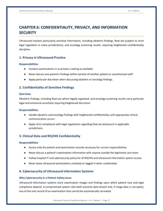
The R-T-A-C-A Framework in Ultrasound
Standard Core Focus in Ultrasound
Respect Every sweep and every examination's completeness honoured equally, regardless of
routine
Timeliness Immediate emergency response; uncompressed systematic scanning rigour
Accuracy Complete anatomical coverage; genuine, verified operator competency
Compassion Genuine warmth in real-time communication; particular care for difficult news
Accountability Every decision owned and documented; every incident honestly reviewed
Daily Quality Standards Checklist for Ultrasound
● Apply systematic, complete anatomical sweep technique to every examination today.
● Maintain verified competency for every scan type performed.
● Apply ALARA-based acoustic output management for every obstetric examination.
● Document every examination completely and accurately.
● Communicate findings honestly and compassionately with every patient.
● Report any incident or safety concern through the quality reporting system.
Ultrasound Services Excellence Manual Dr J L Meena
Ultrasound Services Excellence Manual – Dr J L Meena | Page 26
In Ultrasound, patient safety is measured against an examination that exists only because it was
genuinely created — a patient experiences it as a complete, systematic sweep performed by a skilled
hand that never rushed past a plane it should have examined.
Ultrasound Services Excellence Manual Dr J L Meena
Ultrasound Services Excellence Manual – Dr J L Meena | Page 27
CHAPTER 6: CONFIDENTIALITY, PRIVACY, AND INFORMATION
SECURITY
Ultrasound involves particularly sensitive information, including obstetric findings, fetal sex (subject to strict
legal regulation in many jurisdictions), and oncology screening results, requiring heightened confidentiality
discipline.
1. Privacy in Ultrasound Practice
Responsibilities
● Conduct examinations in as private a setting as available.
● Never discuss one patient's findings within earshot of another patient or unauthorized staff.
● Apply particular discretion when discussing obstetric or oncology findings.
2. Confidentiality of Sensitive Findings
Overview
Obstetric findings, including fetal sex where legally regulated, and oncology screening results carry particular
legal and emotional sensitivity requiring heightened discretion.
Responsibilities
● Handle obstetric and oncology findings with heightened confidentiality until appropriate clinical
communication occurs.
● Apply strict compliance with legal regulations regarding fetal sex disclosure in applicable
jurisdictions.
3. Clinical Data and RIS/HIS Confidentiality
Responsibilities
● Access only the patient and examination records necessary for current responsibilities.
● Never discuss a patient's examination information with anyone outside the legitimate care team.
● Follow hospital IT and cybersecurity policy for all RIS/HIS and ultrasound information system access.
● Never leave ultrasound workstations unlocked or logged in when unattended.
4. Cybersecurity of Ultrasound Information Systems
Why Cybersecurity Is a Patient Safety Issue
Ultrasound information systems store examination images and findings upon which patient care and legal
compliance depend. A compromised system risks both sensitive data breach and, if image data is corrupted,
loss of the only record of an examination that cannot be automatically recreated.
Ultrasound Services Excellence Manual Dr J L Meena
Ultrasound Services Excellence Manual – Dr J L Meena | Page 28
Responsibilities
● Use only authorized, unique login credentials for all RIS/HIS and ultrasound information system
access.
● Never share login credentials with colleagues, regardless of workload pressure.
● Never connect personal devices to hospital clinical networks without IT authorization.
● Report any suspected unauthorized access or cybersecurity anomaly to IT security immediately.
5. Confidentiality for High-Profile and Vulnerable Patients
Responsibilities
● Maintain the same confidentiality standard for high-profile or notorious patients as for every other
patient.
● Never disclose patient identity, diagnosis, or condition to media or unauthorized persons.
Confidentiality Checklist for the Ultrasound Team
● Conduct examinations with maximum available privacy.
● Handle obstetric and oncology findings with heightened discretion.
● Use only authorized credentials for RIS/HIS and ultrasound information systems.
● Never connect personal devices to hospital clinical networks without IT authorization.
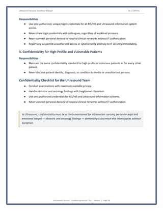
In Ultrasound, confidentiality must be actively maintained for information carrying particular legal and
emotional weight — obstetric and oncology findings — demanding a discretion this team applies without
exception.
Ultrasound Services Excellence Manual Dr J L Meena
Ultrasound Services Excellence Manual – Dr J L Meena | Page 29
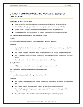
CHAPTER 7: STANDARD OPERATING PROCEDURES (SOPs) FOR
ULTRASOUND
Objectives of Ultrasound SOPs
● Ensure consistent, systematic scanning and honest documentation for every examination.
● Minimize harm through systematic, documented protocols and competency verification.
● Support NABH/JCI/AIUM accreditation compliance for ultrasound service standards.
● Provide a defensible clinical framework for incident investigation and quality improvement.
SOP 1: Systematic Scanning Protocol and Anatomical Sweep
Purpose
To ensure every diagnostically relevant anatomical region is genuinely examined.
Procedure
● Step 1: Apply Standardized Protocol — apply the protocol matched to examination type and clinical
indication.
● Step 2: Apply Complete Anatomical Sweep — sweep systematically through every relevant plane.
● Step 3: Pursue Any Abnormal Finding — investigate and fully document any ambiguous or abnormal
finding.
● Step 4: Document — document the complete examination and findings.
Quality Standards
● 100% of examinations follow documented, systematic scanning protocols.
SOP 2: Operator Competency Verification and Credentialing
Purpose
To ensure diagnostic accuracy is built on genuine, verified skill.
Procedure
● Step 1: Verify Current Credentialing — verify credentialing status before performing any examination
type.
● Step 2: Apply Ongoing Skill Assessment — apply periodic competency reassessment.
● Step 3: Apply Appropriate Supervision — apply supervision for any examination type outside
independent competency.
● Step 4: Document — document the complete credentialing and competency record.
Ultrasound Services Excellence Manual Dr J L Meena
Ultrasound Services Excellence Manual – Dr J L Meena | Page 30
Quality Standards
● 100% of scanning operators maintain verified, current competency credentials.
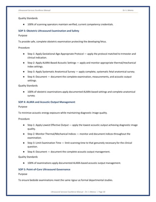
SOP 3: Obstetric Ultrasound Examination and Safety
Purpose
To provide safe, complete obstetric examination protecting the developing fetus.
Procedure
● Step 1: Apply Gestational-Age-Appropriate Protocol — apply the protocol matched to trimester and
clinical indication.
● Step 2: Apply ALARA-Based Acoustic Settings — apply and monitor appropriate thermal/mechanical
index settings.
● Step 3: Apply Systematic Anatomical Survey — apply complete, systematic fetal anatomical survey.
● Step 4: Document — document the complete examination, measurements, and acoustic output
settings.
Quality Standards
● 100% of obstetric examinations apply documented ALARA-based settings and complete anatomical
survey.
SOP 4: ALARA and Acoustic Output Management
Purpose
To minimize acoustic energy exposure while maintaining diagnostic image quality.
Procedure
● Step 1: Apply Lowest Effective Output — apply the lowest acoustic output achieving diagnostic image
quality.
● Step 2: Monitor Thermal/Mechanical Indices — monitor and document indices throughout the
examination.
● Step 3: Limit Examination Time — limit scanning time to that genuinely necessary for the clinical
question.
● Step 4: Document — document the complete acoustic output management.
Quality Standards
● 100% of examinations apply documented ALARA-based acoustic output management.
SOP 5: Point-of-Care Ultrasound Governance
Purpose
To ensure bedside examinations meet the same rigour as formal departmental studies.
Ultrasound Services Excellence Manual Dr J L Meena
Ultrasound Services Excellence Manual – Dr J L Meena | Page 31
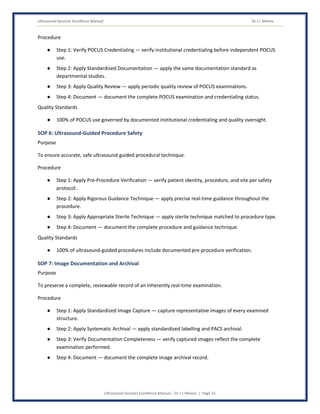
Procedure
● Step 1: Verify POCUS Credentialing — verify institutional credentialing before independent POCUS
use.
● Step 2: Apply Standardized Documentation — apply the same documentation standard as
departmental studies.
● Step 3: Apply Quality Review — apply periodic quality review of POCUS examinations.
● Step 4: Document — document the complete POCUS examination and credentialing status.
Quality Standards
● 100% of POCUS use governed by documented institutional credentialing and quality oversight.
SOP 6: Ultrasound-Guided Procedure Safety
Purpose
To ensure accurate, safe ultrasound-guided procedural technique.
Procedure
● Step 1: Apply Pre-Procedure Verification — verify patient identity, procedure, and site per safety
protocol.
● Step 2: Apply Rigorous Guidance Technique — apply precise real-time guidance throughout the
procedure.
● Step 3: Apply Appropriate Sterile Technique — apply sterile technique matched to procedure type.
● Step 4: Document — document the complete procedure and guidance technique.
Quality Standards
● 100% of ultrasound-guided procedures include documented pre-procedure verification.
SOP 7: Image Documentation and Archival
Purpose
To preserve a complete, reviewable record of an inherently real-time examination.
Procedure
● Step 1: Apply Standardized Image Capture — capture representative images of every examined
structure.
● Step 2: Apply Systematic Archival — apply standardized labelling and PACS archival.
● Step 3: Verify Documentation Completeness — verify captured images reflect the complete
examination performed.
● Step 4: Document — document the complete image archival record.
Ultrasound Services Excellence Manual Dr J L Meena
Ultrasound Services Excellence Manual – Dr J L Meena | Page 32
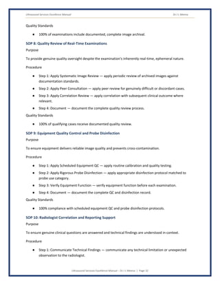
Quality Standards
● 100% of examinations include documented, complete image archival.
SOP 8: Quality Review of Real-Time Examinations
Purpose
To provide genuine quality oversight despite the examination's inherently real-time, ephemeral nature.
Procedure
● Step 1: Apply Systematic Image Review — apply periodic review of archived images against
documentation standards.
● Step 2: Apply Peer Consultation — apply peer review for genuinely difficult or discordant cases.
● Step 3: Apply Correlation Review — apply correlation with subsequent clinical outcome where
relevant.
● Step 4: Document — document the complete quality review process.
Quality Standards
● 100% of qualifying cases receive documented quality review.
SOP 9: Equipment Quality Control and Probe Disinfection
Purpose
To ensure equipment delivers reliable image quality and prevents cross-contamination.
Procedure
● Step 1: Apply Scheduled Equipment QC — apply routine calibration and quality testing.
● Step 2: Apply Rigorous Probe Disinfection — apply appropriate disinfection protocol matched to
probe use category.
● Step 3: Verify Equipment Function — verify equipment function before each examination.
● Step 4: Document — document the complete QC and disinfection record.
Quality Standards
● 100% compliance with scheduled equipment QC and probe disinfection protocols.
SOP 10: Radiologist Correlation and Reporting Support
Purpose
To ensure genuine clinical questions are answered and technical findings are understood in context.
Procedure
● Step 1: Communicate Technical Findings — communicate any technical limitation or unexpected
observation to the radiologist.
Ultrasound Services Excellence Manual Dr J L Meena
Ultrasound Services Excellence Manual – Dr J L Meena | Page 33
● Step 2: Support Additional Scanning When Needed — support additional views per genuine
radiologist consultation.
● Step 3: Verify Image Adequacy — verify diagnostic adequacy before releasing the study.
● Step 4: Document — document the complete correlation and reporting support process.
Quality Standards
● 100% of studies verified for diagnostic adequacy before release.
Ultrasound SOP Daily Compliance Checklist
● Every examination applies documented, systematic scanning protocol today.
● Every operator's competency verified current for the examination type performed.
● Any incident or safety concern reported through the appropriate system.
SOPs in Ultrasound are the operating instructions for creating a genuinely complete, real-time diagnostic
examination. When followed consistently, they are the systematic foundation of scanning completeness
and verified competency for every patient this team serves.
Ultrasound Services Excellence Manual Dr J L Meena
Ultrasound Services Excellence Manual – Dr J L Meena | Page 34
CHAPTER 8: COMMUNICATION WITH SPECIAL GROUPS
The Ultrasound team communicates across a distinctive range of circumstances — from expecting parents
watching a screen in real time, to a trauma team awaiting an urgent FAST examination, to a non-radiologist
clinician requesting POCUS credentialing, to a patient receiving an unexpected oncology finding. Excellent
ultrasound practice requires adapted communication for each of these circumstances.
1. Expecting Parents
Communication Guidelines
● Communicate with genuine warmth, recognizing the profound emotional significance of the
moment.
● Communicate reassuring findings directly while deferring speculative interpretation to radiologist
correlation.
2. Trauma and Emergency Teams
Communication Guidelines
● Communicate rapidly and clearly, matching the team's genuine urgency.
● Communicate critical findings immediately upon identification.
3. Non-Radiologist POCUS Practitioners
Communication Guidelines
● Communicate credentialing requirements and quality standards clearly.
● Support genuine skill development through constructive feedback.
4. Patients Receiving Difficult Oncology or Obstetric Findings
Overview
Findings suggesting malignancy or significant fetal anomaly carry profound emotional weight, requiring
particular compassion in both real-time and follow-up communication.
Communication Guidelines
● Communicate honestly and compassionately, avoiding both false alarm and false reassurance.
● Coordinate closely with the referring clinician for consistent, supportive follow-up.
5. Referring Clinicians
Communication Guidelines
● Communicate examination findings and any genuine limitation clearly and completely.
Ultrasound Services Excellence Manual Dr J L Meena
Ultrasound Services Excellence Manual – Dr J L Meena | Page 35
● Communicate promptly when a finding requires urgent clinical attention.
6. Patients Requiring Ultrasound-Guided Procedures
Communication Guidelines
● Communicate the procedure's purpose and process clearly before beginning.
● Communicate genuine reassurance and guidance throughout the procedure.
7. Patients with Body Habitus or Technical Scanning Limitations
Communication Guidelines
● Communicate any genuine technical limitation honestly and without causing unnecessary
embarrassment.
● Recommend appropriate alternative imaging when limitations genuinely affect diagnostic
completeness.
8. Hospital Administration and Quality/Accreditation Bodies
Communication Guidelines
● Present scanning protocol compliance, competency credentialing, and quality review data in
management-accessible formats.
● Advocate for necessary resources (staffing, equipment, credentialing support) with clear patient
safety justification.
Universal Ultrasound Communication Principles
● Communicate with exact, unambiguous language, given the diagnostic decisions built on every word.
● Respond to every genuine emergency with maximum speed, without compromising systematic
scanning rigour.
● Always confirm genuine understanding, particularly for difficult findings and follow-up
recommendations.
● Document every significant clinical communication completely.
The Ultrasound team that communicates with equal skill toward an expecting parent's joy and a trauma
team's urgent need serves every stakeholder at maximum impact — honest, real-time communication
delivered alongside the genuine compassion an examination this personal and this immediate deserves.
Ultrasound Services Excellence Manual Dr J L Meena
Ultrasound Services Excellence Manual – Dr J L Meena | Page 36
CHAPTER 9: HANDLING DIFFICULT SITUATIONS
Common Difficult Situations for the Ultrasound Team
● A systematic sweep is genuinely difficult due to patient body habitus or fetal position.
● An unexpected, significant finding is identified during a routine examination.
● A colleague's scanning technique appears genuinely incomplete.
● A referring clinician requests an examination type beyond the operator's current competency.
● A patient asks a question the operator should not answer before radiologist correlation.
● A POCUS practitioner requests informal consultation on a difficult finding.
● An obstetric examination reveals a significant fetal anomaly.
● Equipment malfunction is identified during an active examination.
● A trauma FAST examination must be performed under extreme time pressure.
● A patient requests information about fetal sex where legally restricted.
Core Principles for Handling Difficult Situations
1. Systematic Completeness and Genuine Competency Are Never Compromised
In every difficult situation, genuine systematic scanning and verified competency drive every decision — no
time or clinical pressure, however genuine, justifies an incomplete sweep or a scan beyond genuine skill.
2. Apply Standardized Protocols Under Pressure
Technical difficulty and time pressure are precisely when standardized protocols matter most — resist the
temptation to improvise when a proven protocol exists.
3. Escalate Immediately and Persistently
A genuine safety or competency concern is not resolved until it reaches someone who can act on it — escalate
through alternative channels if the first attempt does not produce a response.
4. Document Everything
Every difficult situation must be contemporaneously documented — what was found, what was
communicated, what decisions were made, and by whom.
Handling a Technically Difficult Sweep
Steps
● Apply additional technique or patient positioning to achieve genuine completeness where possible.
● Document any genuine, unavoidable limitation honestly rather than implying false completeness.
Ultrasound Services Excellence Manual Dr J L Meena
Ultrasound Services Excellence Manual – Dr J L Meena | Page 37
Handling an Unexpected Significant Finding
Steps
● Pursue the finding thoroughly with additional views and measurements.
● Communicate the finding promptly to the radiologist and, per protocol, the referring clinician.
Handling Concern About a Colleague's Scanning Completeness
Steps
● Address the concern directly and professionally with the colleague.
● Escalate to department leadership if the concern involves a pattern or remains unresolved.
Handling a Request Beyond Current Competency
Steps
● Decline to proceed independently; arrange appropriate supervision or referral instead.
● Communicate the genuine competency limitation professionally to the requesting clinician.
Handling a Premature Patient Question During Scanning
Steps
● Respond honestly about what can and cannot yet be confirmed.
● Explain that full interpretation will follow radiologist review.
Handling a Significant Fetal Anomaly Finding
Steps
● Complete a thorough, systematic evaluation of the finding.
● Coordinate closely with the referring clinician and, where appropriate, maternal-fetal medicine for
compassionate, coordinated communication.
Handling Equipment Malfunction Mid-Examination
Steps
● Apply immediate contingency protocol to complete the examination safely.
● Communicate any resulting delay transparently to the patient and clinical team.
Handling Extreme Time Pressure in Trauma Scanning
Steps
● Apply the pre-established, standardized FAST protocol rather than improvising.
● Communicate findings immediately to the trauma team.
Ultrasound Services Excellence Manual Dr J L Meena
Ultrasound Services Excellence Manual – Dr J L Meena | Page 38
Handling a Request for Legally Restricted Fetal Sex Disclosure
Steps
● Decline the request professionally, citing the applicable legal restriction clearly.
● Maintain the same standard for every patient without exception.
The CALM Model for Ultrasound
● C – Control the immediate scanning or safety risk.
● A – Acknowledge the clinical reality accurately, including genuine technical limitation.
● L – Lead with evidence — scanning protocols, competency standards, and safety guidelines.
● M – Manage through documented escalation channels when needed.
Difficult situations in Ultrasound are always, ultimately, tests of whether systematic completeness and
genuine competency hold firm under time or clinical pressure. The team that never compromises either,
escalates persistently, and communicates honestly even when uncertain is fulfilling the highest
professional obligation of ultrasound practice.
Ultrasound Services Excellence Manual Dr J L Meena
Ultrasound Services Excellence Manual – Dr J L Meena | Page 39
CHAPTER 10: EMERGENCY RESPONSE — CRITICAL ULTRASOUND
PRESENTATIONS
Overview
Certain presentations demand the Ultrasound team's most immediate, standardized, and rehearsed response
— scenarios where the difference between a coordinated, protocol-driven response and an improvised one is
measured directly in whether a life-threatening finding is identified in time.
Critical Presentations Requiring Immediate Response
● Trauma requiring immediate FAST examination.
● Suspected ectopic pregnancy.
● Suspected ruptured ovarian cyst or ovarian torsion.
● Suspected acute cholecystitis or biliary obstruction.
● Suspected deep vein thrombosis in a patient with acute symptoms.
● Suspected testicular torsion.
● Fetal distress or absent fetal cardiac activity in a viable pregnancy.
● Suspected abdominal aortic aneurysm.
● Equipment malfunction during an active emergency examination.
● Critical incidental finding requiring immediate notification.
1. Trauma FAST Examination
Immediate Response
● Apply immediate, standardized FAST protocol.
● Communicate findings immediately to the trauma team.
2. Suspected Ectopic Pregnancy
Steps
● Apply immediate, systematic pelvic and adnexal examination.
● Communicate findings immediately given the time-critical nature of this diagnosis.
3. Suspected Ovarian Torsion
Steps
● Apply immediate examination including Doppler assessment.
● Communicate findings immediately given the time-critical nature of ovarian salvage.
Ultrasound Services Excellence Manual Dr J L Meena
Ultrasound Services Excellence Manual – Dr J L Meena | Page 40
4. Suspected Acute Cholecystitis
Steps
● Apply immediate, systematic examination of the gallbladder and biliary system.
● Communicate findings promptly to the requesting clinical team.
5. Suspected Testicular Torsion
Steps
● Apply immediate examination including Doppler assessment of testicular blood flow.
● Communicate findings immediately given the extremely narrow window for surgical salvage.
6. Absent Fetal Cardiac Activity
Steps
● Apply careful, complete confirmation before any communication.
● Communicate findings with particular compassion, coordinating with the referring clinician for
support.
7. Suspected Abdominal Aortic Aneurysm
Steps
● Apply immediate examination and measurement.
● Communicate findings immediately given the risk of rupture.
8. Suspected Deep Vein Thrombosis
Steps
● Apply immediate, systematic compression ultrasound examination.
● Communicate findings promptly given the risk of pulmonary embolism.
9. Equipment Malfunction During Emergency Examination
Steps
● Apply immediate contingency protocol or equipment substitution.
● Communicate any resulting delay transparently to the emergency team.
10. Critical Incidental Finding
Steps
● Apply immediate notification to the ordering clinician per critical result protocol.
Ultrasound Services Excellence Manual Dr J L Meena
Ultrasound Services Excellence Manual – Dr J L Meena | Page 41
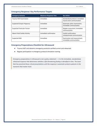
Emergency Response: Key Performance Targets
Emergency Scenario Maximum Response Time Key Action
Trauma FAST Examination Immediate Standardized protocol; immediate
trauma team communication
Suspected Ectopic Pregnancy Immediate Systematic pelvic examination;
immediate communication
Suspected Testicular Torsion Immediate Doppler assessment; immediate
communication
Absent Fetal Cardiac Activity Immediate confirmation Careful confirmation;
compassionate communication
Suspected AAA Immediate Examination and measurement;
immediate communication
Emergency Preparedness Checklist for Ultrasound
● Trauma FAST and obstetric emergency protocols verified current and rehearsed.
● Regular participation in emergency protocol simulation training.
Emergency preparedness in Ultrasound is not a policy statement — it is the immediate, standardized,
rehearsed response that determines whether a life-threatening finding is identified in time. The team
that has practiced these critical presentations until the response is automatic protects patients in the
moments that matter most.
Ultrasound Services Excellence Manual Dr J L Meena
Ultrasound Services Excellence Manual – Dr J L Meena | Page 42
CHAPTER 11: DIGITAL COMPETENCY FOR THE ULTRASOUND TEAM
Objectives
● Enable the Ultrasound team to use scanning, archival, and reporting systems accurately, securely,
and efficiently.
● Leverage technology to improve image documentation and quality review.
● Manage the cybersecurity of ultrasound information systems as a core patient safety function.
Core Digital Competencies for the Ultrasound Team
1. Radiology Information System (RIS) / PACS
Essential Skills
● Use RIS/PACS accurately for scheduling, image archival, and reporting.
2. Ultrasound Console and Protocol Software
Essential Skills
● Use scanner console and protocol software accurately, applying correct examination-specific
settings.
3. Credentialing and Competency Tracking Systems
Essential Skills
● Use credentialing tracking systems accurately to maintain verified, current competency records.
4. Electronic Medical Record (EMR) / Hospital Information System (HIS)
Essential Skills
● Document examination findings accurately and in real time.
5. AI-Assisted Image Quality and Measurement Tools
Awareness
● AI-assisted tools are increasingly applied to support measurement automation and image quality
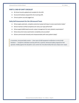
assessment.
● Understand and appropriately leverage these tools while maintaining independent clinical judgment
— AI tools are decision-support, not a substitute for genuine sonographer skill.
6. Telesonography and Remote Consultation
Essential Skills
● Use telesonography platforms for remote consultation where clinically appropriate.
Ultrasound Services Excellence Manual Dr J L Meena
Ultrasound Services Excellence Manual – Dr J L Meena | Page 43
7. Cybersecurity and Information Security
Personal Digital Security Responsibilities
● Use unique, strong credentials for all RIS/HIS and ultrasound information system logins.
● Lock or log out of workstations whenever stepping away, even briefly.
● Never connect personal devices to hospital clinical networks.
● Report all cybersecurity anomalies to IT security immediately.
Digital Competency Checklist
● Navigate RIS/PACS, scanner console, and credentialing tracking systems for all core functions without
assistance.
● Apply accurate, complete examination and image documentation.
● Apply basic cybersecurity practices to all clinical system access.
● Know the downtime procedure for RIS/PACS system failure.
Digital competency for the Ultrasound team is the ability to integrate genuine real-time scanning skill
with the archival, credentialing, and reporting systems that modern ultrasound increasingly depends
upon — using technology to extend, never replace, the systematic completeness that defines safe
ultrasound practice.
Ultrasound Services Excellence Manual Dr J L Meena
Ultrasound Services Excellence Manual – Dr J L Meena | Page 44
CHAPTER 12: DAILY ULTRASOUND CHECKLIST
Objectives
● Provide a structured daily framework for systematic scanning discipline.
● Ensure every scanning, competency, and documentation obligation is addressed every shift.
● Support accurate clinical documentation and departmental performance data quality.
PART A: START-OF-SHIFT CHECKLIST
Personal and Professional Readiness
● Appropriate attire and ID badge displayed.
● RIS/PACS systems functional and accessible.
Equipment and Readiness Review
● Verified scanner functional and probes disinfected per protocol.
● Verified emergency and procedural equipment functional.
PART B: SCANNING CHECKLIST
● Systematic, complete anatomical sweep applied to every examination today.
● Any ambiguous or abnormal finding pursued fully and documented.
PART C: COMPETENCY CHECKLIST
● Every examination performed within current, verified competency.
● Any examination outside independent competency appropriately supervised.
PART D: OBSTETRIC SAFETY CHECKLIST
● ALARA-based acoustic output settings applied and documented for every obstetric examination.
PART E: DOCUMENTATION CHECKLIST
● Complete, standardized image documentation captured for every examination.
● Any significant finding communicated promptly to the radiologist and clinical team.
PART F: SAFETY AND INCIDENT CHECKLIST
● Any incident, near-miss, or scanning completeness concern reported through the appropriate
system.
● CAPA actions from previous incidents reviewed for completion.
Ultrasound Services Excellence Manual Dr J L Meena
Ultrasound Services Excellence Manual – Dr J L Meena | Page 45
PART G: END-OF-SHIFT CHECKLIST
● All clinical records updated and complete for the shift.
● Structured handover prepared for the incoming shift.
● Clinical systems securely logged out.
Daily Self-Assessment for the Ultrasound Team
● Did we apply systematic, complete anatomical sweep technique to every examination today?
● Did we maintain verified competency for every scan type performed?
● Did we apply ALARA-based acoustic output management for every obstetric examination?
● Did we document every examination completely and accurately?
● Did we communicate honestly and compassionately with every patient today?
A consistent, structured daily routine — from start-of-shift equipment verification to end-of-shift
handover — transforms individual scanning skill into a systemic ultrasound safety programme that
operates reliably against the discipline's own central risk: the abnormality that was simply never swept.
Ultrasound Services Excellence Manual Dr J L Meena
Ultrasound Services Excellence Manual – Dr J L Meena | Page 46
CHAPTER 13: KEY PERFORMANCE INDICATORS (KPIs) FOR
ULTRASOUND
Objectives
● Measure scanning completeness, competency, and safety performance objectively.
● Enable data-driven continuous improvement in Ultrasound Services practice.
● Support NABH/JCI/AIUM accreditation standards for ultrasound services.
● Demonstrate the value and patient safety impact of the Ultrasound team to institutional leadership.
Categories of Ultrasound KPIs
1. Scanning Completeness KPIs
KPI Definition Target
Protocol Compliance Rate % of examinations following documented, systematic
scanning protocols
100%
Documented Limitation Rate % of examinations with any genuine technical limitation
honestly documented
Track and trend;
ensure honest
reporting
2. Competency KPIs
KPI Definition Target
Operator Credentialing Compliance % of scanning operators with verified, current
competency credentials
100%
Continuing Education Compliance % of staff meeting required continuing education
targets
100%
3. Obstetric Safety KPIs
KPI Definition Target
ALARA Documentation Compliance % of obstetric examinations with documented
acoustic output management
100%
Fetal Anomaly Detection Rate Rate of anomalies detected on systematic anatomical
survey
At or above
institutional/national
benchmark
4. POCUS Governance KPIs
KPI Definition Target
POCUS Credentialing Compliance % of POCUS use governed by documented
institutional credentialing
100%
Ultrasound Services Excellence Manual Dr J L Meena
Ultrasound Services Excellence Manual – Dr J L Meena | Page 47
KPI Definition Target
POCUS Quality Review Completion % of POCUS examinations receiving periodic quality
review
≥ 90%
5. Outcome and Governance KPIs
KPI Definition Target
RCA Completion Rate % of significant adverse events with completed RCA 100%
Missed Diagnosis Rate on Correlation
Review
Rate of confirmed missed findings identified on
quality/correlation review
At or below
institutional/national
benchmark
Patient Satisfaction Score % positive patient feedback on ultrasound service ≥ 85%
Using KPIs for Continuous Improvement
● Review all Ultrasound KPIs regularly at departmental quality and governance meetings.
● Analyse scanning completeness and missed diagnosis trends to identify targeted process
improvements.
● Use credentialing data to identify training and continuing education needs.
● Present KPI data to hospital management to demonstrate the patient safety impact of Ultrasound
Services excellence.
Ultrasound KPIs translate the discipline of creating a genuinely complete real-time examination into
institutional accountability — demonstrating that the investment in systematic scanning and verified
operator competency is a measurable, trackable programme that ensures the abnormality a probe
should have swept across is never the one that gets away.
Ultrasound Services Excellence Manual Dr J L Meena
Ultrasound Services Excellence Manual – Dr J L Meena | Page 48
CHAPTER 14: TOP 20 DO'S & TOP 20 DON'TS FOR ULTRASOUND
Introduction
The Ultrasound team creates a diagnostic image that does not exist until a skilled hand and trained eye bring
it into being, sweep by sweep, in real time. Their daily professional decisions — about systematic scanning,
verified competency, and honest documentation — determine whether every examination genuinely finds
what it needs to find. These Do's and Don'ts provide the practical daily architecture of Ultrasound Services
excellence.
TOP 20 DO'S FOR ULTRASOUND
● 1. Apply the Complete Systematic Sweep — Every examination, every time.
● 2. Pursue Every Ambiguous Finding — Never pass over uncertainty.
● 3. Verify Your Own Competency Before Every Scan Type — Never assume, always confirm.
● 4. Apply ALARA to Every Obstetric Examination — The lowest acoustic output that answers the
question.
● 5. Document Every Structure Examined — Complete, standardized image capture, every time.
● 6. Communicate Honestly in Real Time — Neither false alarm nor false reassurance.
● 7. Deliver Difficult News with Genuine Compassion — Obstetric and oncology findings carry real
weight.
● 8. Apply the Standardized Trauma Protocol — Speed without abandoning completeness.
● 9. Seek Peer Consultation for Genuine Uncertainty — Humility protects the patient.
● 10. Apply Rigorous Probe Disinfection — Every use, every time.
● 11. Debrief After Every Significant Event — Consolidate learning while it is fresh.
● 12. Provide Structured Handover for Every Transfer — SBAR or equivalent, every time.
● 13. Communicate with Exact, Unambiguous Language — A diagnosis depends on every word.
● 14. Report Every Near-Miss Honestly — Including your own, especially in scanning completeness.
● 15. Document Any Genuine Technical Limitation — Honestly, never implying false completeness.
● 16. Coordinate Proactively with Radiologists — For correlation and difficult case support.
● 17. Apply the Same Rigour to Every Patient — Regardless of setting or volume.
● 18. Govern POCUS with the Same Standards — Credentialing and documentation, without exception.
● 19. Verify Patient Identity Before Every Procedure — Ultrasound-guided procedures included.
● 20. Continue Learning — Ultrasound evidence and technique evolve. The team that does not learn
continuously treats today's patients with yesterday's skills.
Ultrasound Services Excellence Manual Dr J L Meena
Ultrasound Services Excellence Manual – Dr J L Meena | Page 49
TOP 20 DON'TS FOR ULTRASOUND
● 1. Don't Abbreviate the Systematic Sweep — Regardless of time pressure.
● 2. Don't Pass Over an Ambiguous Finding — Pursue it fully, every time.
● 3. Don't Perform a Scan Type Beyond Verified Competency — Without appropriate supervision.
● 4. Don't Apply Higher Acoustic Output Than Genuinely Necessary — ALARA, every obstetric scan.
● 5. Don't Skip Documentation of Any Examined Structure — Complete capture, every time.
● 6. Don't Offer Premature Interpretation — Defer to radiologist correlation appropriately.
● 7. Don't Deliver Difficult News Abruptly — Genuine compassion, always.
● 8. Don't Improvise Under Trauma Time Pressure — Use the standardized protocol.
● 9. Don't Let Ego Prevent Genuine Peer Consultation — Humility protects the patient.
● 10. Don't Skip Probe Disinfection Protocol — Ever, for any reason.
● 11. Don't Skip Team Debriefing After a Critical Event — Unprocessed events repeat their gaps.
● 12. Don't Hand Over Complex Cases Informally — Structure protects against critical omissions.
● 13. Don't Use Ambiguous Language in Any Report — Every word carries diagnostic weight.
● 14. Don't Hide or Minimize a Near-Miss — Suppressed incidents cannot drive the improvement that
prevents recurrence.
● 15. Don't Imply False Completeness — Document genuine technical limitations honestly.
● 16. Don't Delay Radiologist Consultation on Genuine Uncertainty — Ask, don't guess.
● 17. Don't Let Volume Erode Scanning Rigour — Every patient deserves the same standard.
● 18. Don't Allow Ungoverned POCUS Use — Credentialing and oversight, without exception.
● 19. Don't Skip Pre-Procedure Verification — Patient, procedure, and site, every time.
● 20. Don't Practice Outside Current Ultrasound Evidence — Personal habit or outdated method must
never override current best practice.
Ultrasound Golden Rules
● Sweep every plane completely — the image exists only because you create it.
● Pursue every ambiguous finding — never pass over uncertainty.
● Verify your own competency — never assume it.
● Apply ALARA to every obstetric scan — the lowest output that answers the question.
● Document every structure examined — completely, every time.
● Communicate honestly in real time — neither alarm nor false reassurance.
● Deliver difficult news with genuine compassion — always.
● Seek peer consultation for genuine uncertainty — humility protects the patient.
● Report every near-miss — honestly, including your own.
Ultrasound Services Excellence Manual Dr J L Meena
Ultrasound Services Excellence Manual – Dr J L Meena | Page 50
● Never stop learning — today's ultrasound evidence is not yesterday's.
Daily Commitment Statement
“Today, I will sweep every relevant plane systematically, pursue every ambiguous finding, work only within my
verified competency, and document completely and honestly — because this image exists only because I create
it, and what I don't sweep, I will never see.”
The Top 20 Do's and Top 20 Don'ts are the daily professional architecture of Ultrasound Services
excellence — each shaped by the understanding that this discipline's diagnostic image exists only
because its operator creates it, and that there are no minor lapses when the plane never swept is the
abnormality never seen.
Ultrasound Services Excellence Manual Dr J L Meena
Ultrasound Services Excellence Manual – Dr J L Meena | Page 51
CHAPTER 15: ULTRASOUND PROFESSIONAL PLEDGE
Introduction
An Ultrasound Professional Pledge is a formal, collective commitment to uphold the highest standards of
systematic scanning completeness, verified operator competency, and compassionate communication. It
serves as a daily affirmation that every real-time examination is genuinely, completely created, because the
image does not exist until it is.
Official Ultrasound Professional Pledge
“We solemnly pledge that as the Ultrasound team, we will fulfil our collective and individual responsibilities
with the highest standards of scanning discipline, verified competency, and unwavering commitment to a
diagnostic image that exists only because we, personally, create it in real time.
We will apply systematic, complete anatomical sweep technique to every examination, pursuing any
ambiguous or abnormal finding fully rather than moving past it, and we will maintain verified, current
competency for every scan type we perform, never assuming skill we have not genuinely confirmed.
We will apply ALARA-based acoustic output management to every obstetric examination, protecting the
developing fetus without compromising diagnostic image quality, and we will apply complete, standardized
image documentation to every examination, preserving a genuine record of what was truly seen.
We will communicate honestly and compassionately in real time, particularly during obstetric and oncology
examinations that carry profound emotional weight, seek genuine peer consultation whenever uncertainty
remains, and govern point-of-care ultrasound use with the same rigour as formal departmental studies.
We will report every incident and near-miss honestly, resist any pressure to compromise scanning
completeness or competency integrity, and continuously expand our knowledge and skill, knowing that
ultrasound evidence and technique evolve and that the team that does not learn continuously treats today's
patients with yesterday's skills.
With unwavering scanning discipline, genuine verified competency, and profound respect for an image that
exists only because we create it, we pledge to be the Ultrasound team that finds what needs to be found,
because we chose to look carefully enough to see it.”
Daily Commitment Statement
“Today, we commit to sweeping every relevant plane systematically, pursuing every ambiguous finding,
working only within verified competency, and documenting completely and honestly — because this image
exists only because we create it, and what we don't sweep, we will never see.”
Ultrasound Services Excellence Manual Dr J L Meena
Ultrasound Services Excellence Manual – Dr J L Meena | Page 52
Core Values Reflected in the Pledge
1. Systematic Completeness
Every anatomical sweep applied fully, never abbreviated for time or convenience.
2. Verified Competency
Every scan type performed within genuinely confirmed skill, never assumed.
3. Obstetric Safety
ALARA principles applied genuinely, protecting the developing fetus without compromising diagnostic value.
4. Honest, Compassionate Communication
Real-time communication that is neither alarmist nor falsely reassuring, delivered with genuine warmth.
5. Complete Documentation
Every examined structure captured, preserving a genuine record of an inherently real-time examination.
6. Accountability
Every decision owned and documented; every incident honestly reviewed.
7. Continuous Learning
Ultrasound evidence and technique evolve. Professional competence requires perpetual learning.
When the Pledge Should Be Recited
● At the beginning of every new Ultrasound team member's appointment.
● At quality governance and scanning completeness review meetings.
● At NABH/JCI/AIUM accreditation preparation events.
● As a periodic collective affirmation, particularly following a significant adverse event.
Ultrasound Oath of the Created Image
“We pledge that the image we create in real time will be genuinely complete — because what we don't sweep,
we will never see, and a patient's diagnosis depends on us looking carefully enough to see it.”
Ultrasound Signature Commitment
This pledge is recited collectively by the multidisciplinary Ultrasound team and may be individually signed by:
Name: ___________________________
Role: Sonographer / Radiologist / Point-of-Care Ultrasound Practitioner
Department: Ultrasound Services
Registration/Employee ID: _______________
Ultrasound Services Excellence Manual Dr J L Meena
Ultrasound Services Excellence Manual – Dr J L Meena | Page 53
Signature: ________________________
Date: ____________________________
Ultrasound Services Excellence Manual Dr J L Meena
Ultrasound Services Excellence Manual – Dr J L Meena | Page 54
CONCLUSION
There is a discipline in diagnostic imaging unlike any other: the ultrasound image does not exist before the
sonographer creates it. This manual has returned, again and again, to why this reality demands a discipline that
never relaxes — a systematic, complete anatomical sweep that leaves no relevant plane unexamined,
genuinely verified operator competency that is the direct source of diagnostic accuracy rather than a
supporting factor, and honest, complete documentation that preserves a genuine record of an examination
that otherwise exists only in the moment it was performed.
Throughout this manual, we have explored the full scope of Ultrasound Services professional practice — from
systematic scanning protocols and verified operator competency, through obstetric safety and ALARA-based
acoustic output management, to point-of-care governance, procedural guidance, and the professional conduct
that defines excellence across every category of ultrasound practice. These responsibilities collectively define
a discipline that demands both extraordinary real-time technical skill and the humility to seek consultation
whenever genuine uncertainty remains.
The Importance of the Ultrasound Team in Healthcare Quality
● The systematic scanner — sweeping every relevant plane completely, leaving nothing simply passed
over.
● The genuinely competent operator — performing every examination within real, verified skill, never
assumed.
● The fetal protector — applying ALARA principles that protect the developing fetus without
compromising diagnostic value.
● The compassionate communicator — delivering real-time findings, joyful or difficult, with genuine
warmth.
● The honest documenter — preserving a genuine record of an examination that exists only in the
moment it was created.
● The honest reviewer — examining every incident and near-miss to protect the next patient whose
diagnosis depends on a sweep that was never skipped.
Final Commitment
“As the Ultrasound team, we commit to sweeping every relevant plane systematically, pursuing every
ambiguous finding, working only within verified competency, and documenting completely and honestly —
because this image exists only because we create it, and what we don't sweep, we will never see.”
Ultrasound Services Excellence Manual Dr J L Meena
Ultrasound Services Excellence Manual – Dr J L Meena | Page 55
Final Motto
“The Ultrasound Image Exists Only Because a Skilled Hand Creates It in Real Time — What We Don't Sweep,
We Will Never See.”
Together, We Create the Examination That Finds What Needs to Be Found, One Careful Sweep at a
Time.
Ultrasound Services Excellence Manual Dr J L Meena
Ultrasound Services Excellence Manual – Dr J L Meena | Page 56
REFERENCES
Global Accreditation and Regulatory Standards
● World Health Organization (WHO) – Manual of Diagnostic Ultrasound and Safety Guidance.
● Joint Commission International (JCI) – JCI Accreditation Standards for Hospitals: Assessment of
Patients (AOP) Chapter, including Diagnostic Imaging Requirements.
● National Accreditation Board for Hospitals & Healthcare Providers (NABH, India) – Standards for
Diagnostic Imaging and Ultrasound Service Standards (5th Edition).
Ultrasound Practice Standards
● American Institute of Ultrasound in Medicine (AIUM) – Practice Parameters and Technical Standards
for Ultrasound Examinations.
● International Society of Ultrasound in Obstetrics and Gynecology (ISUOG) – Practice Guidelines for
Obstetric and Gynaecological Ultrasound.
Operator Competency and Credentialing Standards
● American Registry for Diagnostic Medical Sonography (ARDMS) – Credentialing and Competency
Standards.
● American College of Emergency Physicians (ACEP) – Point-of-Care Ultrasound Guidelines.
Acoustic Output and ALARA Standards
● American Institute of Ultrasound in Medicine (AIUM) – Official Statements on Bioeffects and
Acoustic Output Safety.
● British Medical Ultrasound Society (BMUS) – Guidelines for the Safe Use of Diagnostic Ultrasound
Equipment.
Emergency and Procedural Ultrasound Standards
● American College of Surgeons (ACS) – FAST Examination Protocol Guidelines.
● Society of Interventional Radiology (SIR) – Guidelines for Ultrasound-Guided Procedures.
Indian Regulatory and National Framework
● Pre-Conception and Pre-Natal Diagnostic Techniques (PCPNDT) Act, India – Regulation of Fetal Sex
Determination and Obstetric Ultrasound.
● Indian Radiological and Imaging Association (IRIA) – Clinical Practice Guidelines for Ultrasound.
● National Medical Commission (NMC) – Code of Medical Ethics Regulations Applicable to Radiology
Practice.
Ultrasound Services Excellence Manual Dr J L Meena
Ultrasound Services Excellence Manual – Dr J L Meena | Page 57
Quality Improvement and Patient Safety Frameworks
● Institute for Healthcare Improvement (IHI) – Framework for Patient Safety and Quality Improvement
in Diagnostic Imaging.
● World Health Organization – Global Patient Safety Action Plan 2021–2030.
Cybersecurity and Digital Health Records
● NIST Cybersecurity Framework – Applied to Healthcare Radiology Information Systems.
● HL7 International / DICOM Standards Committee – Medical Imaging Data Interchange Standards.
This manual has been developed using internationally recognized principles and best practices from WHO
Manual of Diagnostic Ultrasound, JCI Assessment of Patients Standards, NABH Diagnostic Imaging Standards,
AIUM Practice Parameters and Technical Standards, ISUOG Obstetric and Gynaecological Guidelines, ARDMS
Credentialing Standards, ACEP POCUS Guidelines, AIUM Bioeffects and Acoustic Output Statements, BMUS
Safety Guidelines, ACS FAST Protocol Guidelines, SIR Ultrasound-Guided Procedure Guidelines, India's PCPNDT
Act, IRIA Clinical Practice Guidelines, and applicable Indian statutory and regulatory requirements. Institutions
should verify current versions of all cited guidelines and regulatory requirements, as clinical protocols and
accreditation standards are subject to periodic revision.
ॐ जय माता द( ॐ
Ultrasound Services Excellence Manual Dr J L Meena
Ultrasound Services Excellence Manual – Dr J L Meena | Page 58
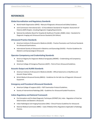
END OF MANUAL
Thank You
This manual has been prepared with the vision to empower the multidisciplinary Ultrasound team with global
standards, best practices, and professional guidance to create genuinely complete, real-time diagnostic
examinations for every patient.
Our Commitment Continues
The journey of learning never ends. As ultrasound technology and evidence evolve, our commitment to
systematic scanning, verified competency, and honest documentation must remain unwavering — because an
image never created cannot later be recovered.
Together, We Build Trust
Every plane swept systematically, every finding pursued fully, every competency genuinely verified, and every
examination documented honestly contributes to one greater purpose — finding what needs to be found,
because someone chose to look carefully enough to see it.
“The Ultrasound Image Exists Only Because a Skilled Hand Creates It in Real Time — What We
Don't Sweep, We Will Never See.”
REMEMBER: Systematic sweep is our discipline • Competency is our foundation • ALARA protects the
smallest patient • Compassion is our voice • Learning is our journey
Thank you for being a part of this vital mission. Your dedication makes a difference every day.
Dr J L Meena