Processed by Luminess, 75001 PARIS (FR)
(19)
EP
4
427
750
A1
(Cont. next page)
*EP004427750A1*
(11) EP 4 427 750 A1
(12) EUROPEAN PATENT APPLICATION
(43) Date of publication:
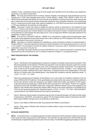
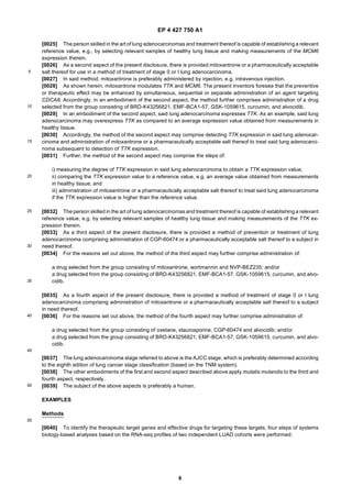
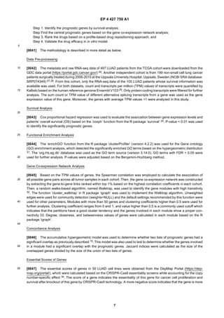
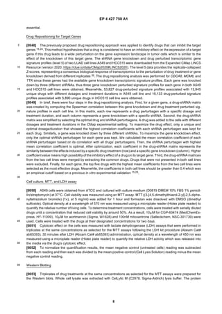
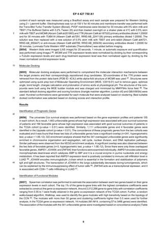
11.09.2024 Bulletin 2024/37
(21) Application number: 23160777.1
(22) Date of filing: 08.03.2023
(51) International Patent Classification (IPC):
A61K 31/506 (2006.01)
A61P 35/00 (2006.01)
A61K 31/136 (2006.01)
A61K 31/585 (2006.01)
(52) Cooperative Patent Classification (CPC):
(C-Sets available)
A61P 35/00; A61K 31/136; A61K 31/506;
A61K 31/585 (Cont.)
(84) Designated Contracting States:
AL AT BE BG CH CY CZ DE DK EE ES FI FR GB
GR HR HU IE IS IT LI LT LU LV MC ME MK MT NL
NO PL PT RO RS SE SI SK SM TR
Designated Extension States:
BA
Designated Validation States:
KH MA MD TN
(71) Applicant: Bash Biotech Inc
San Diego, CA 92101 (US)
(72) Inventors:
• MARDINOGLU, Adil
412 60 Göteborg (SE)
• LI, Xiangyu
113 46 Stockholm (SE)
(74) Representative: Kransell & Wennborg KB
P.O. Box 27834
115 93 Stockholm (SE)
(54) TREATMENT OF LUNG ADENOCARCINOMA
(57) The present disclosure provides CGP-60474 for
use in a method of prevention or treatment of lung ade-
nocarcinoma and mitoxantrone for use in a method of
treatment of stage o or I lung adenocarcinoma.
(Cont. next page)
2
EP 4 427 750 A1
(52) Cooperative Patent Classification (CPC): (Cont.)
C-Sets
A61K 31/136, A61K 2300/00;
A61K 31/506, A61K 2300/00;
A61K 31/585, A61K 2300/00
EP 4 427 750 A1
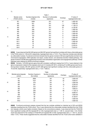
3
5
10
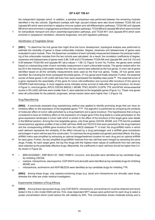
15
20
25
30
35
40
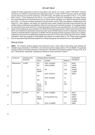
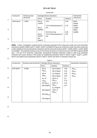
45
50
55
Description
TECHNICAL FIELD
[0001] The present disclosure relates to the field of treatment of adenocarcinoma.
BACKGROUND
[0002] Lung cancer is the most lethal cancer worldwide, causing more than 1.7 million deaths in 20201. Lung adeno-
carcinoma (LUAD) is the most prevalent histologic subtype in lung cancer, and accounts for half of all lung cancer deaths
2. LUAD has a strong association with smoking history, while it also showed an increasing occurrence in non-smokers
2. Patients with localized and early-stage tumor receive standard surgical resection, but the majority of patients are
usually diagnosed at an advanced stage and recommended the conventional therapies including chemoradiotherapy,
targeted therapy and immunotherapy. Various mutations in genes EGFR, ALK, KRAS, ROS1, BRAF, NTRK1/2/3, MET,
RET, and ERBB2 are observed in LUAD patients with relatively high coverages and these genes are involved in the
molecular testing for patients’ targeted therapy recommendation in clinical practice 3. For instance, the first-line treatment
for patients with EGFR activating mutations includes tyrosine kinase inhibitors osimertinib, erlotinib, afatinib, gefitinib
and dacomitinib. Despite these different therapy regimens, 5-year overall survival rate ranges from 4 to 17% depending
on different tumor stages and regions 4. Thus, there is need to discover new drug targets and develop effective drugs
for LUAD treatment, and thus improve the survival outcomes of patients.
[0003] Numerous studies have focused on the identification of biomarker genes based on the bioinformatics analysis
of transcriptomics profiles of LUAD patients 5-13. These studies validated the importance of the identified biomarkers by
proving the prognostic value of these genes or linking these genes with some meaningful tumor hallmarks, e.g., immune
infiltration, cell differentiation or glycolysis. However, none of these studies attempted to find the potential drug candidates
for targeting the marker genes. Computational drug repositioning (CDR), aiming to identify new uses of existing drugs
based on data driven analysis, has now been widely used in drug discovery for different human diseases. Traditional
drug discovery utilizes a de novo design approach, which requires high cost and many years of drug development before
it reaches the market. In contrast, CDR can dramatically decrease the overall cost, time duration and development risks
of bringing the drug to market because it aims to reuse the clinical drugs or compounds under medical investigation,
and the safety, pharmacokinetic profiles, formulation development and even bulk manufacturing of these drugs have
been well-characterized in early medical investigation and these processes can be therefore by-passed. The most
meaningful examples were observed from the recent outbreak of Covid-19. JAK inhibitor baricitinib and antiviral medicine
paxlovid (nirmatrelvir /ritonavir) have been authorized for emergency use by the FDA for the treatment of Covid-19
patients. Baricitinib and paxlovidwerepreviously used for treatment of rheumatoid arthritisand HIV infection, respectively.
[0004] CDR, including various data-driven approaches such as drug/protein structure-based similarity analysis, gene
expression profile-based signature matching or pathway modulating, provides a systematic way to discover all possible
pairs between existed drugs and disease targets or pathways14-17. It is also helpful to explain the mechanisms of action
of drugs for disease treatment. Especially, profile-based CDR does not rely on the prior knowledge, and it increases the
ability to discovery new drug-disease-target pairs 18. It has been reported that drug repositioning accounts for approxi-
mately 30% of the newly FDA-approved drugs and vaccines in recent years 19,20. Huang et al. identified 1,597 LUAD-
related genes and applied a network-overlap and -distance based drug repositioning method to detect the potential drug
candidates for targeting these genes 21. However, this study did not provide any experimental validation for the drug
efficacy. Bastiani et al. 22 and Kwon et al. 23 employed a profile-based drug repositioning approach by mapping the drug-
induced and LUAD-associated gene expression profiles based on the hypothesis that a drug is considered to have a
therapeutic worth if it can reverse the disease-associated molecular dysregulation. However, a drug identified by this
method is supposed to have multiple gene targets, mixed with driver oncogenes and passenger genes, which limits the
understanding and validation of the drug mechanism of action.
SUMMARY
[0005] To solve the above-mentioned problem, the present inventors recently proposed a computational drug repo-
sitioning method to predict the drugs that can target a specific gene instead of general regulation of a set of disease-
related genes 15. Moreover, the present inventors have validated the feasibility of the drug repositioning approach for
the treatment of kidney and liver cancers as well as non-alcoholic fatty liver disease 15,24,25.
[0006] In the study presented herein, the hub genes that drives the LUAD development were identified and promising
drugs that can target these genes were repurposed. First, the consensus prognostic genes were identified in two inde-
pendent LUAD cohorts based on survival analysis. Secondly, cohort-specific gene co-expression networks were con-
structed based on their transcriptomics profiles and a set of gene modules associated with the patients’ prognoses was
EP 4 427 750 A1
4
5
10
15
20
25
30
35
40
45
50
55
identified. Further, potentially promising drugs for these targets were identified and the drug efficacy was validated by
performing in vitro cell line experiments.
[0007] This study demonstrates that the inventors’ method is feasible in the therapeutic target identification and drug
repositioning in LUAD. Nine targetable genes (KIF23, CDCA8, MAD2L1, MCM6, TPX2, NCAPH, CLSPN, TTK, and
KIF4A) that are associated with patients’ survival outcomes were identified. According to the study results, these genes
play a central role in key functional modules and are also essential for tumor cell growth. Moreover, one clinically used
drug (i.e. mitoxantrone) and two drugs under medical investigation (i.e. CGP-60474 and wortmannin) that target MCM6
or TTK were identified for the treatment of LUAD patients.
[0008] Two phase II studies have failed to validate the antitumor activity of mitoxantrone in the treatment for lung
cancer 50,51. However, both studies had small sample sizes (each recruited 24 patients), and only advanced or metastatic
non-small-cell lung cancer were involved. Based on the analysis of the present inventors, mitoxantrone should be used
for the treatment of LUAD patients with early-stage tumors, since its target gene MCM6 is particularly powerful for the
classification of stage I tumors 5.6.
[0009] In the prior art, CGP-60474 (CAS No.: 164658-13-3) is described as a highly potent anti-endotoxemic agent
and a potent cyclin-dependent kinase (CDK) inhibitor that is also a selective and ATP-competitive PKC inhibitor. It has
not been connected to lung cancer in the prior art.
[0010] Wortmannin is a fungal metabolite that is identified as a potent and selective inhibitor for phosphoinositide 3-
kinases (PI3Ks) 52. It has been reported that wortmannin inhibits the growth of non-small cell lung cancer in in vitro and
in vivo models 53 and its treatment reverses the cisplatin resistance in lung cancer cells 54. Wortmannin has thus been
connected to lung cancer in the prior art.
[0011] Accordingly, the present disclosure provides CGP-60474 for use in a method of prevention or treatment of lung
adenocarcinoma and mitoxantrone for use in a method of treatment of stage 0 or I lung adenocarcinoma.
BRIEF DESCRIPTION OF THE FIGURES
[0012]
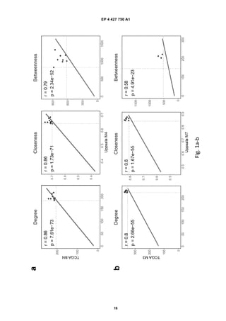
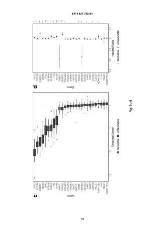
Figure 1. Identification of the targetable genes. Spearman correlation of centrality measurements (a) between TCGA
M4 and Uppsala M4, and (b) between TCGA M3 and Uppsala M7. Each dot (black) represents a gene that is ranked
within the top 20 based on a descending order of the corresponding centrality measurement. The diagonal lines
show a linear fit with the shaded region showing the 95% confidence intervals. In addition, r and p represent the
Spearman correlation coefficient and p-value, respectively. (c) Essential scores of the selected genes in 50 LUAD
cell lines. (d) Hazard ratios of the selected genes. Lines indicate 95% confidence intervals. Significance levels: * p
< 0.05, ** p < 0.01, *** p < 0.001.
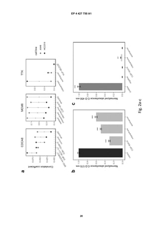
Figure Drug repositioning and validation of drug efficacy in an in vitro model. (a) Correlation coefficients of the top
drugs in A549 and HCC515 cell lines. The drug candidates were ranked by the mean values of correlation coefficients
from the two cell lines. (b) Bar plot showing the cell viability of A549 cell line after drug treatment in MTT assays.
(c) Bar plot showing the cytotoxicity on A549 cell lines after drug treatment in LDH assays. "Positive" and "negative"
represent the lysis cells and DMSO treated cells, respectively. Difference between groups was estimated by the
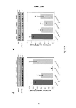
Student’s t-test. Western blots showing the inhibitory effect of drugs on the protein expression of (d) MCM6 and (e)
TTK. Error bar represents standard error. Significance levels: * p< 0.05, ** p< 0.01, *** p< 0.001. 10mM CGP-60474,
10mM wortmannin and 100nM mitoxantrone were used for cell treatment.
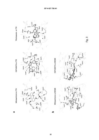
Figure 3. Molecular docking analysis of predicted drugs and their corresponding targets. (a) Putative binding modes
of mitoxantrone, CGP-60474, and wortmannin against TTK. (b) Putative binding modes of mitoxantrone and CGP-
60474 against MCM6. Hydrogen bonds are presented as dashed lines.
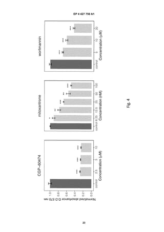
Figure 4. Cell viability of A549 cell line after drug treatment with different concentrations.
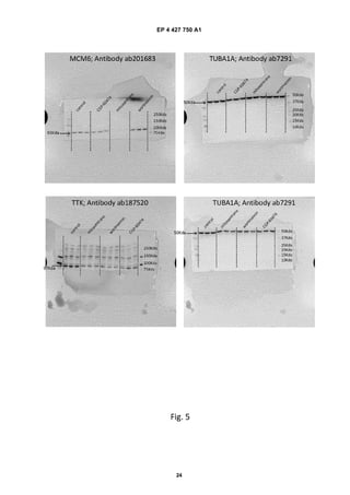
Figure 5. Raw images of Western blots showing the inhibitory effect of drugs on the protein expression levels of
MCM6 and TTK.
DETAILED DESCRIPTION
[0013] As described herein, an integrated approach has been employed to identify therapeutic targets and previously
known drugs have been repurposed for LUAD treatment. The treatment may help patients who cannot benefit from the
chemotherapies or targeted therapies that are used in the clinic today. The identified druggable gene targets are CDCA8,
EP 4 427 750 A1
5
5
10
15
20
25
30
35
40
45
50
55
MCM6 and TTK.
[0014] CDCA8 encodes a component of the chromosomal passenger complex which is an essential regulator for
mitosis and cell division. It has been reported that the phosphorylation and activation of CDCA8 plays a key role in lung
carcinogenesis and the suppression of CDCA8 significantly inhibits the growth of lung cancer cells 43, which supports
the findings presented herein.
[0015] MCM6 encodes one of the highly conserved mini-chromosome maintenance proteins that are essential for the
imitation of eukaryotic genome replication. MCM6 is involved in the prognostic signatures for indicating the survival
outcomes for particularly early-stage LUAD patients 5,6.
[0016] TTK encodes a dual specificity protein kinase with the ability to phosphorylate tyrosine, serine and threonine,
which is a critical mitotic checkpoint protein for accurate segregation of chromosomes during mitosis 44. It has been
reported that TTK is overexpressed in tumor tissues compared to normal lung tissue and its selective inhibitor CFI-
402257 shows significant antineoplastic activity in LUAD mouse models 45.
[0017] Further, the present inventors predicted that oxetane, mitoxantrone, staurosporine, CGP-60474 and alvocidib
had a potential inhibitory effect on the expression of MCM6, and that mitoxantrone, wortmannin, and NVP-BEZ235 had
a potential inhibitory effect on the expression of TTK. Among these drugs, the drug efficacy of mitoxantrone and CGP-
60474 for targeting MCM6, and mitoxantrone and wortmannin for targeting TTK was validated in an in vitro model.
Interestingly, CGP-60474 also suppressed the protein level of TTK. In the drug repositioning analysis, the present
inventors set that the correlation coefficient in both lung cancer cell line A549 and HCC515 should be higher than 0.4
based on previously empirical setting in kidney cancer and liver diseases 15,24,25. However, it was observed that the
correlation coefficient of interaction between CGP-60474 and TTK was 0.3895 in the cell line A549, which was in border
line. Thus, unfortunately, this interaction was skipped based on above criterion during the prediction. CGP-60474 is a
potent inhibitor of CDK1 which is one of the cyclin-dependent kinases 46. Both CDK1 and TTK are involved the control
of G1/S and G2/M phases 47,48 and these two genes showed a high correlation in expression across patients from LUAD
TCGA cohort in the analysis (Spearman correlation coefficient = 0.88), which might explain that CGP-60474 can target
TTK. Mitoxantrone is an inhibitor of DNA topoisomerase II alpha (TOP2A), and it leads to cell deaths by the induction
of double stranded DNA breaks 49. It is currently applied in the treatment of breast and prostate cancers, lymphomas
and leukemias. There were two phase II studies that failed to validate the antitumor activity of mitoxantrone in the
treatment for lung cancer 50,51. Both studies had small sample sizes (each recruited 24 patients), and only advanced or
metastatic non-small-cell lung cancer were involved. Based on the present inventors’ analysis, mitoxantrone should
however be used for the treatment of LUAD patients with early-stage tumors, since its target gene MCM6 is particularly
powerful for the classification of stage I tumors 5,6.
[0018] In the study presented herein, two different LUAD cohorts in which the patients had different geographic char-
acteristics and cultural backgrounds were used. Despite these heterogeneities, repeatable prognostic genes, functional
gene modules and druggable targets were found.
[0019] As a first aspect of the present disclosure, there is provided CGP-60474 or a pharmaceutically acceptable salt
thereof for use in a method of prevention or treatment of lung adenocarcinoma.
[0020] As shown herein, CGP-60474 modulates MCM6. The present inventors foresee that the preventive or thera-
peutic effect may be enhanced by simultaneous, sequential or separate administration of an agent modulating TTK.
Accordingly, in an embodiment of the first aspect, the method further comprises administration of a drug selected from
the group consisting of mitoxantrone, wortmannin and NVP-BEZ235.
[0021] The present inventors also foresee that the preventive or therapeutic effect may be enhanced by simultaneous,
sequential or separate administration of an agent modulating CDCA8. Accordingly, in an alternative or complimentary
embodiment of the first aspect, the method further comprises administration of a drug selected from the group consisting
of BRD-K43256821, EMF-BCA1-57, GSK-1059615, curcumin, and alvocidib.
[0022] In an embodiment of the first aspect, said lung adenocarcinoma expresses MCM6. As an example, said lung
adenocarcinoma may overexpress MCM6 as compared to an average expression value obtained from measurements
in healthy tissue.
[0023] Accordingly, the method of the first aspect may comprise detecting MCM6 expression in said lung adenocar-
cinoma and administration of CGP-60474 or apharmaceuticallyacceptable salt thereof to treat said lung adenocarcinoma
subsequent to detection of MCM6 expression.
[0024] Further, the method of the first aspect may comprise the steps of:
i) measuring the degree of MCM6 expression in said lung adenocarcinoma to obtain a MCM6 expression value;
ii) comparing the MCM6 expression value to a reference value, e.g. an average value obtained from measurements
in healthy tissue; and
iii) administration of CGP-60474 or a pharmaceutically acceptable salt thereof to treat said lung adenocarcinoma if
the MCM6 expression value is higher than the reference value.
EP 4 427 750 A1
6
5
10
15
20
25
30
35
40
45
50
55
[0025] The person skilled in the art of lung adenocarcinomas and treatment thereof is capable of establishing a relevant
reference value, e.g., by selecting relevant samples of healthy lung tissue and making measurements of the MCM6
expression therein.
[0026] As a second aspect of the present disclosure, there is provided mitoxantrone or a pharmaceutically acceptable
salt thereof for use in a method of treatment of stage 0 or I lung adenocarcinoma.
[0027] In said method, mitoxantrone is preferably administered by injection, e.g. intravenous injection.
[0028] As shown herein, mitoxantrone modulates TTK and MCM6. The present inventors foresee that the preventive
or therapeutic effect may be enhanced by simultaneous, sequential or separate administration of an agent targeting
CDCA8. Accordingly, in an embodiment of the second aspect, the method further comprises administration of a drug
selected from the group consisting of BRD-K43256821, EMF-BCA1-57, GSK-1059615, curcumin, and alvocidib.
[0029] In an embodiment of the second aspect, said lung adenocarcinoma expresses TTK. As an example, said lung
adenocarcinoma may overexpress TTK as compared to an average expression value obtained from measurements in
healthy tissue.
[0030] Accordingly, the method of the second aspect may comprise detecting TTK expression in said lung adenocar-
cinoma and administration of mitoxantrone or a pharmaceutically acceptable salt thereof to treat said lung adenocarci-
noma subsequent to detection of TTK expression.
[0031] Further, the method of the second aspect may comprise the steps of:
i) measuring the degree of TTK expression in said lung adenocarcinoma to obtain a TTK expression value;
ii) comparing the TTK expression value to a reference value, e.g. an average value obtained from measurements
in healthy tissue; and
iii) administration of mitoxantrone or a pharmaceutically acceptable salt thereof to treat said lung adenocarcinoma
if the TTK expression value is higher than the reference value.
[0032] The person skilled in the art of lung adenocarcinomas and treatment thereof is capable of establishing a relevant
reference value, e.g. by selecting relevant samples of healthy lung tissue and making measurements of the TTK ex-
pression therein.
[0033] As a third aspect of the present disclosure, there is provided a method of prevention or treatment of lung
adenocarcinoma comprising administration of CGP-60474 or a pharmaceutically acceptable salt thereof to a subject in
need thereof.
[0034] For the reasons set out above, the method of the third aspect may further comprise administration of:
a drug selected from the group consisting of mitoxantrone, wortmannin and NVP-BEZ235; and/or
a drug selected from the group consisting of BRD-K43256821, EMF-BCA1-57, GSK-1059615, curcumin, and alvo-
cidib.
[0035] As a fourth aspect of the present disclosure, there is provided a method of treatment of stage 0 or I lung
adenocarcinoma comprising administration of mitoxantrone or a pharmaceutically acceptable salt thereof to a subject
in need thereof.
[0036] For the reasons set out above, the method of the fourth aspect may further comprise administration of:
a drug selected from the group consisting of oxetane, staurosporine, CGP-60474 and alvocidib; and/or
a drug selected from the group consisting of BRD-K43256821, EMF-BCA1-57, GSK-1059615, curcumin, and alvo-
cidib.
[0037] The lung adenocarcinoma stage referred to above is the AJCC stage, which is preferably determined according
to the eighth edition of lung cancer stage classification (based on the TNM system).
[0038] The other embodiments of the first and second aspect described above apply mutatis mutandis to the third and
fourth aspect, respectively.
[0039] The subject of the above aspects is preferably a human.
EXAMPLES
Methods
[0040] To identify the therapeutic target genes and effective drugs for targeting these targets, four steps of systems
biology-based analyses based on the RNA-seq profiles of two independent LUAD cohorts were performed:
EP 4 427 750 A1
7
5
10
15
20
25
30
35
40
45
50
55
Step 1. Identify the prognostic genes by survival analysis;
Step Find the central prognostic genes based on the gene co-expression network analysis;
Step 3. Rank the drugs based on a profile-based drug repositioning approach; and
Step 4. Validate the drug efficacy in in vitro model.
[0041] The methodology is described in more detail as below.
Data Pre-processing
[0042] The metadata and raw RNA-seq data of 497 LUAD patients from the TCGA cohort were downloaded from the
GDC data portal (https://portal.gdc.cancer.gov/) 26. Another independent cohort is from 199 non-small cell lung cancer
patients surgically treated during 2006-2010 at the Uppsala University Hospital, Uppsala, Sweden (NCBI SRA database:
SRP074349) 27,28. From this cohort, only the RNA-seq data of the 105 LUAD patients whose survival information was
available was used. For both datasets, count and transcripts per million (TPM) values of transcripts were quantified by
Kallisto based on the human reference genome Ensembl V103 29. Only protein-coding transcripts were filtered for further
analysis. The sum count or TPM value of different alternative splicing transcripts from a gene was used as the gene
expression value of this gene. Moreover, the genes with average TPM values >1 were analyzed in this study.
Survival Analysis
[0043] Cox proportional hazard regression was used to evaluate the association between gene expression levels and
patients’ overall survival (OS) based on the ’coxph’ function from the R package ’survival’ 30. P-value < 0.01 was used
to identify the significantly prognostic genes.
Functional Enrichment Analysis
[0044] The ‘enrichGO’ function from the R package ’clusterProfiler’ (version 4.2.2) was used for the Gene ontology
(GO) enrichment analysis, which detected the significantly enriched GO terms based on the hypergeometric distribution
31. The ’org.Hs.eg.db’ database was used as the GO term source (version 3.14.0). GO terms with FDR < 0.05 were
used for further analysis. P-values were adjusted based on the Benjamini-Hochberg method.
Gene Co-expression Network Analysis
[0045] Based on the TPM values of genes, the Spearman correlation was employed to calculate the association of
all possible gene pairs across all tumor samples in each cohort. Then, the gene co-expression network was constructed
by extracting the gene-to-gene links ranked within top 1% based on the highest correlation coefficients in each cohort.
Then, a random walks-based algorithm, named Walktrap, was used to identify the gene modules with high transitivity
32. The function ’cluster_walktrap’ in R package ‘igraph’ was used to implement the Walktrap algorithm. Unweighted
edges were used for community detection (weights=NULL) and the default settings recommended by this function were
used for other parameters. Modules with more than 50 genes and clustering coefficients higher than 0.5 were used for
further analysis. Clustering coefficient ranges from 0 and 1, and value higher than 0.5 is a commonly used cutoff which
indicates that the partitions have a good cluster tendency and the genes involved in each module show a proper con-
nectivity 33. Degree, closeness, and betweenness values of genes were calculated in each module based on the R
package ‘igraph’.
Concordance Analysis
[0046] The accumulative hypergeometric model was used to determine whether two lists of prognostic genes had a
significant overlap as previously described 15. This model was also used to test to determine whether the genes involved
in a module had a significant overlap with the prognostic genes. Jaccard indices were calculated as the size of the
overlapped genes divided by the size of the union of two lists of genes.
Essential Scores of Genes
[0047] The essential scores of genes in 50 LUAD cell lines were obtained from the DepMap Portal (https://dep-
map.org/portal/), which were calculated based on the CRISPR-Cas9 essentiality screens while accounting for the copy
number-specific effect 34. The score of a gene indicates the essentiality of this gene for cancer cell proliferation and
survival after knockout of this gene by CRISPR-Cas9 technology. A more negative score indicates that the gene is more
EP 4 427 750 A1
8
5
10
15
20
25
30
35
40
45
50
55
essential.
Drug Repositioning for Target Genes
[0048] The previously proposed drug repositioning approach was applied to identify drugs that can inhibit the target
genes 15,24. This method hypothesizes that a drug is considered to have an inhibitory effect on the expression of a target
gene if this drug leads to a wide perturbation on the gene expression landscape in tumor cells which is similar to the
effect of the knockdown of this target gene. The shRNA gene knockdown and drug perturbed transcriptomic gene
signature profiles (level 5) of two LUAD cell lines A549 and HCC515 were downloaded from the Expanded CMap LINCS
Resource (version 2020, https://clue.io/data/CMap2020#LINCS2020). The level 5 data provides the replicate-collapsed
Z-scores, representing a consensus biological response of transcriptomics to the perturbation of drug treatment or gene
knockdown derived from different replicates 35. The drug repositioning analysis was performed for CDCA8, MCM6, and
TTK since these genes had the available gene knockdown transcriptomic signature profiles. Each gene was knocked
down by three different shRNAs, thus three gene knockdown perturbed signature profiles for each gene in both A549
and HCC515 cell lines were obtained. Meanwhile, 53,827 drug-perturbed signature profiles associated with 13,945
unique drugs with different dosages and treatment durations in A549 cell line and 16,133 drug-perturbed signature
profiles associated with 5,890 unique drugs in HCC515 cell line were obtained.
[0049] In brief, there were four steps in the drug repositioning analysis. First, for a given gene, a drug-shRNA matrix
was created by computing the Spearman correlation between this gene knockdown and drug treatment perturbed sig-
nature profiles in each cell line. In this matrix, each row represents a drug perturbagen with a specific dosage and
treatment duration, and each column represents a gene knockdown with a specific shRNA. Second, the drug-shRNA
matrix was simplified by selecting the optimal drug and shRNA perturbagens. A drug was added to the cells with different
dosages and treatment durations in the CMap experimental setting. To maximize the drug effect, only a unique and
optimal dosage/duration that showed the highest correlation coefficients with each shRNA perturbagen was kept for
each drug. Similarly, a gene was knocked down by three different shRNAs. To maximize the gene knockdown effect,
only the optimal shRNA perturbagen for each gene was kept. We calculated the mean correlation coefficient of each
shRNA perturbagen based on its correlation with all drugs’ perturbagens. Then, the shRNA perturbagen with highest
mean correlation coefficient is optimal. After optimization, each coefficient in the drug-shRNA matrix represents the
similarity between the effects induced by a specific drug treatment (row) and a specific gene knockdown (column). Higher
coefficient value means high possibility of the inhibitory effect of a drug on its target gene. Third, the drug-shRNA matrices
from the two cell lines were merged by extracting the common drugs. Drugs that were not presented in both cell lines
were excluded. Finally, for each gene, the top five drugs with the highest mean coefficients from the two cell lines were
selected as the most effective drugs. Meanwhile, the coefficients in both cell lines should be greater than 0.4 which was
an empirical cutoff based on a previous in vitro experimental validation 15,25.
Cell culture, MTT, and LDH assay
[0050] A549 cells were obtained from ATCC and cultured with culture medium (D0819 DMEM 10% FBS 1% penicil-
lin/streptomycin) at 37°C. Cell viability was measured using an MTT assay. MTT ((3-[4,5-dimethylthiazol-2-yl]-2,5-diphe-
nyltetrazolium bromide) (1x), at 5 mg/ml) was added for 1 hour and formazan was dissolved with DMSO (dimethyl
sulfoxide). Optical density at a wavelength of 570 nm was measured using a microplate reader (Hidex plate reader) to
quantify the relative number of living cells. To determine treatment concentrations, cells were treated with serially diluted
drugs until a concentration that reduced cell viability by around 50%. As a result, 10mM for CGP-60474 (MedChemEx-
press, HY-11009), 10mM for wortmannin (Sigma, W1628) and 100nM mitoxantrone (Selleckchem, NSC-301739) were
used. Cells were treated with the drugs at their designated concentrations for two days.
[0051] Cytotoxic effect on the cells was measured with lactate dehydrogenase (LDH) assays that were performed in
triplicates at the same concentrations as selected for the MTT assays following the LDH kit procedure (Abeam Cat#
ab65393). 30 minutes after LDH (Abcam Cat# ab65393) administration, optical density at a wavelength of 450 nm was
measured using a microplate reader (Hidex plate reader) to quantify the relative LDH activity which was released into
the media via the drug’s cytotoxic effect.
[0052] To normalize the quantification results, the mean negative control (untreated cells) reading was subtracted
from each reading and then each was divided by the mean positive control (Cell Lysis Solution) reading minus the mean
negative control reading.
Western Blotting
[0053] Triplicates of drug treatments at the same concentrations as selected for the MTT assays were prepared for
the Western blots. Whole cell lysate was extracted with CelLytic M (C2978, Sigma-Aldrich) lysis buffer. The protein
EP 4 427 750 A1
9
5
10
15
20
25
30
35
40
45
50
55
content of each sample was measured using a Bradford assay and each sample was prepared for Western blotting
using 23 Laemmli buffer. Electrophoresis was run at 150 V for 40 minutes and membrane transfer was performed with
the Trans-Blot Turbo Transfer System (Biorad). PVDF membranes were blocked for 30 minutes with 5% skim milk and
TBST (Tris Buffered Saline with 0.2% Tween 20) and then treated overnight on a shaker plate at 4°C with 0.5% skim
milk withTBST and MCM6 (Abcam Cat# ab201683) and TTK (Abcam Cat# ab187520) primary antibodies diluted 1:20000
and for 30 minutes with TUBA1A (Abeam Cat# ab7291, RRID:AB_2241126) primary antibodies diluted 1:20000. The
solution was then replaced with a new solution of 0.5% skim milk with TBST and anti-rabbit (Abeam Cat# ab6721,
RRID:AB_955447) or anti-mouse (Abeam Cat# ab6789, RRID:AB_955439) secondary antibodies diluted 1:20000 for
30 minutes. Luminata Forte Western HRP substrate (Thermofisher) was added before imaging.
[0054] Western blots were imaged (LAS image) for 20 seconds, 1 minute, or automatic exposure and quantification
was performed using ImageJ 36. MCM6 and TTK expression levels were normalized by dividing by their corresponding
α-Tubulin expression levels and each drug treatment expression level was then normalized again by dividing by the
mean normalized control expression level.
Molecular Docking
[0055] Molecular docking analyses were performed to comprehend the molecular interaction mechanisms between
the target proteins and their correspondingly repositioned drug candidates. 3D-coordinates of the TTK protein were
retrieved from the protein data bank (PDB ID: 4C4J) while alpha-fold structure of MCM6 was used 37. Structures were
optimized using quick prep tool of Molecular Operating Environment (MOE, V2019.01) suit to remove gaps, optimize
angle, bond length, charges calculation and protonation of amino acids at physiological pH. The structures of the com-
pounds were built using the MOE builder module and was charged and minimized by MMFF94x force field 38. The
standard default docking algorithm and scoring functions (triangle matcher algorithm, London dG and GBVI/WSA) were
used. Hundred conformations were generated for each molecule that were analyzed based on clustering. Best suitable
docked conformation was selected based on docking scores and interaction profile.
Results
Identification of Prognostic Genes
[0056] The univariate Cox survival analysis was performed based on the gene expression profiles and patients’ OS
in each cohort. As a result, 1,463 unfavorable genes whose high expression was associated with poor survival outcomes
of patients and 166 favorable gene whose high expression was associated with good survival outcomes of patients in
the TCGA cohort (p-value < 0.01) were identified. Similarly, 1,111 unfavorable genes and 4 favorable genes were
identified in the Uppsala cohort (p-value < 0.01). The concordance of these prognostic genes from the two cohorts was
evaluated and it was found that these two lists of unfavorable genes have a significant overlap (n=341, hypergeometric
test, p-value < 1.0E-12). GO enrichment analysis showed that the 341 overlapped unfavorable genes were significantly
enriched in chromosome organization and segregation, cell cycle, nuclear division, and DNA replication pathways.
Similar pathways were observed from the KEGG enrichment analysis. A significant overlap was also observed between
the two lists of favorable genes (n=3, hypergeometric test, p-value < 1.0E-12). Since there were only three overlapped
favorable genes, AMPD1, JCHAIN, and ZNF540, their functions were examined individually. AMPD1 encodes adenosine
monophosphate deaminase which catalyzes AMP to IMP and it is a crucial enzyme in purine nucleotide and energy
metabolism. It has been reported that AMPD1 expression level is positively correlated to immune cell infiltration level in
LUAD 39. JCHAIN encodes immunoglobulin J-chain which is essential in the formation and stabilization of polymeric
IgA and IgM structures. The transcription of JCHAIN in the lungs substantially decreases during tumorigenesis, which
can be explained by the immunosuppressing effect of tumor cells 40. ZNF540 acts as a transcriptional repressor, and it
is associated with CD8+ T cells infiltrating in LUAD 41.
Identification of Functional Modules
[0057] Spearman correlation was performed to estimate the association between each two genes based on their gene
expression levels in each cohort. The top 1% of the gene-to-gene links with the highest correlations coefficients were
extracted to construct the gene co-expression network. Around 2,012,288 gene-to-gene links with correlation coefficients
ranging from 0.55 to 1 were finally obtained in the gene co-expression network of the TCGA cohort. Further, a random
walks-based algorithm, Walktrap, was used to identify the gene modules with high transitivity based on the topology of
the CCN 32. The modules with more than 50 genes and clustering coefficients higher than 0.5 were used for further
analysis. In the TCGA gene co-expression network, 14 modules (M1-M14, containing 57 to 3466 genes) were identified.
The association of the modules with the 341 unfavorable genes were investigated based on concordance analysis (Table
EP 4 427 750 A1
10
5
10
15
20
25
30
35
40
45
50
55
1).
[0058] It was observed that M3 (455 genes) and M4 (357 genes) had significant overlaps with these unfavorable genes
in the TCGA cohort (n= 126 and 20, respectively, hypergeometric test, p < 0.01). Thus, these two modules were denoted
as unfavorable modules. Functional enrichment showed that the genes involved in M3 were significantly enriched in the
chromosome segregation, DNA replication and repair, nuclear division, and cell phase transition related pathways. The
genes involved in the M4 were significantly enriched in the extracellular organization and angiogenesis pathways. Similar
pathways were obtained by KEGG enrichment analysis.
[0059] Similarly, 1,979,922 gene-to-gene links with correlation coefficient ranging from 0.57 to 1 were obtained in the
gene co-expression network of the Uppsala cohort and 12 modules (M1-M12, containing 51 to 2664 genes) were found.
Among these modules, M4 (391 genes) and M7 (269 genes) had significant overlaps with the unfavorable genes (n=
19 and 99, respectively, hypergeometric test, p < 0.01, Table 2).
[0060] Functional enrichment analysis showed that the two modules exhibited an identical set of GO and KEGG
pathways resulting from the TCGA cohort. Thus, it was inferred that the unfavorable modules identified from the TCGA
and Uppsala cohorts were highly consistent. Based on the concordance analysis, it was observed that the TCGA M3
had a significant overlap with the Uppsala M7 (n= 242, hypergeometric test: p < 1.0E-12, Jaccard index = 0.5). Similar
result was observed for the overlaps of TCGA M4 and Uppsala M4 (n=239, hypergeometric test: p < 1.0E-12, Jaccard
index = 0.47). These results suggested that the unfavorable modules identified from the TCGA cohort were validated in
Table 1.
Module name
(TCGA cohort)
Number of genes inthe
module
Number of unfavorable
signatures
Overlaps
P value of
hypergeometric test
M1 3466 341 47 1,00E+00
M2 831 341 1 1,00E+00
M3 455 341 126 < 1.0E-12
M4 357 341 20 4,12E-04
M5 286 341 0 1,00E+00
M6 243 341 0 1,00E+00
M7 227 341 0 1,00E+00
M8 122 341 0 1,00E+00
M9 89 341 0 1,00E+00
M10 82 341 1 8,65E-01
M11 80 341 0 1,00E+00
M12 75 341 1 8,40E-01
M13 70 341 3 2,37E-01
M14 57 341 1 7,51E-01
Table
Modulename (Uppsala
cohort)
Number of genes in
the module
Number of unfavorable
signatures
Overlaps
P value of
hypergeometric test
M1 2664 341 42 1,00E+00
M2 1031 341 22 7,64E-01
M3 579 341 1 1,00E+00
M4 391 341 19 3,24E-03
M5 372 341 7 8,02E-01
M6 324 341 11 1,65E-01
M7 269 341 99 < 1.0E-12
M8 264 341 1 9,99E-01
M9 90 341 0 1,00E+00
M10 58 341 2 4,12E-01
M11 55 341 0 1,00E+00
M12 51 341 0 1,00E+00
EP 4 427 750 A1
11
5
10
15
20
25
30
35
40
45
50
55
the independent Uppsala cohort. In addition, a pairwise comparison was performed between the remaining modules
identified in the two cohorts. Significant overlaps with high Jaccard indices were also found between TCGA M2 and
Uppsala M3 which were enriched in adaptive immune system and cell differentiation pathways, TCGA M5 and Uppsala
M8 which were enriched in angiogenesis and blood circulation pathways, TCGA M8 and Uppsala M9 which were enriched
for extracellular transport and cilium assembly/organization pathways, and TCGA M11 and Uppsala M10 which were
enriched in cytoplasmic translation, ribosome biogenesis, and p53 regulation pathways.
Identification of Targetable Genes
[0061] To determine the hub genes that might drive the tumor development, topological analysis was performed to
estimate the centrality of genes in these unfavorable modules. Degree, closeness and betweenness of genes were
evaluated in each module. Then, the Spearman correlation of each centrality measurement between the pairwise match-
ing of the unfavorable modules from the two cohorts was calculated. As a result, the correlation coefficients of degree,
closeness and betweenness of genes were 0.86, 0.86 and 0.79 between TCGA M4 and Uppsala M4, and 0.8, 0.8 and
0.58 between TCGA M3 and Uppsala M7 (all p-values < 1.0E-12, Figure 1a and 1b). Further, the genes were ranked
based on a descending order of each centrality measurement in each unfavorable module. The genes ranked within the
top 20 in the matching unfavorable modules from the two cohorts were selected as the hub genes. A union set of genes
identified based on all the centrality measurements were used as the final hub genes. Totally, 24 hub genes were
identified. By including the three overlapped favorable genes, 27 hub genes were finally obtained. Further, the essential
scores of these genes in 50 LUAD cell lines from were downloaded the DepMap data portal 34. The essential score of
a gene represents the essentiality of this gene for tumor cell proliferation and survival after knockout of this gene by
CRISPR-Cas9 technology. A more negative score indicates worse survival of cells after the gene knockout. As shown
in Figure 1c, ninehub genes, KIF23, CDCA8, MAD2L1, MCM6, TPX2, NCAPH, CLSPN, TTK, and KIF4A, whoseessential
scores in 50 LUAD cell lines were smaller than 0, were selected as the targetable genes (Figure 1c). These nine genes
were all unfavorable for the patients’ prognoses, whose hazard ratios were higher than 1 (Figure 1d).
Drug Repositioning
[0062] A previously proposed drug repositioning method was applied to identify promising drugs that can have an
inhibitory effect on the expression of the targetable genes 15,24. The algorithm is performed by comparing the similarity
of molecular effect on tumor cells perturbed by a drug treatment and a gene knockout. This hypothesizes that a drug is
considered to have an inhibitory effect on the expression of a target gene if this drug leads to a wide perturbation on the
gene expression landscape in tumor cells which is similar to the effect of the knockout of this target gene (see details
in the Method section). Among the nine targetable genes, only three genes CDCA8, MCM6, and TTK had the available
transcriptomics signature profiles of two LUAD cell lines A549 and HCC515 that were perturbed by the drug treatment
and the corresponding shRNA gene knockout from the LINCS database 42. In brief, the drug-shRNA matrix in which
each element represents the similarity of the effect induced by a drug perturbagen and a shRNA gene knockdown
perturbagen in each cell line was first constructed. To maximize the drug treated and genetic perturbed effects, the drug-
shRNA matrix was simplified by extracting an optimal dosage/treatment duration for each drug and an optimal shRNA
for each gene. Further, the two simplified drug-shRNA matrices from two cell lines were merged by extracting the same
drugs. Finally, for each target gene, the top five drugs with the highest mean values of coefficients from two cell lines
were selected as the potentially effective drugs. Meanwhile, the coefficients in each cell lines should be higher than 0.4.
As shown in Figure 2a:
- BRD-K43256821, EMF-BCA1-57, GSK-1059615, curcumin, and alvocidib were identified as top candidate drugs
for inhibiting CDCA8;
- oxetane, mitoxantrone, staurosporine, CGP-60474 and alvocidib were identified as top candidate drugs for inhibiting
MCM6; and
- mitoxantrone, wortmannin and NVP-BEZ235 were identified as top candidate drugs for inhibiting TTK.
[0063] Among these drugs, only oxetane-containing drugs (e.g. taxol) and mitoxantrone are clinically used drugs,
whereas the other are under medical investigation.
Experimental Validation of Drug Efficacy
[0064] Among these repurposed drugs, only CGP-60474, mitoxantrone, and wortmannin could be obtained and hence
tested in the in vitro model A549 cell line. First, dose dependent MTT assays were performed for each drug to select a
proper concentration which could reduce the cell viability by 50%. This concentration shows moderate toxicity and is
EP 4 427 750 A1
12
5
10
15
20
25
30
35
40
45
50
55
suitable for further experiments to study the drug effect in the cell line. As a result, 10mM for CGP-60474, 10mM for
wortmannin and 100nM for mitoxantrone were determined (Figure 4). Further, the MTT assay was used for cell viability
and the LDH assay was used for cytotoxicity. In the MTT assay, cell viability was decreased to 29.1% 60.7 by CGP-
60474, 52.2% 61.4 by mitoxantrone and, 62.3% 62.4 by wortmannin (Figure 2b). Interestingly, LDH assays showed
that 10mM CGP-60474 and 10mM wortmannin did not induce necrotic cell death, and1 00nM of mitoxantrone showed
only 3.45% 60.4 necrotic cell death compared to the lysis positive control (100%) and negative untreated group (0%)
(Figure 2c). Taken together, cell viability and cytotoxicity assay results indicated that these drugs decreased the cell
viability mainly by inhibiting the cell proliferation rather than inducing necrotic cell death procedure. To investigate whether
the repurposed drugs could inhibit their corresponding genes (that they were repurposed for), the protein expression
levels of these target genes were evaluated in the A549 cell lines after drug treatment. Western blots showed that CGP-
60474 and mitoxantrone significantly reduced the protein level of MCM6 (Figures 2d and 5). Especially, mitoxantrone
showed an extremely effective suppression on MCM6, with 95% decrease of protein expression (Figure 2d). In addition,
mitoxantrone and wortmannin significantly inhibited the protein level of TTK by around 50% (Figures 2e and 5). Notably,
CGP-60474, which was predicted to inhibit MCM6, also showed an inhibitory effect on TTK. These results suggested
that the repurposed drugs effectively targeted the corresponding genes and reduced the tumor cell proliferation.
Molecular Docking
[0065] The molecular docking analyses were performed to gain a deep insight of the binding mode between the
validated drugs (mitoxantrone, CGP-60474, and wortmannin) and the target proteins (TTK and MCM6). The predicted
docking scores for the binding interactions of mitoxantrone, CGP-60474 and wortmannin against TTK were -9.39, -8.18
and -7.28 (kcal/mol), respectively, representing moderate to strong interactions (Table 3).
Table 3.
Compound Docking scores
(Kcal/mol)
Hydrogen Bonds interactions Hydrophobic
Interactions
Donor Acceptor Distance
Mitoxantrone -9.3935 OH COO- of Glu603 2.11 Ile531
NH COO-of Gly605 3.13 Val539
Ala551
NH of
Gly605
OH 2.39 Asp608
Leu654
COO- of Ser611 2.69 Ile663
OH
COO- of Ala651 1.9
NH
N3 3.2
NH of
Gln670
O3 2.82
NH of
Met671
CGP-60474 -8.1757 NH COO- of Gly605 2.36 Ile531
Val539
NH of
Gly605
N of pyrimidine ring 2.27 Ala551
Asp608
Leu654
OH COO- of Asp608 2.71 Ile663
EP 4 427 750 A1
13
5
10
15
20
25
30
35
40
45
50
55
[0066] Further, investigation revealed that the comparative potential of the compounds mostly due to the hydrophilic
interactions (Glu603, Gly605, Ser611, Ala651, Gln671 and Met674) (Figure 3a), while the aromatic ring of the compounds
confers stability by forming hydrophobic interactions with the active site residues (Ile531, Val539, Ala551, Leu654, Ile663
and Asp664). The predicted docking scores for the binding interactions of mitoxantrone and CGP-60474 against MCM6
were - 7.54 and -6.37 (kcal/mol), respectively, representing moderate interactions (Table 4). Both drug candidates
interacted with crucial residues of the protein by establishing hydrophilic (Gln212, Ala213, Arg217, Arg217, Lys241,
Ser413 and Arg573) and hydrophobic (Gln212, Ala213, Glu410 and Glu411) interaction (Figure 3b).
(continued)
Compound Docking scores
(Kcal/mol)
Hydrogen Bonds interactions Hydrophobic
Interactions
Donor Acceptor Distance
Wortmannin -7.2825 NH of
Lys553
=CO 2.39 Ile531
Val539
=CO of tetrahydropyran-2-
one
3.18 Ala551
NH of
Asp608,
Leu654
Ile663
O2 of furan ring 3.36 Asp664
NH of
Gln670
=CO of tetrahydropyran-2-
one
2.97
NH
Asp674
Table 4.
Compounds Docking scores (Kcal/mol) Hydrogen Bonds interactions Hydrophobic Interactions
Donor Acceptor Distance
CGP-60474 -6.3695 NH of
Arg217
Npl of ligand 3.08 Gln212
Npl of ligand 2.42 Ala213
NH of
Arg217
O3 of ligand 2.93 Glu410
Npl of ligand 2.90 Glu411
N3+ of
Lys241
COO- of
Glu410
3.0
2.16
OH Glu410 COO- of
Glu411
3.45
Npl of ligand 2.07
O2 of ligand
Npl of ligand COO- of
Ala416
NH of Ala416
OH of ligand
EP 4 427 750 A1
14
5
10
15
20
25
30
35
40
45
50
55
REFERENCES
[0067]
1. Sung H, Ferlay J, Siegel RL, et al. Global Cancer Statistics 2020: GLOBOCAN Estimates of Incidence and
Mortality Worldwide for 36 Cancers in 185 Countries. CA Cancer J Clin 2021; 71(3): 209-49.
2. Behrend SJ, Giotopoulou GA, Spella M, Stathopoulos GT. A role for club cells in smoking-associated lung ade-
nocarcinoma. Eur Respir Rev 2021; 30(162).
3. Ettinger DS, Wood DE, Aisner DL, et al. Non-Small Cell Lung Cancer, Version 3.2022, NCCN Clinical Practice
Guidelines in Oncology. J Natl Compr Canc Netw 2022; 20(5): 497-530.
4. Hirsch FR, Scagliotti GV, Mulshine JL, et al. Lung cancer: current therapies and new targeted treatments. Lancet
2017; 389(10066): 299-311.
5. Melocchi V, Dama E, Mazzarelli F, et al. Aggressive early-stage lung adenocarcinoma is characterized by epithelial
cell plasticity with acquirement of stem-like traits and immune evasion phenotype. Oncogene 2021; 40(31): 4980-91.
6. Kadara H, Behrens C, Yuan P, et al. A five-gene and corresponding protein signature for stage-I lung adenocar-
cinoma prognosis. Clin Cancer Res 2011; 17(6): 1490-501.
7. Feng Z, Zhang Y, He M, et al. AMICA1 is a diagnostic and prognostic biomarker and induces immune cells
infiltration by activating cGAS-STING signaling in lung adenocarcinoma. Cancer Cell Int 2022; 22(1): 111.
8. He M, Han Y, Cai C, et al. CLEC10A is a prognostic biomarker and correlated with clinical pathologic features
and immune infiltrates in lung adenocarcinoma. J Cell Mol Med 2021; 25(7): 3391-9.
9. Liu XS, Zhou LM, Yuan LL, et al. NPM1 Is a Prognostic Biomarker Involved in Immune Infiltration of Lung
Adenocarcinoma and Associated With m6A Modification and Glycolysis. Front Immunol 2021; 12: 724741.
10.He Y, LiuR, Yang M, etal.Identificationof VWFasa Novel Biomarker in LungAdenocarcinoma byComprehensive
Analysis. Front Oncol 2021; 11: 639600.
(continued)
Compounds Docking scores (Kcal/mol) Hydrogen Bonds interactions Hydrophobic Interactions
Donor Acceptor Distance
Mitoxantrone -7.5429 OH of ligand COO- of
Gln140
2.0 Gln212
2.63 Ala213
NH of
Gln212
O2 of ligand 3.25 Glu410
N3 of ligand 2.26 Glu411
NH of Ala213 N3 of ligand 2.99
N3 of ligand 1.91
NH of
Arg217
N3 of ligand 2.14
COO- of
Ser413
3.53
NH of
Arg217 =CO of ligand
NH of
Lys241
NH of ligand
NH of
Arg573
EP 4 427 750 A1
15
5
10
15
20
25
30
35
40
45
50
55
11. Lu M, Fan X, Liao W, et al. Identification of significant genes as prognostic markers and potential tumor sup-
pressors in lung adenocarcinoma via bioinformatical analysis. BMC Cancer 2021; 21(1): 616.
12. Bian T, Zheng M, Jiang D, et al. Prognostic biomarker TUBA1C is correlated to immune cell infiltration in the
tumor microenvironment of lung adenocarcinoma. Cancer Cell Int 2021; 21(1): 144.
13. Jiawei Z, Min M, Yingru X, et al. Identification of Key Genes in Lung Adenocarcinoma and Establishment of
Prognostic Mode. Front Mol Biosci 2020; 7: 561456.
14. Pushpakom S, Iorio F, Eyers PA, et al. Drug repurposing: progress, challenges and recommendations. Nat Rev
Drug Discov 2019; 18(1): 41-58.
15. Li X, Shong K, Kim W, et al. Prediction of drug candidates for clear cell renal cell carcinoma using a systems
biology-based drug repositioning approach. EBioMedicine 2022; 78: 103963.
16. Lamb J, Crawford ED, Peck D, et al. The Connectivity Map: using gene-expression signatures to connect small
molecules, genes, and disease. Science 2006; 313(5795): 1929-35.
17. Iskar M, Campillos M, Kuhn M, Jensen LJ, van Noort V, Bork P. Drug-induced regulation of target expression.
PLoS ComputBiol 2010; 6(9).
18. Nagaraj AB, Wang QQ, Joseph P, et al. Using a novel computational drug-repositioning approach (DrugPredict)
to rapidly identify potent drug candidates for cancer treatment. Oncogene 2018; 37(3): 403-14.
19. Pillaiyar T, Meenakshisundaram S, Manickam M, Sankaranarayanan M. A medicinal chemistry perspective of
drug repositioning: Recent advances and challenges in drug discovery. Eur JMed Chem 2020; 195: 112275.
20. Jin G, Wong ST. Toward better drug repositioning: prioritizing and integrating existing methods into efficient
pipelines. Drug Discov Today 2014; 19(5): 637-44.
21. Huang RX, Siriwanna D, Cho WC, et al. Lung adenocarcinoma-related target gene prediction and drug reposi-
tioning. Front Pharmacol 2022; 13: 936758.
22. De Bastiani MA, Klamt F. Integrated transcriptomics reveals master regulators of lung adenocarcinoma and
novel repositioning of drug candidates. Cancer Med 2019; 8(15): 6717-29.
23. Kwon OS, Lee H, Kong HJ, et al. Connectivity map-based drug repositioning of bortezomib to reverse the
metastatic effect of GALNT14 in lung cancer. Oncogene 2020; 39(23): 4567-80.
24. Zhang C, Shi M, Kim W, et al. Discoveryof therapeutic agentstargeting PKLR for NAFLD using drug repositioning.
EBioMedicine 2022; 83: 104214.
25. Yuan M, Shong K, Li X, et al. A Gene Co-Expression Network-Based Drug Repositioning Approach Identifies
Candidates for Treatment of Hepatocellular Carcinoma. Cancers (Basel) 2022; 14(6).
26. Cancer Genome Atlas Research N. Comprehensive molecular profiling of lung adenocarcinoma. Nature 2014;
511(7511): 543-50.
27. Mezheyeuski A, Bergsland CH, Backman M, et al. Multispectral imaging for quantitative and compartment-
specific immune infiltrates reveals distinct immune profiles that classify lung cancer patients. JPathol 2018; 244(4):
421-31.
28. Goldmann T, Marwitz S, Nitschkowski D, et al. PD-L1 amplification is associated with an immune cell rich
phenotype in squamous cell cancer of the lung. Cancer Immunol Immunother 2021; 70(9): 2577-87.
29. Bray NL, Pimentel H, Melsted P, Pachter L. Erratum: Near-optimal probabilistic RNA-seq quantification. NatBi-
otechnol 2016; 34(8): 888.
EP 4 427 750 A1
16
5
10
15
20
25
30
35
40
45
50
55
30. Therneau T, Lumley T. R survival package. R Core Team 2013.
31. Yu G, Wang LG, Han Y, He QY. clusterProfiler: an R package for comparing biological themes among gene
clusters. OMICS 2012; 16(5): 284-7.
32. Pons P, Latapy M. Computing Communities in Large Networks Using Random Walks. International Symposium
on Computer and Information Sciences 2005; 3733: 284-93.
33. Nascimento M, Carvalho A. A graph clustering algorithm based on a clustering coefficient for weighted graphs.
Journal of the Brazilian Computer Society 2011; 17: 19.
34. Meyers RM, Bryan JG, McFarland JM, et al. Computational correction of copy number effect improves specificity
of CRISPR-Cas9 essentiality screens in cancer cells. Nat Genet 2017; 49(12): 1779-84.
35. Subramanian A, Narayan R, Corsello SM, et al. A Next Generation Connectivity Map: L1000 Platform and the
First 1,000,000 Profiles. Cell 2017; 171(6): 1437-52 e17.
36. Schneider CA, Rasband WS, Eliceiri KW. NIH Image to ImageJ: 25 years of image analysis. Nat Methods 2012;
9(7): 671-5.
37. Naud S, Westwood IM, Faisal A, et al. Structure-based design of orally bioavailable 1H-pyrrolo[3,2-c]pyridine
inhibitors of mitotic kinase monopolar spindle 1 (MPS1). J Med Chem 2013; 56(24): 10045-65.
38. Halgren TA. Merck molecular force field. I. Basis, form, scope, parameterization, and performance of MMFF94.
Journal of computational chemistry 1996; 17: 490.
39. Xu ZY, Zhao M, Chen W, et al. Analysis of prognostic genes in the tumor microenvironment of lung adenocar-
cinoma. PeerJ 2020; 8: e9530.
40. Slizhikova DK, Zinov’eva MV, Kuz’min DV, et al. [Decrease in expression of human J-chain in lung squamous
cell cancer and adenocarcinoma]. Mol Biol (Mosk) 2007; 41(4): 659-65.
41. Zhang M, Ma J, Guo Q, Ding S, Wang Y, Pu H. CD8(+) T Cell-Associated Gene Signature Correlates With
Prognosis Risk and Immunotherapy Response in Patients With Lung Adenocarcinoma. Front Immunol 2022; 13:
806877.
42. Stathias V, Turner J, Koleti A, et al. LINCS Data Portal 2.0: next generation accesspoint for perturbation-response
signatures. Nucleic Acids Res 2020; 48(D1): D431-D9.
43. Hayama S, Daigo Y, Yamabuki T, et al. Phosphorylation and activation of cell division cycle associated 8 by
aurora kinase B plays a significant role in human lung carcinogenesis. Cancer Res 2007; 67(9): 4113-22.
44. Lauze E, Stoelcker B, Luca FC, Weiss E, Schutz AR, Winey M. Yeast spindle pole body duplication gene MPS1
encodes an essential dual specificity protein kinase. EMBO J 1995; 14(8): 1655-63.
45. Zheng L, Chen Z, Kawakami M, et al. Tyrosine Threonine Kinase Inhibition Eliminates Lung Cancers by Aug-
menting Apoptosis and Polyploidy. Mol Cancer Ther 2019; 18(10): 1775-86.
46. Furet P, Zimmermann J, Capraro HG, Meyer T, Imbach P. Structure-based design of potent CDK1 inhibitors
derived from olomoucine. J Comput Aided Mol Des 2000; 14(5): 403-9.
47. Hogg D, Guidos C, Bailey D, et al. Cell cycle dependent regulation of the protein kinase TTK. Oncogene 1994;
9(1): 89-96.
48. Bashir T, Pagano M. Cdk1: the dominant sibling of Cdk2. Nat Cell Biol 2005; 7(8): 779-81.
49. Evison BJ, Sleebs BE, Watson KG, Phillips DR, Cutts SM. Mitoxantrone, More than Just Another Topoisomerase
II Poison. Med Res Rev 2016; 36(2): 248-99.
EP 4 427 750 A1
17
5
10
15
20
25
30
35
40
45
50
55
50. Suga J, Saijo N, Shinkai T, et al. Phase II study of mitoxantrone in patients with non-small cell lung cancer. Jpn
J Clin Oncol 1986; 16(2): 147-51.
51. Feun LG, Savaraj N, Solomon J, Liebmann A, Hurley J. Phase II trial of mitoxantrone and cisplatin in advanced
non-small-cell lung cancer. Am J Clin Oncol 1996; 19(2): 190-2.
52. Powis G, Bonjouklian R, Berggren MM, et al. Wortmannin, a potent and selective inhibitor of phosphatidylinositol-
3-kinase. Cancer Res 1994; 54(9): 2419-23.
53. Boehle AS, Kurdow R, Boenicke L, et al. Wortmannin inhibits growth of human non-small-cell lung cancer in
vitro and in vivo. Langenbecks Arch Surg 2002; 387(5-6): 234-9.
54. Zhang Y, Bao C, Mu Q, et al. Reversal of cisplatin resistance by inhibiting PI3K/Akt signal pathway in human
lung cancer cells. Neoplasma 2016; 63(3): 362-70.
Claims
1. CGP-60474 or a pharmaceutically acceptable salt thereof for use in a method of prevention or treatment of lung
adenocarcinoma.
2. CGP-60474 or a pharmaceutically acceptable salt thereof for use according to claim 1, wherein said method further
comprises administration of a drug selected from the group consisting of BRD-K43256821, EMF-BCA1-57, GSK-
1059615, curcumin and alvocidib.
3. CGP-60474 or a pharmaceutically acceptable salt thereof for use according to claim 1 or 2, wherein said method
further comprises administration of a drug selected from the group consisting of mitoxantrone, wortmannin and NVP-
BEZ235.
4. CGP-60474 or a pharmaceutically acceptable salt thereof for use according to any one of the preceding claims,
wherein said lung adenocarcinoma expresses MCM6.
5. CGP-60474 or a pharmaceutically acceptable salt thereof for use according to any one of the preceding claims,
wherein said lung adenocarcinoma overexpresses MCM6 as compared to an average expression value obtained
from measurements in healthy tissue.
6. Mitoxantrone or a pharmaceutically acceptable salt thereof for use in a method of treatment of stage 0 or I lung
adenocarcinoma.
7. Mitoxantrone or a pharmaceutically acceptable salt thereof for use according to claim 6, wherein said method further
comprises administration of a drug selected from the group consisting of BRD-K43256821, EMF-BCA1-57, GSK-
1059615, curcumin and alvocidib.
8. Mitoxantrone or a pharmaceutically acceptable salt thereof for use according to claim 6 or 7, wherein said method
further comprises administration of a drug selected from the group consisting of oxetane, staurosporine, CGP-60474
and alvocidib.
9. Mitoxantrone or a pharmaceutically acceptable salt thereof for use according to any one of claims 6-8, wherein said
lung adenocarcinoma expresses TTK.
10. Mitoxantrone or a pharmaceutically acceptable salt thereof for use according to any one of claims 6-9, wherein said
lung adenocarcinoma overexpresses TTK as compared to an average expression value obtained from measure-
ments in healthy tissue.
EP 4 427 750 A1
18
EP 4 427 750 A1
19
EP 4 427 750 A1
20
EP 4 427 750 A1
21
EP 4 427 750 A1
22
EP 4 427 750 A1
23
EP 4 427 750 A1
24
EP 4 427 750 A1
25
5
10
15
20
25
30
35
40
45
50
55
EP 4 427 750 A1
26
5
10
15
20
25
30
35
40
45
50
55
EP 4 427 750 A1
27
5
10
15
20
25
30
35
40
45
50
55
EP 4 427 750 A1
28
5
10
15
20
25
30
35
40
45
50
55
EP 4 427 750 A1
29
REFERENCES CITED IN THE DESCRIPTION
This list of references cited by the applicant is for the reader’s convenience only. It does not form part of the European
patent document. Even though great care has been taken in compiling the references, errors or omissions cannot be
excluded and the EPO disclaims all liability in this regard.
Non-patent literature cited in the description
• CHEMICAL ABSTRACTS, 164658-13-3 [0009]
• SUNG H ; FERLAY J ; SIEGEL RL et al. Global Can-
cer Statistics 2020: GLOBOCAN Estimates of Inci-
dence and Mortality Worldwide for 36 Cancers in 185
Countries. CA Cancer J Clin, 2021, vol. 71 (3), 209-49
[0067]
• BEHREND SJ ; GIOTOPOULOU GA ; SPELLA M ;
STATHOPOULOS GT. A role for club cells in smok-
ing-associated lung adenocarcinoma. Eur Respir
Rev, 2021, vol. 30 (162 [0067]
• ETTINGER DS ; WOOD DE ; AISNER DL et al.
Non-Small Cell Lung Cancer, Version 3.2022, NCCN
Clinical Practice Guidelines in Oncology. J Natl Com-
pr Canc Netw, 2022, vol. 20 (5), 497-530 [0067]
• HIRSCH FR ; SCAGLIOTTI GV ; MULSHINE JL et
al. Lung cancer: current therapies and new targeted
treatments. Lancet, 2017, vol. 389 (10066), 299-311
[0067]
• MELOCCHI V ; DAMA E ; MAZZARELLI F et al. Ag-
gressive early-stage lung adenocarcinoma is char-
acterized by epithelial cell plasticity with acquirement
of stem-like traits and immune evasion phenotype.
Oncogene, 2021, vol. 40 (31), 4980-91 [0067]
• KADARA H ; BEHRENS C ; YUAN P et al. A
five-gene and corresponding protein signature for
stage-I lung adenocarcinoma prognosis. Clin Cancer
Res, 2011, vol. 17 (6), 1490-501 [0067]
• FENG Z ; ZHANG Y ; HE M et al. AMICA1 is a diag-
nosticandprognosticbiomarker and inducesimmune
cells infiltration by activating cGAS-STING signaling
in lung adenocarcinoma. Cancer Cell Int, 2022, vol.
22 (1 [0067]
• HE M ; HAN Y ; CAI C et al. CLEC10A is a prognostic
biomarker and correlated with clinical pathologic fea-
tures and immune infiltrates in lung adenocarcinoma.
J Cell Mol Med, 2021, vol. 25 (7), 3391-9 [0067]
• LIU XS ; ZHOU LM ; YUAN LL et al. NPM1 Is a Prog-
nostic Biomarker Involved in Immune Infiltration of
Lung Adenocarcinoma and Associated With m6A
Modification and Glycolysis. Front Immunol, 2021,
vol. 12, 724741 [0067]
• HE Y ; LIU R ; YANG M et al. Identification of VWF
as a Novel Biomarker in Lung Adenocarcinoma by
Comprehensive Analysis. Front Oncol, 2021, vol. 11,
639600 [0067]
• LU M ; FAN X ; LIAO W et al. Identification of signif-
icant genes as prognostic markers and potential tu-
mor suppressors in lung adenocarcinoma via bioin-
formatical analysis. BMC Cancer, 2021, vol. 21 (1),
616 [0067]
• BIAN T ; ZHENG M ; JIANG D et al. Prognostic bi-
omarker TUBA1C is correlated to immune cell infil-
tration in the tumor microenvironment of lung aden-
ocarcinoma. Cancer Cell Int, 2021, vol. 21 (1), 144
[0067]
• JIAWEI Z ; MIN M ; YINGRU X et al. Identification of
Key Genes in Lung Adenocarcinoma and Establish-
ment of Prognostic Mode. Front Mol Biosci, 2020, vol.
7, 561456 [0067]
• PUSHPAKOM S ; IORIO F ; EYERS PA et al. Drug
repurposing: progress, challenges and recommen-
dations. Nat Rev DrugDiscov, 2019, vol. 18(1), 41-58
[0067]
• SHONG K ; KIM W et al. Prediction of drug candi-
dates for clear cell renal cell carcinoma using a sys-
tems biology-based drug repositioning approach.
EBioMedicine, 2022, vol. 78, 103963 [0067]
• LAMB J ; CRAWFORD ED ; PECK D et al. The Con-
nectivity Map: using gene-expression signatures to
connect small molecules, genes, and disease. Sci-
ence, 2006, vol. 313 (5795), 1929-35 [0067]
• ISKARM ; CAMPILLOSM ; KUHNM ;JENSENLJ ;
VAN NOORT V ; BORK P. Drug-induced regulation
of target expression. PLoS ComputBiol, 2010, vol. 6
(9 [0067]
• NAGARAJ AB ; WANG QQ ; JOSEPH P et al. Using
a novel computational drug-repositioning approach
(DrugPredict) to rapidly identify potent drug candi-
dates for cancer treatment. Oncogene, 2018, vol. 37
(3), 403-14 [0067]
• PILLAIYAR T ; MEENAKSHISUNDARAM S ; MAN-
ICKAM M ; SANKARANARAYANANM. A medicinal
chemistry perspective of drug repositioning: Recent
advances and challenges in drug discovery. Eur
JMed Chem, 2020, vol. 195, 112275 [0067]
• JIN G ; WONG ST. Toward better drug repositioning:
prioritizing and integrating existing methods into effi-
cient pipelines. Drug Discov Today, 2014, vol. 19 (5),
637-44 [0067]
• HUANG RX ; SIRIWANNA D ; CHO WC et al. Lung
adenocarcinoma-related target gene prediction and
drug repositioning. Front Pharmacol, 2022, vol. 13,
936758 [0067]
EP 4 427 750 A1
30
• DE BASTIANI MA ; KLAMT F. Integrated transcrip-
tomics reveals master regulators of lung adenocar-
cinoma and novel repositioning of drug candidates.
Cancer Med, 2019, vol. 8 (15), 6717-29 [0067]
• KWON OS ; LEE H ; KONG HJ et al. Connectivity
map-based drug repositioning of bortezomib to re-
verse the metastatic effect of GALNT14 in lung can-
cer. Oncogene, 2020, vol. 39 (23), 4567-80 [0067]
• ZHANG C ; SHI M ; KIM W et al. Discovery of ther-
apeutic agents targeting PKLR for NAFLD using drug
repositioning. EBioMedicine, 2022, vol. 83, 104214
[0067]
• YUAN M ; SHONG K ; LI X et al. A Gene Co-Expres-
sion Network-Based Drug Repositioning Approach
Identifies Candidates for Treatment of Hepatocellular
Carcinoma. Cancers (Basel), 2022, vol. 14 (6 [0067]
• Comprehensive molecular profiling of lung adenocar-
cinoma. Nature, 2014, vol. 511 (7511), 543-50 [0067]
• MEZHEYEUSKI A ; BERGSLAND CH ; BACKMAN
M et al. Multispectral imaging for quantitative and
compartment-specific immune infiltrates reveals dis-
tinct immune profiles that classify lung cancer pa-
tients. JPathol, 2018, vol. 244 (4), 421-31 [0067]
• GOLDMANN T ; MARWITZ S ; NITSCHKOWSKI D
et al. PD-L1 amplification is associated with an im-
mune cell rich phenotype in squamous cell cancer of
the lung. Cancer Immunol Immunother, 2021, vol. 70
(9), 2577-87 [0067]
• BRAY NL ; PIMENTEL H ; MELSTED P ; PACHTER
L. Erratum: Near-optimal probabilistic RNA-seq
quantification. NatBiotechnol, 2016, vol. 34 (8), 888
[0067]
• THERNEAU T ; LUMLEY T. R survival package. R
Core Team, 2013 [0067]
• YU G ; WANG LG ; HAN Y ; HE QY. clusterProfiler:
an R package for comparing biological themes
among gene clusters. OMICS, 2012, vol. 16 (5),
284-7 [0067]
• PONS P ; LATAPY M. Computing Communities in
Large Networks Using Random Walks. International
Symposium on Computer and Information Sciences,
2005, vol. 3733, 284-93 [0067]
• NASCIMENTO M ; CARVALHO A. A graph cluster-
ing algorithm based on a clustering coefficient for
weighted graphs. Journal of the Brazilian Computer
Society, 2011, vol. 17, 19 [0067]
• MEYERS RM ; BRYAN JG ; MCFARLAND JM et al.
Computational correction of copy number effect im-
proves specificity of CRISPR-Cas9 essentiality
screens in cancer cells. Nat Genet, 2017, vol. 49 (12),
1779-84 [0067]
• SUBRAMANIAN A ; NARAYAN R ; CORSELLO
SM et al. ANext GenerationConnectivity Map: L1000
Platform and the First 1,000,000 Profiles. Cell, 2017,
vol. 171 (6), 1437-52 [0067]
• SCHNEIDER CA ; RASBAND WS ; ELICEIRI KW.
NIH Image to ImageJ: 25 years of image analysis.
Nat Methods, 2012, vol. 9 (7), 671-5 [0067]
• NAUD S ; WESTWOOD IM ; FAISAL A et al. Struc-
ture-based design of orally bioavailable 1H-pyrro-
lo[3,2-c]pyridine inhibitors of mitotic kinase monopo-
lar spindle 1 (MPS1). J Med Chem, 2013, vol. 56 (24),
10045-65 [0067]
• HALGREN TA. Merck molecular force field. I. Basis,
form, scope, parameterization, and performance of
MMFF94. Journal of computational chemistry, 1996,
vol. 17, 490 [0067]
• XU ZY ; ZHAO M ; CHEN W et al. Analysis of prog-
nostic genes in the tumor microenvironment of lung
adenocarcinoma. PeerJ, 2020, vol. 8, e9530 [0067]
• SLIZHIKOVA DK ; ZINOV’EVA MV ; KUZ’MIN DV
et al. Decrease in expression of human J-chain in
lungsquamous cell cancer and adenocarcinoma. Mol
Biol (Mosk), 2007, vol. 41 (4), 659-65 [0067]
• ZHANG M ; MA J ; GUO Q ; DING S ; WANG Y ; PU
H. CD8(+) T Cell-Associated Gene Signature Corre-
lates With Prognosis Risk and Immunotherapy Re-
sponsein Patients WithLungAdenocarcinoma. Front
Immunol, 2022, vol. 13, 806877 [0067]
• STATHIAS V ; TURNER J ; KOLETI A et al. LINCS
Data Portal 2.0: next generation access point for per-
turbation-response signatures. Nucleic Acids Res,
2020, vol. 48 (D1), D431-D9 [0067]
• HAYAMA S ; DAIGO Y ; YAMABUKI T et al. Phos-
phorylation and activation of cell division cycle asso-
ciated 8 by aurora kinase B plays a significant role in
human lung carcinogenesis. Cancer Res, 2007, vol.
67 (9), 4113-22 [0067]
• LAUZE E ; STOELCKER B ; LUCA FC ; WEISS E ;
SCHUTZ AR ; WINEY M. Yeast spindle pole body
duplication gene MPS1 encodes an essential dual
specificity protein kinase. EMBO J, 1995, vol. 14 (8),
1655-63 [0067]
• ZHENG L ; CHEN Z ; KAWAKAMI M et al. Tyrosine
Threonine Kinase InhibitionEliminates Lung Cancers
by Augmenting Apoptosis and Polyploidy. Mol Can-
cer Ther, 2019, vol. 18 (10), 1775-86 [0067]
• FURET P ; ZIMMERMANN J ; CAPRARO HG ;
MEYER T ; IMBACH P. Structure-based design of
potent CDK1 inhibitors derived from olomoucine. J
Comput Aided Mol Des, 2000, vol. 14 (5), 403-9
[0067]
• HOGG D ; GUIDOS C ; BAILEY D et al. Cell cycle
dependent regulation of the protein kinase TTK. On-
cogene, 1994, vol. 9 (1), 89-96 [0067]
• BASHIR T ; PAGANO M. Cdk1: the dominant sibling
of Cdk2. Nat Cell Biol, 2005, vol. 7 (8), 779-81 [0067]
• EVISON BJ ; SLEEBS BE ; WATSON KG ; PHIL-
LIPS DR ; CUTTS SM. Mitoxantrone, More than Just
Another Topoisomerase II Poison. Med Res Rev,
2016, vol. 36 (2), 248-99 [0067]
• SUGA J ; SAIJO N ; SHINKAI T et al. Phase II study
of mitoxantrone in patients with non-small cell lung
cancer. Jpn J Clin Oncol, 1986, vol. 16 (2), 147-51
[0067]
EP 4 427 750 A1
31
• FEUN LG ; SAVARAJ N ; SOLOMON J ; LIEB-
MANN A ; HURLEY J. Phase II trial of mitoxantrone
and cisplatin in advanced non-small-cell lung cancer.
Am J Clin Oncol, 1996, vol. 19 (2), 190-2 [0067]
• POWIS G ; BONJOUKLIAN R ; BERGGREN MM et
al. Wortmannin, a potent and selective inhibitor of
phosphatidylinositol-3-kinase. Cancer Res, 1994,
vol. 54 (9), 2419-23 [0067]
• BOEHLE AS ; KURDOW R ; BOENICKE L et al.
Wortmannin inhibits growth of human non-small-cell
lung cancer in vitro and in vivo. Langenbecks Arch
Surg, 2002, vol. 387 (5-6), 234-9 [0067]
• ZHANG Y ; BAO C ; MU Q et al. Reversal of cisplatin
resistance by inhibiting PI3K/Akt signal pathway in
human lung cancer cells. Neoplasma, 2016, vol. 63
(3), 362-70 [0067]