The Balance of Immunity: Immune Tolerance and Autoimmunity in Veterinary Health
Explores immune tolerance mechanisms, autoimmunity, T and B cell tolerance, and autoimmune diseases in veterinary science, highlighting central and peripheral tolerance and future prospects.
The Balance of Immunity: Immune Tolerance and Autoimmunity in Veterinary Health
1.
1
Post Graduate CreditSeminar on
The Balance of Immunity: Immune Tolerance
and Autoimmunity in Veterinary Health
Major Guide
Dr. R. A. Mathakiya
Assistant Professor & Head
Dept. of Veterinary Microbiology
College of Vet. Sci. & A. H.
Speaker
Dr. Charmi M. Patel
VMC-591
Reg No.: 2022010160021240
Date: 26/06/2024
Department of Veterinary Microbiology
College of Veterinary Science & AH, KU, Anand-388001
Minor Guide
Dr. S. J. Jakhesara
Assistant Professor
Dept. of Veterinary Biotechnology
College of Vet. Sci. & A. H.
2.
2
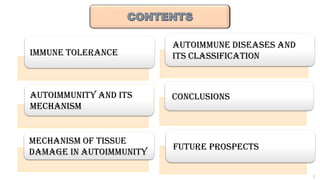
IMMUNE TOLERANCE
AUTOIMMUNITY ANDITS
MECHANISM
MECHANISM OF TISSUE
DAMAGE IN AUTOIMMUNITY
AUTOIMMUNE DISEASES AND
ITS CLASSIFICATION
CONCLUSIONS
FUTURE PROSPECTs
3.
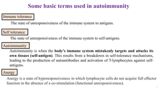
Some basic termsused in autoimmunity
Immune tolerance
3
Autoimmunity
Anergy
The state of unresponsiveness of the immune system to self-antigens.
Self tolerance
The state of unresponsiveness of the immune system to antigens.
Anergy is a state of hyporesponsiveness in which lymphocyte cells do not acquire full effector
function in the absence of a co-stimulation (functional unresponsiveness).
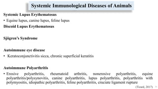
Autoimmunity is when the body's immune system mistakenly targets and attacks its
own tissues (self-antigen). This results from a breakdown in self-tolerance mechanisms,
leading to the production of autoantibodies and activation of T-lymphocytes against self-
antigens.
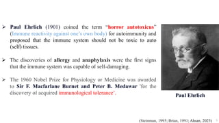
Paul Ehrlich(1901) coined the term “horror autotoxicus”
(Immune reactivity against one’s own body) for autoimmunity and
proposed that the immune system should not be toxic to auto
(self) tissues.
The discoveries of allergy and anaphylaxis were the first signs
that the immune system was capable of self-damaging.
The 1960 Nobel Prize for Physiology or Medicine was awarded
to Sir F. Macfarlane Burnet and Peter B. Medawar 'for the
discovery of acquired immunological tolerance’.
5
(Steinman, 1995; Brian, 1991; Ahsan, 2023)
Paul Ehrlich
6.
IMMUNE TOLERANCE
6
Theimmune system usually does not react to its own antigens due to tolerance
mechanism.
Breach in tolerance can lead to autoimmunity.
Tolerance induction involves educating B and T cells in central (bone marrow,
thymus) and peripheral (spleen, lymph nodes) lymphoid organs.
• Here, Lymphocytes become immune competent or tolerant to encountered
antigens.
Tolerance mechanisms differ between B and T cells and between central and
peripheral organs.
Immune tolerance is not innate; it is established during fetal and postnatal
development, relying on mechanisms controlling lymphocyte development.
(Brent, 1997)
7.
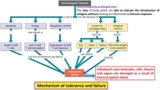
Individual’s own molecules,cells, tissues
and organs are damaged as a result of
immune system attack
Mechanism of tolerance and failure
Immunologically privileged sites:
The sites of body which are able to tolerate the introduction of
antigens without eliciting an inflammatory immune response.
7
8.
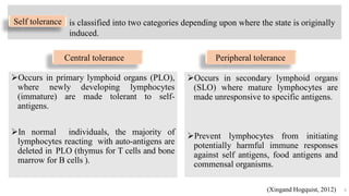
Occurs in primarylymphoid organs (PLO),
where newly developing lymphocytes
(immature) are made tolerant to self-
antigens.
In normal individuals, the majority of
lymphocytes reacting with auto-antigens are
deleted in PLO (thymus for T cells and bone
marrow for B cells ).
Occurs in secondary lymphoid organs
(SLO) where mature lymphocytes are
made unresponsive to specific antigens.
Prevent lymphocytes from initiating
potentially harmful immune responses
against self antigens, food antigens and
commensal organisms.
8
is classified into two categories depending upon where the state is originally
induced.
Central tolerance Peripheral tolerance
(Xingand Hogquist, 2012)
Self tolerance
9.
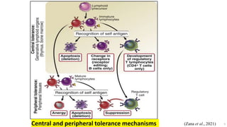
9
(Zana et al.,2021)
Central and peripheral tolerance mechanisms
10.
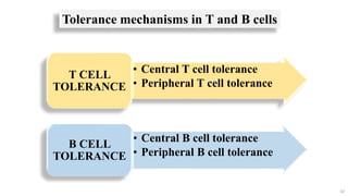
10
• Central Tcell tolerance
• Peripheral T cell tolerance
T CELL
TOLERANCE
• Central B cell tolerance
• Peripheral B cell tolerance
B CELL
TOLERANCE
Tolerance mechanisms in T and B cells
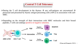
Central T CellTolerance
During the T cell development in the thymus any self-antigens are encountered
processed and presented by thymic antigen-presenting cells (APCs) in association with self-
MHC.
Depending on the strength of their interactions with MHC molecules and their bound
peptides, T cells can undergo positive or negative selection.
12
(Xing and Hogquist, 2012)
13.
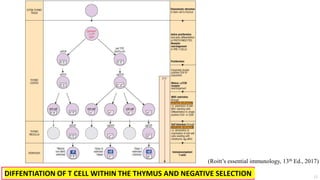
DIFFENTIATION OF TCELL WITHIN THE THYMUS AND NEGATIVE SELECTION
(Roitt’s essential immunology, 13th Ed., 2017)
13
14.
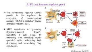
AIRE (autoimmune regulatorgene)
14
The autoimmune regulator (AIRE)
protein is that regulates the
expression of tissue-restricted
antigens (TRAs) in medullary thymic
epithelial cells (MTECs).
AIRE contributes to generating
thymically-derived Foxp3+
regulatory T cells (Treg) by
interacting with medullary thymic
epithelial cells, influencing both
developing and recirculating Treg
populations.
(Anderson and Su, 2016 )
15.
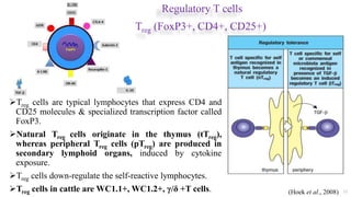
Regulatory T cells
Treg(FoxP3+, CD4+, CD25+)
Treg cells are typical lymphocytes that express CD4 and
CD25 molecules & specialized transcription factor called
FoxP3.
Natural Treg cells originate in the thymus (tTreg),
whereas peripheral Treg cells (pTreg) are produced in
secondary lymphoid organs, induced by cytokine
exposure.
Treg cells down-regulate the self-reactive lymphocytes.
Treg cells in cattle are WC1.1+, WC1.2+, γ/δ +T cells. 15
IL-2R
(Hoek et al., 2008)
16.
Peripheral T CellTolerance
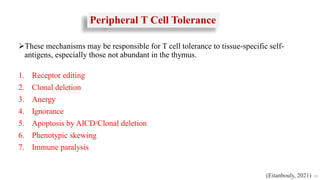
These mechanisms may be responsible for T cell tolerance to tissue-specific self-
antigens, especially those not abundant in the thymus.
1. Receptor editing
2. Clonal deletion
3. Anergy
4. Ignorance
5. Apoptosis by AICD/Clonal deletion
6. Phenotypic skewing
7. Immune paralysis
16
(Eitanbouly, 2021)
17.
Receptor editing
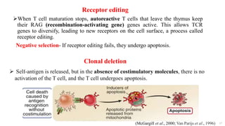
When Tcell maturation stops, autoreactive T cells that leave the thymus keep
their RAG (recombination-activating gene) genes active. This allows TCR
genes to diversify, leading to new receptors on the cell surface, a process called
receptor editing.
Negative selection- If receptor editing fails, they undergo apoptosis.
17
Self-antigen is released, but in the absence of costimulatory molecules, there is no
activation of the T cell, and the T cell undergoes apoptosis.
Clonal deletion
(McGargill et al., 2000; Van Parijs et al., 1996)
18.
18
Ignorance
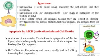
Self-reactive Tcells might never encounter the self-antigen that they
recognize.
Self-antigen with low immunogenicity (low levels of expression or low
binding affinity)
T-cells ignore certain self-antigens because they are located in immune-
privileged sites e.g. corneal proteins, testicular antigens, and antigens from the
brain.
Apoptosis by AICD (Activation-induced Cell Death)
Activation of autoreactive T cells induces upregulation of the Fas
ligand which subsequently interacts with the death receptor Fas
leading (Fas L)to apoptosis.
IL-2 affects the Fas pathway, and can eventually lead to AICD by
increasing FasL expression.
(Van Parijs et al., 1996)
19.
Anergy (functional unresponsiveness)
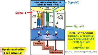
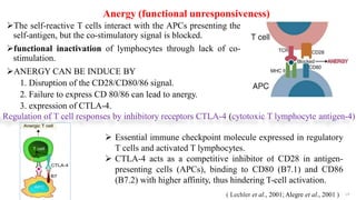
Theself-reactive T cells interact with the APCs presenting the
self-antigen, but the co-stimulatory signal is blocked.
functional inactivation of lymphocytes through lack of co-
stimulation.
ANERGY CAN BE INDUCE BY
1. Disruption of the CD28/CD80/86 signal.
2. Failure to express CD 80/86 can lead to anergy.
3. expression of CTLA-4.
19
Regulation of T cell responses by inhibitory receptors CTLA-4 (cytotoxic T lymphocyte antigen-4)
Essential immune checkpoint molecule expressed in regulatory
T cells and activated T lymphocytes.
CTLA-4 acts as a competitive inhibitor of CD28 in antigen-
presenting cells (APCs), binding to CD80 (B7.1) and CD86
(B7.2) with higher affinity, thus hindering T-cell activation.
( Lechler et al., 2001; Alegre et al., 2001 )
20.
20
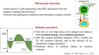
Phenotypic skewing
Self-reactive Tcells interacting with APCs presented with self-
antigens, undergo full activation.
Secrete non-pathogenic cytokines and chemokine receptors profile.
Immune paralysis
Very low or very high doses of an antigen can induce a
form of clonal anergy called immune paralysis.
Here the antigen probably bypasses APCs, reaches the
Th cell receptors directly, and in the absence of co-
stimulation, triggers paralysis.
Moderate doses, in contrast, induce an immune
response.
(Alegre et al., 2001; Baker et al., 1974)
21.
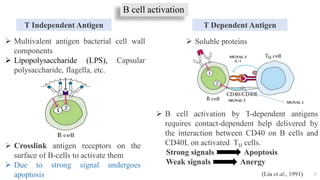
B cell activation
21
TDependent Antigen
T Independent Antigen
Soluble proteins
Multivalent antigen bacterial cell wall
components
Lipopolysaccharide (LPS), Capsular
polysaccharide, flagella, etc.
Crosslink antigen receptors on the
surface of B-cells to activate them
Due to strong signal undergoes
apoptosis
B cell activation by T-dependent antigens
requires contact-dependent help delivered by
the interaction between CD40 on B cells and
CD40L on activated TH cells.
Strong signals Apoptosis
Weak signals Anergy
IL-4
SIGNAL 1
SIGNAL 2
SIGNAL 3
(Liu et al., 1991)
22.
Central B CellTolerance
Central B cell tolerance refers to the process that negatively selects newly
generating immature B cells that react with a self-antigen in the bone marrow.
Central B cell tolerance involve 3 major mechanisms to precent self
reactivity.
22
1. Receptor editing
2. Clonal deletion / clonal abortion
3. Anergy
(Basten and Silveira, 2010)
23.
23
The processby which many of the B cells reactivate the machinery of antigen
receptor gene rearrangement (mainly genes coding for light chains), so that a
different (edited) B cell receptor will be produced that no longer recognizes the
self-antigen.
If receptor editing fails, they undergo apoptosis (negative selection).
Receptor editing
If immature B cell encounters and bind self-antigen, the BCR transmits a signal
that arrests cell development, blocks synapse formation, and triggers apoptosis
leading to termination of B cell development. The clonal abortion occurs in the
early encounter of antigen during B cell development in the bone marrow.
Clonal deletion/clonal abortion
(Nemazee, 2017)
24.
24
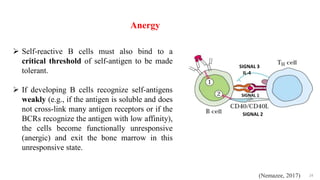
Self-reactive Bcells must also bind to a
critical threshold of self-antigen to be made
tolerant.
If developing B cells recognize self-antigens
weakly (e.g., if the antigen is soluble and does
not cross-link many antigen receptors or if the
BCRs recognize the antigen with low affinity),
the cells become functionally unresponsive
(anergic) and exit the bone marrow in this
unresponsive state.
SIGNAL 1
SIGNAL 3
IL-4
SIGNAL 2
Anergy
(Nemazee, 2017)
25.
25
1. Receptor editing
2.Apoptosis
3. Clonal anergy
4. Clonal exhaustion: repeated exhaustive antigenic stimulation to B cell may differentiate
into short-lived plasma cells. Such plasma cells, no memory B cells will remain to
respond to antigen, and tolerance will result.
5. Blockage of BCRs: polymeric antigens may bind to the B cell receptor irreversibly and
block any further response these cell.
Peripheral B-cell Tolerance
(Getahun, 2022)
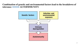
Combination of geneticand environmental factors lead to the breakdown of
tolerance AUTOIMMUNITY
27
28.
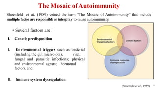
The Mosaic ofAutoimmunity
• Several factors are :
28
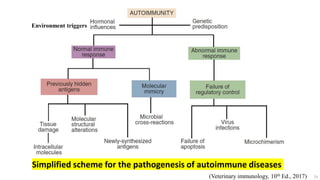
I. Genetic predisposition
I. Environmental triggers such as bacterial
(including the gut microbiota), viral,
fungal and parasitic infections; physical
and environmental agents; hormonal
factors, and
II. Immune system dysregulation
Shoenfeld et al. (1989) coined the term “The Mosaic of Autoimmunity” that include
multiple factor are responsible or interplay to cause autoimmunity.
(Shoenfeld et al., 1989)
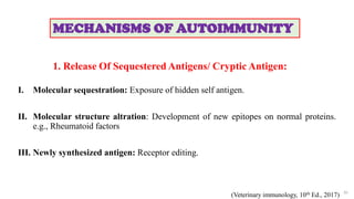
1. Release OfSequestered Antigens/ Cryptic Antigen:
I. Molecular sequestration: Exposure of hidden self antigen.
II. Molecular structure altration: Development of new epitopes on normal proteins.
e.g., Rheumatoid factors
III. Newly synthesized antigen: Receptor editing.
30
MECHANISMS OF AUTOIMMUNITY
(Veterinary immunology, 10th Ed., 2017)
31.
2. Epitope Spreading
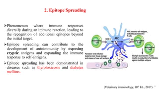
Phenomenonwhere immune responses
diversify during an immune reaction, leading to
the recognition of additional epitopes beyond
the initial target.
Epitope spreading can contribute to the
development of autoimmunity by exposing
cryptic antigens and expanding the immune
response to self-antigens.
Epitope spreading has been demonstrated in
diseases such as thyrotoxicosis and diabetes
mellitus.
31
(Veterinary immunology, 10th Ed., 2017)
32.
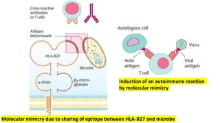
3. Molecular Mimicry
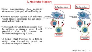
Somemicroorganisms share antigenic
determinants (epitopes) with self-antigens.
Immune response against such microbes
would produce antibodies that can cross-
react with self-antigen.
Cross-reactions with foreign antigens may
be sufficient to trigger a helper T cell
population that will promote an
autoimmune response by B cells.
A helper effect triggered by a foreign
antigen may inadvertently permit an
autoimmune response to occur.
32
(Rojas et al., 2018)
Self antigen alone Foreign antigen
33.
Induction of anautoimmune reaction
by molecular mimicry
Molecular mimicry due to sharing of epitope between HLA-B27 and microbe
34.
34
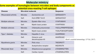
Molecular mimicry
Some examplesof homologies between microbes and body components as
potential cross‐reacting T‐cell epitopes
(Roitt’s essential immunology, 13th Ed., 2017)
35.
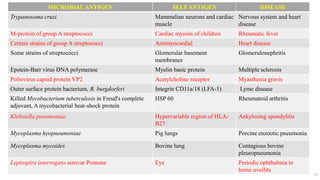
MICROBIALANTIGEN SELFANTIGEN DISEASE
Trypanosomacruzi Mammalian neurons and cardiac
muscle
Nervous system and heart
disease
M-protein of group A streptococci Cardiac myosin of children Rheumatic fever
Certain strains of group A streptococci Antimyocardial Heart disease
Some strains of streptococci Glomerular basement
membranes
Glomerulonephritis
Epstein-Barr virus DNA polymerase Myelin basic protein Multiple sclerosis
Poliovirus capsid protein VP2 Acetylcholine receptor Myasthenia gravis
Outer surface protein bacterium, B. burgdorferi Integrin CD11a/18 (LFA-1) Lyme disease
Killed Mycobacterium tuberculosis in Freud's complete
adjuvant, A mycobacterial heat-shock protein
HSP 60 Rheumatoid arthritis
Klebsiella pneumoniae Hypervariable region of HLA-
B27
Ankylosing spondylitis
Mycoplasma hyopneumoniae Pig lungs Porcine enzootic pneumonia
Mycoplasma mycoides Bovine lung Contagious bovine
pleuropneumonia
Leptospira interrogans serovar Pomona Eye Periodic ophthalmia in
horse uveilits
35
36.
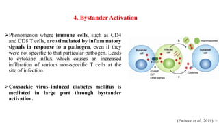
4. BystanderActivation
Phenomenon whereimmune cells, such as CD4
and CD8 T cells, are stimulated by inflammatory
signals in response to a pathogen, even if they
were not specific to that particular pathogen. Leads
to cytokine influx which causes an increased
infiltration of various non-specific T cells at the
site of infection.
Coxsackie virus–induced diabetes mellitus is
mediated in large part through bystander
activation.
36
(Pacheco et al., 2019)
37.
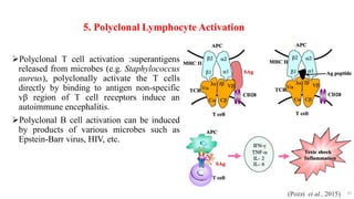
5. Polyclonal LymphocyteActivation
Polyclonal T cell activation :superantigens
released from microbes (e.g. Staphylococcus
aureus), polyclonally activate the T cells
directly by binding to antigen non-specific
vβ region of T cell receptors induce an
autoimmune encephalitis.
Polyclonal B cell activation can be induced
by products of various microbes such as
Epstein-Barr virus, HIV, etc.
37
(Pozzi et al., 2015)
38.
38
6. Breakdown ofT-cell Anergy
In the presence of tissue necrosis and local inflammation express co-stimulatory
molecules (B7) .
• multiple sclerosis, rheumatoid arthritis and psoriasis
7. Failure Of AICD
Failure of the auto-reactive activated T cells to undergo activation-induced cell
death (AICD).
• SLE (systemic lupus erythematosus)
(Lechler et al., 2001; Badami and Quaratino, 2019)
39.
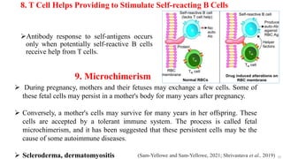
8. T CellHelps Providing to Stimulate Self-reacting B Cells
Antibody response to self-antigens occurs
only when potentially self-reactive B cells
receive help from T cells.
39
9. Microchimerism
During pregnancy, mothers and their fetuses may exchange a few cells. Some of
these fetal cells may persist in a mother's body for many years after pregnancy.
Conversely, a mother's cells may survive for many years in her offspring. These
cells are accepted by a tolerant immune system. The process is called fetal
microchimerism, and it has been suggested that these persistent cells may be the
cause of some autoimmune diseases.
Scleroderma, dermatomyositis (Sam-Yellowe and Sam-Yellowe, 2021; Shrivastava et al., 2019)
40.
Genetic Predisposition toAutoimmunity
Any defect in genes that code for cytokines, cytokine receptors, co-stimulators,
molecules that regulate apoptosis, molecules that regulate antigen clearance, and
members of cytokine or antigen-signaling cascades and inherited complement
deficiencies are responsible for regulation of autoimmunity.
Defect in a single gene:
• e.g., LPR or GLD gene mutations results in destruction of self-reactive T cells.
In their absence, excessive T cell proliferation occurs.
Role of gene complex : More common
• genes influence the severity of disease.
• The genes that most commonly associated with naturally occurring
autoimmune diseases are those in the MHC.
o MHC 1199 molecules regulate the presentation of processed epitopes.
• Studies of humans have shown that almost all autoimmune diseases are linked
to multiple MHC loci.
40
(Veterinary immunology, 10th Ed., 2017)
41.
The Effects ofSpaying or Neutering on Autoimmune Diseases in
Dogs
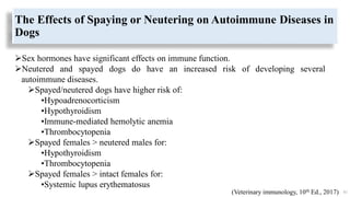
Sex hormones have significant effects on immune function.
Neutered and spayed dogs do have an increased risk of developing several
autoimmune diseases.
Spayed/neutered dogs have higher risk of:
•Hypoadrenocorticism
•Hypothyroidism
•Immune-mediated hemolytic anemia
•Thrombocytopenia
Spayed females > neutered males for:
•Hypothyroidism
•Thrombocytopenia
Spayed females > intact females for:
•Systemic lupus erythematosus
41
(Veterinary immunology, 10th Ed., 2017)
42.
Breed Predispositions toAutoimmunity
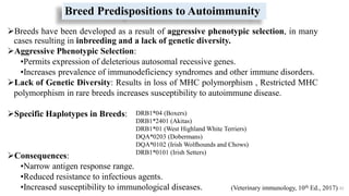
Breeds have been developed as a result of aggressive phenotypic selection, in many
cases resulting in inbreeding and a lack of genetic diversity.
Aggressive Phenotypic Selection:
•Permits expression of deleterious autosomal recessive genes.
•Increases prevalence of immunodeficiency syndromes and other immune disorders.
Lack of Genetic Diversity: Results in loss of MHC polymorphism , Restricted MHC
polymorphism in rare breeds increases susceptibility to autoimmune disease.
Specific Haplotypes in Breeds:
Consequences:
•Narrow antigen response range.
•Reduced resistance to infectious agents.
•Increased susceptibility to immunological diseases. 42
DRB1*04 (Boxers)
DRB1*2401 (Akitas)
DRB1*01 (West Highland White Terriers)
DQA*0203 (Dobermans)
DQA*0102 (Irish Wolfhounds and Chows)
DRB1*0101 (Irish Setters)
(Veterinary immunology, 10th Ed., 2017)
43.
43
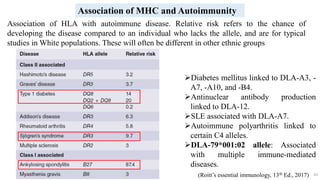
Diabetes mellitus linkedto DLA-A3, -
A7, -A10, and -B4.
Antinuclear antibody production
linked to DLA-12.
SLE associated with DLA-A7.
Autoimmune polyarthritis linked to
certain C4 alleles.
DLA-79*001:02 allele: Associated
with multiple immune-mediated
diseases.
Association of MHC and Autoimmunity
Association of HLA with autoimmune disease. Relative risk refers to the chance of
developing the disease compared to an individual who lacks the allele, and are for typical
studies in White populations. These will often be different in other ethnic groups
(Roitt’s essential immunology, 13th Ed., 2017)
44.
44
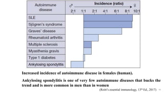
Increased incidence ofautoimmune disease in females (human).
Ankylosing spondylitis is one of very few autoimmune diseases that bucks the
trend and is more common in men than in women
(Roitt’s essential immunology, 13th Ed., 2017)
45.
Intestinal Microbiota
Changes inthe gut microbiota due to antibiotic treatment can exacerbate the
autoimmune disease.
Alterations in the intestinal microbiota also influence autoimmune diseases such
as rheumatoid arthritis, ankylosing spondylitis, and experimental autoimmune
encephalitis.
45
(Veterinary immunology, 10th Ed., 2017)
Mechanisms of TissueDamage in Autoimmunity
Autoimmune disease results when tissues are damaged by autoreactive T
cells or antibodies. This damage is a result of hypersensitivity reactions.
Mechanisms involved in tissue damage are very complex.
In a number of autoimmune diseases more than one mechanism is
involved.
47
(Pedersen, 1999)
48.
48
Mechanisms Of TissueDamage
1. Anaphylactic hypersensitivity /IgE autoantibodies (Type I): Rare. A possible
example: “Milk allergy” in Jersey cattle; IgE autoantibody response to α-casein.
2. Antibody Dependent cell mediated Cytotoxicity ADCC (Type II):
Autoantibodies may cause cell lysis with the assistance of complement or cytotoxic
cells.
Autoantibodies to cell surface, e.g.
I. RBC........................Autoimmune hemolytic anemia
II. Thrombocytes....... Autoimmune thrombocytopenia
III. Thyroid cells………………… Thyroiditis
IV. Acetylcholine receptor…….. Myasthenia gravis
V. Insulin receptor….. Diabetes
VI. β-adrenoceptors… Asthma
(Pedersen, 1999)
49.
3. Immune complexmediated hypersensitivity (Type III):
• Autoantibodies (Ab) + Antigen (Ag) i.e. [Ab-Ag immune complex] e.g. SLE, RA
4. T-Cell Mediated Damage (Type IV):
• T cells are primarily involved in tissue damage, e.g. autoimmune thyroiditis,
ulcerative colitis, insulin-dependent juvenile-onset diabetes, autoimmune
encephalitis.
5. Stimulatory hypersensitivity (Type V):
• Ab against some cell surface component may stimulate the cell instead of
destroying the cell.
• Thyroid hyper-reactivity in Grave’s disease due to thyroid stimulating
antibodies.
49
(Pedersen, 1999)
51
Autoimmune diseases arerelatively common. They occur in about 5% of domestic
mammals.
Autoimmune diseases often target a specific organ or tissue due to an abnormal
response to a few self-antigens, without necessarily indicating a loss of control of
the entire immune system.
Many factors influence susceptibility to autoimmunity.
• These include sex and age, genetic background, the microbiota, and virus
infections.
Autoimmune diseases can be classified by body systems that are affected. Some
conditions influence multiple body systems, whereas others affect only one organ.
(Tizard, 2017)
AUTOIMMUNE DISEASES AND
ITS CLASSIFICATION
Immune-Mediated Hemolytic Anemia(IMHA)
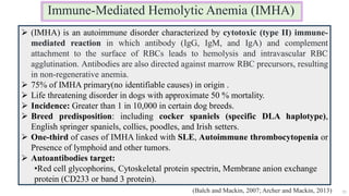
55
(IMHA) is an autoimmune disorder characterized by cytotoxic (type II) immune-
mediated reaction in which antibody (IgG, IgM, and IgA) and complement
attachment to the surface of RBCs leads to hemolysis and intravascular RBC
agglutination. Antibodies are also directed against marrow RBC precursors, resulting
in non-regenerative anemia.
75% of IMHA primary(no identifiable causes) in origin .
Life threatening disorder in dogs with approximate 50 % mortality.
Incidence: Greater than 1 in 10,000 in certain dog breeds.
Breed predisposition: including cocker spaniels (specific DLA haplotype),
English springer spaniels, collies, poodles, and Irish setters.
One-third of cases of IMHA linked with SLE, Autoimmune thrombocytopenia or
Presence of lymphoid and other tumors.
Autoantibodies target:
•Red cell glycophorins, Cytoskeletal protein spectrin, Membrane anion exchange
protein (CD233 or band 3 protein).
(Balch and Mackin, 2007; Archer and Mackin, 2013)
56.
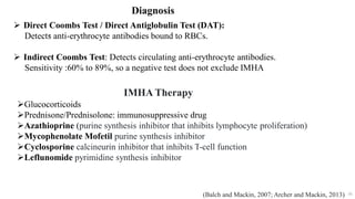
56
Diagnosis
.
Direct CoombsTest / Direct Antiglobulin Test (DAT):
Detects anti-erythrocyte antibodies bound to RBCs.
Indirect Coombs Test: Detects circulating anti-erythrocyte antibodies.
Sensitivity :60% to 89%, so a negative test does not exclude IMHA
(Balch and Mackin, 2007; Archer and Mackin, 2013)
IMHA Therapy
Glucocorticoids
Prednisone/Prednisolone: immunosuppressive drug
Azathioprine (purine synthesis inhibitor that inhibits lymphocyte proliferation)
Mycophenolate Mofetil purine synthesis inhibitor
Cyclosporine calcineurin inhibitor that inhibits T-cell function
Leflunomide pyrimidine synthesis inhibitor
57.
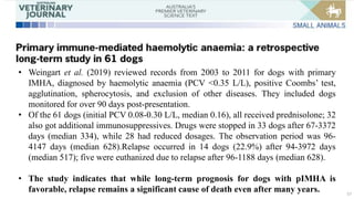
57
• Weingart etal. (2019) reviewed records from 2003 to 2011 for dogs with primary
IMHA, diagnosed by haemolytic anaemia (PCV <0.35 L/L), positive Coombs’ test,
agglutination, spherocytosis, and exclusion of other diseases. They included dogs
monitored for over 90 days post-presentation.
• Of the 61 dogs (initial PCV 0.08-0.30 L/L, median 0.16), all received prednisolone; 32
also got additional immunosuppressives. Drugs were stopped in 33 dogs after 67-3372
days (median 334), while 28 had reduced dosages. The observation period was 96-
4147 days (median 628).Relapse occurred in 14 dogs (22.9%) after 94-3972 days
(median 517); five were euthanized due to relapse after 96-1188 days (median 628).
• The study indicates that while long-term prognosis for dogs with pIMHA is
favorable, relapse remains a significant cause of death even after many years.
58.
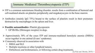
Immune Mediated Thrombocytopenia(ITP)
58
Approximately 30% of the cases ITP and immune-mediated hemolytic anemia (IMHA)
occur together in one animal (Evans’ syndrome).
Commonly observed in association with
• IMHA and SLE
• Multiple myeloma or other lymphoid tumors,
• Ehrlichiosis and leishmaniasis, or following certain drug treatments.
ITP is a common autoimmune bleeding disorder. results from a combination of humoral and
cell‐mediated attacks on platelets peripherally and megakaryocytes in the bone marrow.
Antibodies (mainly IgG 75%) bound to the surface of platelets result in their premature
destruction by macrophages in the spleen and liver.
Possible autoantibodies :Platelet glycoprotein
• GP IIb/IIIa (fibrinogen receptor) in dogs.
(LeVine and Brooks, 2019)
59.
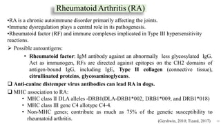
Rheumatoid Arthritis (RA)
59
•RAis a chronic autoimmune disorder primarily affecting the joints.
•Immune dysregulation plays a central role in its pathogenesis.
•Rheumatoid factor (RF) and immune complexes implicated in Type III hypersensitivity
reactions.
Possible autoantigens:
• Rheumatoid factor: IgM antibody against an abnormally less glycosylated IgG.
Act as immunogen, RFs are directed against epitopes on the CH2 domains of
antigen-bound IgG, including IgE, Type II collagen (connective tissue),
citrullinated proteins, glycosaminoglycans.
Anti-canine distemper virus antibodies can lead RA in dogs.
MHC association to RA:
• MHC class II DLA alleles -DRB1(DLA-DRB1*002, DRB1*009, and DRB1*018)
• MHC class III gene C4 allotype C4-4.
• Non-MHC genes; contribute as much as 75% of the genetic susceptibility to
rheumatoid arthritis. (Gershwin, 2010; Tizard, 2017)
60.
60
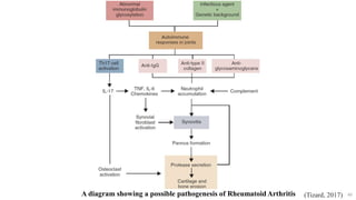
A diagram showinga possible pathogenesis of Rheumatoid Arthritis (Tizard, 2017)
61.
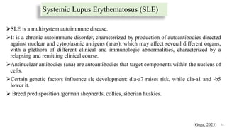
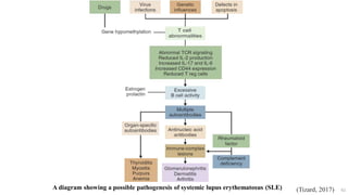
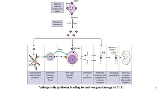
Systemic Lupus Erythematosus(SLE)
SLE is a multisystem autoimmune disease.
It is a chronic autoimmune disorder, characterized by production of autoantibodies directed
against nuclear and cytoplasmic antigens (anas), which may affect several different organs,
with a plethora of different clinical and immunologic abnormalities, characterized by a
relapsing and remitting clinical course.
Antinuclear antibodies (ana) are autoantibodies that target components within the nucleus of
cells.
Certain genetic factors influence sle development: dla-a7 raises risk, while dla-a1 and -b5
lower it.
Breed predisposition :german shepherds, collies, siberian huskies.
61
(Guga, 2023)
62.
62
A diagram showinga possible pathogenesis of systemic lupus erythematosus (SLE) (Tizard, 2017)
65
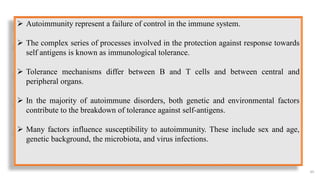
Autoimmunity representa failure of control in the immune system.
The complex series of processes involved in the protection against response towards
self antigens is known as immunological tolerance.
Tolerance mechanisms differ between B and T cells and between central and
peripheral organs.
In the majority of autoimmune disorders, both genetic and environmental factors
contribute to the breakdown of tolerance against self-antigens.
Many factors influence susceptibility to autoimmunity. These include sex and age,
genetic background, the microbiota, and virus infections.
66.
66
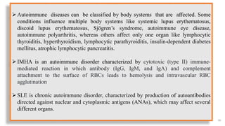
Autoimmune diseases canbe classified by body systems that are affected. Some
conditions influence multiple body systems like systemic lupus erythematosus,
discoid lupus erythematosus, Sjögren’s syndrome, autoimmune eye disease,
autoimmune polyarthritis, whereas others affect only one organ like lymphocytic
thyroiditis, hyperthyroidism, lymphocytic parathyroiditis, insulin-dependent diabetes
mellitus, atrophic lymphocytic pancreatitis.
IMHA is an autoimmune disorder characterized by cytotoxic (type II) immune-
mediated reaction in which antibody (IgG, IgM, and IgA) and complement
attachment to the surface of RBCs leads to hemolysis and intravascular RBC
agglutination
SLE is chronic autoimmune disorder, characterized by production of autoantibodies
directed against nuclear and cytoplasmic antigens (ANAs), which may affect several
different organs.
There is aneed to understand the pathophysiology, diagnosis and treatment
strategies of autoimmune diseases in detail.
There is a need for development of new treatment protocol for autoimmune
diseases, as it has wide variability, different causative and pathogenic process,
which is highly dynamic, making the current treatment strategies to fall short of
expected complete remission.
68

 Most read