TYPE Original Research
PUBLISHED 01 July 2026
DOI 10.3389/fphar.2026.1823203
Targeting PKLR and lipogenic
enzymes through JNK inhibition
to develop a therapeutic strategy
for MASLD and MASH
Woonghee Kim1†
, Mengzhen Li1†
, Xinmeng Liao1
, Sevilay Özmen2
,
Edanur Yıldız3
, Melik Saraçoğlu3
, Cem Baba4
,
Fatih Caglar Celikezen5
, Said Ali Atalay6
, Ahmet Tuğrul Akkuş6
,
Fatih Alper6
, Han Jin1
, Hong Yang1
, Shazia Iqbal7
,
Jihad Sebhaoui7,8
, Sajda Ashraf7
, Burcu Belmen7
, Jan Boren9
,
Mathias Uhlen1
, Cheng Zhang1
, Hasan Turkez10
and
Adil Mardinoglu � 1,11
*
1
Science for Life Laboratory, KTH – Royal Institute of Technology, Stockholm, Sweden, 2
Department of
Pathology, Faculty of Medicine, Atatürk University, Erzurum, Türkiye, 3
Institute of Science, Atatürk
University, Erzurum, Türkiye, 4
Institute of Science, Erzurum Technical University, Erzurum, Türkiye,
5
Department of Chemistry, Science and Letter Faculty, Bitlis Eren University, Bitlis, Türkiye, 6
Department of
Radiology, Faculty of Medicine, Atatürk University, Erzurum, Türkiye, 7
Trustlife Labs Drug Research &
Development Center, Istanbul, Türkiye, 8
Life and Health Sciences Laboratory, FMP, Abdelmalek Essaadi
University, Tetouan, Morocco, 9
Department of Molecular and Clinical Medicine, University of Gothenburg,
Sahlgrenska University Hospital, Gothenburg, Sweden, 10
Department of Medical Biology, Faculty of
Medicine, Atatürk University, Erzurum, Türkiye, 11
Centre for Host-Microbiome Interactions, Faculty of
Dentistry, Oral & Craniofacial Sciences, King’s College London, London, United Kingdom
Background: Pyruvate kinase liver and red blood cells (PKLR) is linked to metabolic
dysfunction-associated steatotic liver disease (MASLD). Previous study, we
identified JNK-IN-5A, a c-Jun N-terminal kinase (JNK) inhibitor that
suppresses PKL expression in HepG2 cells using computational drug
repurposing and screened out four hit JNK-IN-5A derivatives (SET-151, SET-
152, SET-162, SET-130).
Materials and Methods: We validated therapeutic efficacy of JNK-IN-5A and four
derivative (SET-151, SET-152, SET-162, SET-130). HepG2 de novo lipogenesis
(DNL) steatosis model was used in vitro validation. RNA sequencing data were
analysed using systems biology approaches, including transcriptomic profiling
and COMPASS analysis. GLP-like toxicity assessment in rat model shows in vivo
safety and MASLD rat model revealed in vivo therapeutic effect to MASLD
and MASH.
Results: In a HepG2 DNL steatosis model, all compounds reduced intracellular
triacylglycerol (TAG) and inhibited key DNL proteins (PKL, FASN, ACACA, SCD1,
SREBP1-c, ChREBP). Transcriptomic profiling revealed stronger anti-steatotic
effects with SET-151, SET-152, and SET-162, which uniquely downregulated
genes in pyruvate metabolism, bile acid synthesis, fatty acid metabolism, and
glycolysis. Compass analysis showed these derivatives significantly altered lipid-
related metabolic reactions, unlike JNK-IN-5A. In a high-sucrose, high-fat diet-
induced MASLD rat model, JNK-IN-5A and SET-152 reduced hepatic lipid
accumulation, liver stiffness, and MASLD biomarkers.
OPEN ACCESS
EDITED BY
Joseph Nickels,
Jr, Genesis Biotechnology Group,
United States
REVIEWED BY
Wei Wang,
Xuzhou Medical University, China
Yansong Fu,
Central South University, China
*CORRESPONDENCE
Adil Mardinoglu,
adilm@scilifelab.se
†
These authors have contributed equally
to this work and share first authorship
RECEIVED 04 March 2026
REVISED 29 May 2026
ACCEPTED 02 June 2026
PUBLISHED 01 July 2026
CITATION
Kim W, Li M, Liao X, Özmen S, Yıldız E,
Saraçoğlu M, Baba C, Celikezen FC,
Atalay SA, Akkuş AT, Alper F, Jin H, Yang H,
Iqbal S, Sebhaoui J, Ashraf S, Belmen B,
Boren J, Uhlen M, Zhang C, Turkez H and
Mardinoglu A (2026) Targeting PKLR and
lipogenic enzymes through JNK inhibition
to develop a therapeutic strategy for
MASLD and MASH.
Front. Pharmacol. 17:1823203.
doi: 10.3389/fphar.2026.1823203
COPYRIGHT
© 2026 Kim, Li, Liao, Özmen, Yıldız,
Saraçoğlu, Baba, Celikezen, Atalay, Akkuş,
Alper, Jin, Yang, Iqbal, Sebhaoui, Ashraf,
Belmen, Boren, Uhlen, Zhang, Turkez and
Mardinoglu. This is an open-access article
distributed under the terms of the Creative
Commons Attribution License (CC BY).
The use, distribution or reproduction in
other forums is permitted, provided the
original author(s) and the copyright
owner(s) are credited and that the original
publication in this journal is cited, in
accordance with accepted academic
practice. No use, distribution or
reproduction is permitted which does not
comply with these terms.
Frontiers in Pharmacology frontiersin.org
01
Conclusion: Our findings identify PKLR as a promising therapeutic target for MASLD
and MASH. SET-152 suppressing PKLR through JNK inhibition highlights its
potential as a new drug for MASLD and MASH therapy.
KEYWORDS
DNL de novo lipogenesis, hepatic steatosis, JNK (c-Jun N-terminal kinase), MASLD,
new drug
Introduction
Metabolic dysfunction-associated steatotic liver disease
(MASLD) is a chronic metabolic liver disease representing a
major global health concern, driven by underlying metabolic
dysfunction and strongly linked to dysregulated lipid metabolism,
obesity and type 2 diabetes (Mardinoglu and Palsson, 2025). Macro
vesicular steatosis causes chronic inflammation, which can develop
into metabolic dysfunction-associated steatohepatitis (MASH)
(Sinton et al., 2021). Chronic hepatic inflammation leads to
collagen deposition and fibrotic tissue formation, ultimately
progressing to cirrhosis (Dham et al., 2019). Approximately 20%
of MASH patients progress to cirrhosis (Sheka et al., 2020). The
estimated global prevalence of MASLD among adults is
approximately 32%, with a higher prevalence observed in males
(40%) compared to females (26%) (Riazi et al., 2022). Currently,
resmetirom is the only FDA-approved therapeutic agent for MASH
(FDA approves first MASH drug, 2024). Despite this advancement,
there remains an urgent need for the development of additional
effective therapies for MASH, and extensive research efforts are
ongoing in this field.
The liver, as a central regulator of systemic metabolism, takes up
and processes various metabolites circulating in the blood. Free fatty
acids (FFAs) entering the liver from the bloodstream are converted
into triacylglycerols (TAGs) (Bradbury, 2006). Excess carbohydrates
are used in the synthesis of fatty acids through de novo lipogenesis
(DNL) (Browning and Horton, 2004). High-carbohydrate intake,
particularly under conditions of hyperinsulinemia and
hyperglycaemia, activates key transcription factors such as
SREBP-1c and ChREBP (Sanders and Griffin, 2016). These
transcription factors upregulate the expression of critical DNL
enzymes, leading to increased fatty acid and TG synthesis from
glucose (Canbay et al., 2007). To develop effective therapeutic
interventions for steatosis, current research is focused on
targeting various key regulators within these pathways.
Kristine G. et al. (Griffett and Burris, 2023) reported the
development of TLC-2716, an inverse agonist of the liver X
receptor (LXR), which is currently undergoing Phase 1 clinical
trials. LXR is a key transcription factor that forms a heterodimer
with the retinoid X receptor (RXR) and plays a central role in
promoting hepatic lipogenesis and steatosis (Shen et al., 2011). By
binding to LXR as an inverse agonist, TLC-2716 suppresses hepatic
lipogenesis and fibrosis (Griffett and Burris, 2023; Griffett et al.,
2015). Another promising therapeutic approach targets hepatic
thyroid hormone receptor β (THR-β). Resmetirom (MGL-3196),
a selective THR-β agonist, has been developed to activate lipid
catabolism in the liver and has recently received FDA approval
following successful Phase 3 trials (FDA approves first MASH drug,
2024; Harrison et al., 2023; Kelly et al., 2014). THR-β, activated by
triiodothyronine (T3) thyroid hormone, regulates the expression of
genes involved in systemic lipid reduction, increased bile acid
synthesis, and enhanced fat oxidation (Kannt et al., 2021).
Resmetirom has demonstrated therapeutic efficacy in reducing
hepatic fat accumulation by selectively activating THR-β (Kannt
et al., 2021; Hatziagelaki et al., 2022; Zhou et al., 2022; Karim
et al., 2023).
Lee et al. (Lee et al., 2017) applied a systems biology approach to
compare healthy liver tissue with samples from individuals with
MASLD and hepatocellular carcinoma (HCC). This study identified
key genes that were highly upregulated in both MASLD and HCC,
highlighting their potential as therapeutic targets. Among these,
three genes, including pyruvate kinase liver (PKL) and red blood cell
(PKLR), patatin-like phospholipase domain-containing 3
(PNPLA3), and proprotein convertase subtilisin/kexin type 9
(PCSK9), were identified as effective drug targets. PKLR, a key
enzyme in the DNL pathway and lipid metabolism, plays a critical
role in hepatic steatosis (Liu et al., 2019; Yuan et al., 2025). To
advance the development of novel therapeutics for MASLD, we
established an in vitro model of DNL-induced steatosis and
performed computational drug repurposing to identify small-
molecule regulators of PKLR. In our previous study, we
established a HepG2 DNL steatosis model using insulin and the
LXR agonist T0901317 (Kim et al., 2023). The HepG2 cell line is a
well-validated and widely used in vitro model for studying hepatic
metabolism include glucose (Abruscato et al., 2025; Pittala et al.,
2024), insulin (Sefried et al., 2018), and lipid metabolism (Lin et al.,
2025; Rida and Kreydiyyeh, 2023). We induced DNL in HepG2 cells
using insulin and T0901317. This method led to significant increases
in intracellular TAG accumulation, upregulation of key DNL
pathway transcription factors and enzymes, as well as enhanced
glycolytic activity and also showed clinical relevancy compared to
MAFLD phenotype human cohorts (Kim et al., 2023).
To identify the modulators of PKLR, we applied a systems
biology-driven drug repurposing approach and identified JNK-
IN-5A as a small molecule that reduces PKL protein levels in
HepG2 cells, exhibiting promising anti-steatotic effects in the
HepG2 DNL steatosis model (Kim et al., 2023; Zhang et al.,
2022). JNK-IN-5A is a potent and selective ATP-competitive
inhibitor of the c-Jun N-terminal kinase (JNK) family. It has
been reported that there are strong mechanistic links between the
JNK signalling pathways and DNL. It has been widely used to
modulate JNK pathways in the in vitro and in vivo models of
metabolic disorders, certain types of cancer and neurodegeneration.
In this study, we evaluated the effect of the JNK-IN-5A, a PKLR-
suppressing small molecule that suppresses PKLR, using a
HepG2 DNL steatosis model. We observed that JNK-IN-5A
treatment significantly reduced TAG accumulation by
downregulating DNL pathway protein expression in the
HepG2 DNL steatosis model. Furthermore, we identified four
novel derivatives of JNK-IN-5A, including SET-151, SET-152,
Frontiers in Pharmacology frontiersin.org
02
Kim et al. 10.3389/fphar.2026.1823203
SET-162 and SET-130 that exhibited potent inhibitory effects on
DNL-induced steatosis in vitro. These derivatives effectively
decreased TAG accumulation and suppressed DNL pathway
activation. For comparative purposes, we also evaluated the
impact of the THR-β agonist resmetirom (Harrison et al., 2024)
in the HepG2 DNL steatosis model. Additionally, RNA sequencing
was performed to investigate the transcriptomic differences between
the treatment groups. To further elucidate the mechanisms of action
of JNK-IN-5A and its derivatives, we applied systems biology
analyses to identify altered pathways and metabolic reactions.
These analyses provided insights into how the compounds
modulate key metabolic processes relevant to MASLD.
Furthermore, we tested the tolerability of SET-152 in GLP-like
toxicity study in rats and evaluated the therapeutic potential of
SET-152 in an in vivo MASLD model induced by a high-sucrose and
high-fat diet. Treatment with the compounds significantly improved
MASLD-related symptoms, as demonstrated by magnetic resonance
imaging (MRI), two-dimensional shear wave elastography (2D-
SWE), liver histology, and blood biochemical markers.
Materials and Methods
Cell culture and DNL steatosis induction
HepG2 wild-type cells (ATCC, ATCC HB-8065™, USA) were
purchased from the genome engineering company Synthego. Cells
were maintained with RPMI 1640 (R2405, Sigma-Aldrich)
supplemented with 10% fetal bovine serum (FBS, F7524, Sigma-
Aldrich, Germany), 1% P/S (P4333, Sigma-Aldrich, Germany) and
used within under passage 20.6 × 10⁴
cells HepG2 cells were seeded
into a 96-well plate format for assay, and 1 × 10⁶
cells were plated into a
6-well plate for Western blot and image analysis. DMEM high glucose
(D0819, Sigma-Aldrich, Germany) with 10% FBS, 1% P/S supplemented
with 10 μg/mL insulin (I9278, Sigma-Aldrich, Germany), and 10 µM
T0901317 (T2320, Sigma-Aldrich, Germany) was changed to induce
DNL steatosis in HepG2 cells. DNL steatosis media and compounds
were changed for 1 week, with a 3-day, 2-day, and 2-day cycle.
Triacylglycerol (TAG), cell viability (MTT)
assay, and BODIPY™ staining
TAG levels were quantified using the Triacylglycerol Assay
Kit–Quantification (ab65336, Abcam, United Kingdom).
HepG2 cells were lysed and incubated with 50 µL of assay buffer
containing 2 µL of Cholesterol Esterase/Lipase for 30 min at room
temperature to hydrolyse TAGs. The cell lysate was then mixed with
an additional 100 µL of assay buffer and centrifuged using a tabletop
centrifuge. A 50 µL aliquot of the supernatant was combined with
50 µL of assay buffer containing 2 µL of Triglyceride Enzyme Mix
and 2 µL of OxiRed Probe, followed by a 10-min incubation at room
temperature. Absorbance was measured at 570 nm using a
microplate reader (Hidex Sense Beta Plus, Germany).
Cell viability was assessed using the MTT (M6494,
ThermoFisher, USA) according to the manufacturer’s
instructions. BODIPY™ 493/503 (D3922, Invitrogen, USA) was
used for fluorescent staining of intracellular TAGs. After fixation
with 4% formaldehyde, cells were washed with PBS and incubated
with 2 µM BODIPY™ 493/503 in PBS for 15 min in the dark. For
counterstaining, 100 nM Phalloidin Alexa Fluor™ 594 (A12381,
Invitrogen, USA) in PBS was applied following PBS wash.
Western blot analysis and CETSA
HepG2 cells were lysed with CelLytic M (C2978, Sigma-Aldrich,
Germany) buffer. 20µg protein lysate was prepared with 2x Laemmli
Sample Buffer (1,610,737, Biorad, USA). SDS PAGE was conducted
using Mini-PROTEAN® TGX™ Precast Gels (Bio-Rad) and
transferred by Trans-Blot® Turbo™ Transfer System (Bio-Rad,
USA). FASN (ab22759, Abcam, United Kingdom), ACACA
(NBP2-55439, Novus, USA), ChREBP (ab92809, Abcam,
United Kingdom), SREBP-1C (PA1 337, Invitrogen, USA), SCD-
1 (ab236868, abcam, United Kingdom), STAT1 (HPA000982,
Sweden), JNK1 (ab199380, Abcam, United Kingdom), JNK2
(ab76125, Abcam, United Kingdom), PKL (06653, Sigma-Aldrich,
Germany), PKM (4053S, Cell signalling, USA), Tyr-105 p-PKM
(3827S, Cell signalling, USA), and GAPDH (ab8245, Abcam,
United Kingdom) were blotted as a primary antibody for
overnight. Secondary antibodies, Goat Anti-Rabbit HRP
(ab205718, Abcam, United Kingdom) and goat anti-mouse IgG-
HRP (ab67895, Abcam, United Kingdom) were blotted for 1 hour.
The protein band was detected with ImageQuantTMLAS 500 (29-
0050-63, GE, USA). For CETSA, one million HepG2 cells were
treated with 20 µM compounds for 2 h s. Cells were washed and
prepared into 100 µL PBS in 1.5 mL Eppendorf tube. Heat shock was
given by MultiTherm Shaker (H5000-HC, Benchmark scientific) at
50 °C for 3min 300rpm. Proteins were extracted with freeze thaw
method using liquid nitrogen. After three times freeze and thaw,
samples were centrifuged at 4 °C for 20min max speed.
Library preparation and RNA-sequencing
The Illumina Stranded Total RNA Prep, Ligation with Ribo-
Zero Plus kit was used for the construction of NGS libraries. RNA
samples were sequenced with 2 × 100 paired-end reads by the
NovaSeq 6,000 system. Raw sequencing data (.bcl) was converted to
FastQ with DRAGEN Software (v3.9.5). The data was delivered in
FASTQ format using Illumina 1.8 quality scores.
RNA-seq data pre-processing
The fastq files were first explored by FastQC (v0.11.9) for quality
control. Gene expression count data was quantified using the
standard protocol of Kallisto (v0.48.0). (Bray et al., 2016). We
retrieve the reference cDNA (was GRCh38, Ensembl release
110 for Homo sapiens) for alignment and quantification from the
Ensembl website. After filtering out non-protein-coding genes and
genes with an average count of less than 5, the Kallisto data was used
for the downstream analysis. There were 14,444 genes for the
downstream analysis.
Open mechanism of action (MoA)
Open MoA (Liao et al., 2023) was used to predict the potential
MoA for compounds. Open MoA integrated network was used as
the reference network. Genes with TPM values more than 1.00 were
Frontiers in Pharmacology frontiersin.org
03
Kim et al. 10.3389/fphar.2026.1823203
used to build the HepG2 network. MAPK9 (JNK2) was set as the
starting point and FDR values of DEGs were computed to construct
the weighted network. Eventually, specific weighted subnetworks
were built for each of the drugs. In terms of the MoA prediction,
‘most possible path’ function was used to identify the most potential
MoA between JNK2-SREBP1-c and JNK2-ChREBP, respectively.
Differential expression analysis
DESeq2 R package (v1.36.0) (Love et al., 2014) was used to
identify the differentially expressed genes (DEGs). To better
visualize the data, we adapted apeglm method for effect size
shrinkage (Zhu et al., 2019). Adjusted P-value <0.05 was chosen
as the threshold for the significance of DEGs, with log2 fold
change >1 for upregulated genes and log2 fold change < −1 for
downregulated genes. The Benjamini–Hochberg (BH) correction
was used for multiple testing corrections. Jaccard index was used to
assess the similarity among the DEGs across various treatment
groups, which is defined as the size of the intersection divided by
the size of the union of two gene sets.
Principal component analysis
Gene expression profiles after variance stabilizing
transformation were used in Principal Component Analysis
(PCA) to explore the sample distribution using the R package of
pcaMethods (v1.92.0) (Stacklies et al., 2007).
Gene set functional analysis
Gene set overrepresentation analysis (GSOA) was applied to
determine whether known biological functions or processes were
overrepresented in the DEGs induced by different treatments. Up
and downregulated genes of different treatment groups were
extracted for genes of interest and all detected genes were
extracted as the background genes. Then, GSOA was applied to
determine whether a list of DEGs of interest was significantly
associated with specific Gene Ontology (GO) biological
process terms.
We also performed gene set enrichment analysis (GSEA)
(Subramanian et al., 2005). For this, genes were sorted by log2
fold change in descending order, and disease-related genes from
DisGeNET (Piñero et al., 2015) were tested for their significance.
The R package clusterProfiler (v4.4.4) (Wu et al., 2021; Yu et al.,
2012) was used for both GSOA and GSEA.
Metabolic activity analysis
We used Compass, a flux balance-based algorithm for metabolic
model analysis (Wagner et al., 2021). Gene expression levels of all
the samples were used as input. The model was created using
Recon3D, which was downloaded from the BiGG Models
platform (King et al., 2016).
GLP-like toxicity study in rats
A 7-day oral toxicity study was conducted in Wistar rats
(supplier: Envigo, Venray, Netherlands) to preliminarily assess
the tolerability of SET-152 at dose levels of 30, 100, and
300 mg/kg body weight. The study included a vehicle control
group (4 males and four females) and three SET-152-treated
groups, each consisting of three male and three female rats. The
vehicle consisted of 1.5% (w/w) hydroxypropyl methylcellulose
(HPMC) and 1.5% (w/w) polysorbate 80 (PS80) in 10 mM
phosphate-buffered saline (PBS, pH 7), administered at a dose
volume of 5 mL/kg. Dosing was performed once daily in the
morning for seven consecutive days.
0.5 mL of blood was collected into K2EDTA tubes per animal for
haematology analysis, and 0.6 mL of blood was collected into lithium
heparin tubes for plasma chemistry analysis. All samples were
analysed within 60 min of collection. The Exigo haematology
analyser was calibrated against a reference sample provided by
the manufacturer, and a complete control sample analysis cycle
was performed before analysis. All animal procedures and
ethical reviews were performed in accordance with the 2010/63/
EU Directive on the protection of animals used for
biomedical research.
Animal management and MASH induction
in vivo
Sprague Dawley rats (age: 6–8 weeks; weight: 250 ± 17 g) were
obtained from Experimental Research and Application Center of
Atatürk University (ATADEM) with ethical approval. A total of four
groups were established. The control group consisted of eight rats
and continued to receive standard feed used under normal
conditions throughout the study (Bayramoğlu Yem, Erzurum,
TURKIYE). The remaining three groups, in which the MASH
model was to be induced, were fed a specialized diet
(MD.88137), the composition of high sucrose (34% by weight),
high fat (21% by weight; 42% kcal from fat), cholesterol (0.2% total
cholesterol). All groups had access to clean drinking water (Doyum
Su, Erzurum, TURKIYE). The detailed composition of the diet is
provided in Supplementary Figure S3. All experimental groups were
weighed weekly.
2D shear wave elastography and magnetic
resonance imaging (MRI)
Ultrasonography and 2D Shear Wave Elastography (2D-SWE)
imaging were performed using the EPIQ Elite (Philips, Amsterdam,
the Netherlands) device by two well-experienced radiologists. Five
measurements with a 1 mm ROI diameter were taken from the liver
with a high-frequency eL18-4 linear probe. The average stiffness
values in kilopascals (kPa) of these five measurements were
recorded. For the MRI measurement, animals were anesthetized
intraperitoneally while lying supine with their hind limbs extended
parallel to their body. Magnetic resonance imaging (MRI) was
performed using a 3 T clinical scanner (Magnetom Skyra;
Siemens Healthineers, Erlangen, Germany), with the rats
positioned in a prone position. T1 FL2D (Fast Low-Angle Shot
Two-dimensional) sequences were used to obtain in-phase and out-
of-phase images for evaluating and calculating fat fraction. The
T1 FL2D sequence parameters included a TR (repetition time) of
110 m, TE (echo time) of 1.40 m, voxel size of 1.1 × 1.1 × 3.5 mm,
30 slices with a slice thickness of 3.5 mm. Manual measurements
Frontiers in Pharmacology frontiersin.org
04
Kim et al. 10.3389/fphar.2026.1823203
were conducted to calculate fat fraction. Signal intensities were
detected with a 0.11 mm2
ROI diameter from liver and with a
0.3–0.5 mm2
ROI diameter from spleen, both on in-phase and out-
of-phase images. Fat percentage was measured by 100 × (liver SIIP/
spleen SIIP - liver SIOP/spleen SIOP)/(2 × liver SIIP/spleen SIIP).
Histopathological analysis
At the end of the experiment, tissue samples were fixed in 10%
neutral buffered formaldehyde for 48 h and processed using
standard histological techniques. Following paraffin embedding,
FIGURE 1
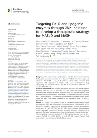
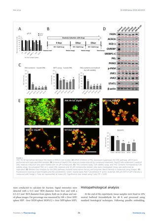
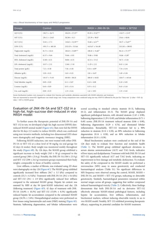
JNK-IN-5A derivatives decrease TAG levels in MASLD cell models. (A) siRNA inhibition of PKL expression suppresses the DNL pathway. qPCR were
performed with triplicated RNA samples. (B) Scheme of HepG2 DNL steatosis model induced by compound treatment. HepG2 cells underwent 1 week of
DNL steatosis induction and were treated with 10 µM compounds. (C) TAG contents assay, Cell viability assay, and TAG contents normalized by Cell
viability assay. Accumulated TAG on 1-week DNL steatosis and 10 µM compounds-treated cells were investigated. Assay was performed as
triplicated. (D) Western blot analysis for the DNL pathway involved transcription factors and enzymes. Band intensity is measured using ImageJ. (E)
Fluorescence staining of lipid droplets and the cytoskeleton. Green: neutral lipids, Red: Cytoskeleton (F-actin), Scale bar, 100 µm. ROI of GFP intensity is
measured with Image J. Data are represented as mean±SD. Significance was tested using t-test (*P < 0.05).
Frontiers in Pharmacology frontiersin.org
05
Kim et al. 10.3389/fphar.2026.1823203
4 µm-thick sections were prepared and stained with hematoxylin
and eosin (H&E) for histopathological evaluation under a light
microscope (Nikon Eclipse Ci). Histopathological assessment was
performed based on characteristic morphological features. The
severity of hepatic steatosis was graded separately, considering
both histological activity and fibrosis stage. The activity level was
assessed using the MAFLD Activity Score (NAS), calculated as the
sum of three histological components—steatosis (0–3), lobular
inflammation (0–3), and hepatocellular ballooning (0–2)—
yielding a total score ranging from 0 to 8, as described by
Kleiner et al. (Kleiner et al., 2005).
Micronucleus assay in bone marrow smears
Femur bones were isolated from sacrificed rats, and surrounding
muscle tissue was removed. Bone marrow was flushed using a sterile
syringe with 0.5 mL FBS and 0.5 mL DMEM into Falcon tubes. The
suspension was centrifuged at 2000 rpm for 5 min, and the
supernatant was discarded. The pellet was resuspended in a drop
of FBS, homogenized, and smeared onto slides. After air-drying,
slides were fixed in absolute methanol for 10 min. Staining was
performed using a commercial kit (ChemBio Laboratory Research,
Turkiye) following the manufacturer’s instructions. Slides were
examined under a light microscope (100× oil immersion), and
1,000 polychromatic erythrocytes (PCEs) per slide were scored to
determine the frequency of micronuclei (MN).
Statistical analysis for RNA-seq and
experimental data
Error bars in the graphs are presented as mean ± standard
deviation (SD) for cell-based experiments. Box plots display the
median and interquartile range (IQR) for hematology and plasma
chemistry measurements. For RNA-seq data, P-values were adjusted
using the Benjamini–Hochberg procedure where applicable.
Statistical significance was defined as P < 0.05 (or adjusted P <
0.05) and is indicated by an asterisk (*).
Results
JNK-IN-5A derivatives effectively inhibit the
DNL pathway in a HepG2 steatosis model
A systems biology approach previously identified PKL as one of
the genes strongly associated with MASLD and HCC (Lee et al.,
2017). To explore the functional role of PKL in hepatic steatosis via
DNL, we examined the expression of key genes involved in the DNL
pathway following siRNA-mediated PKL knockdown in the
HepG2 in vitro steatosis model (Figures 1A,B). PKL knockdown
significantly reduced the expression of PKL (9.5%), ChREBP
(14.4%), FASN (63%). While not statistically significant, the level
of ACACA trended downward by approximately 51.6%.
Building on these findings, we identified JNK-IN-5A as a small
molecule that reduces PKL protein levels in HepG2 cells (Kim et al.,
2023; Zhang et al., 2022). To enhance therapeutic efficacy, we
synthesized a series of JNK-IN-5A derivatives and performed
screening at HepG2 DNL steatosis model with 10 µM (Iqbal
et al., 2024). We identified four compounds (SET-151, SET-152,
SET-162, and SET-130) with more potent anti-steatotic activity. We
evaluated these compounds in the 1-week HepG2 DNL steatosis
model (Figure 1B). Treatment with 10 μM of each compound
significantly reduced TAG accumulation (32.9% for JNK-IN-5A,
11.5% for SET-151, 10.7% for SET-152, 12.3% for SET-162, and
16.7% for SET-130). After normalization to cell viability, TAG
reductions were 58.4% for JNK-IN-5A, 18.6% for SET-151, 24.5%
for SET-152, 25.1% for SET-162, and 26.1% for SET-130
(Figure 1C). Western blot analysis revealed that all compounds
significantly decreased the expression of key DNL pathway enzymes,
including fatty acid synthase (FASN), acetyl-CoA carboxylase
(ACACA), and stearoyl-CoA desaturase 1 (SCD-1), which
catalyses monounsaturated fatty acid (MUFA) production and
promotes hepatic TAG storage and VLDL secretion (Figure 1D).
Additionally, the expression of DNL-regulating transcription
factors, ChREBP and SREBP-1c, was markedly reduced in all
treatment groups.
Ito et al. (2013) suggest that JNK2 plays a dominant role over
JNK1 in regulating SREBP-1c activation. Consistent with this, JNK-IN-
5A and its derivatives did not alter JNK1 protein expression, except for
a modest reduction by SET-151 (0.65-fold). Interestingly, SET-151
(0.57-fold), SET-152 (0.66-fold), and SET-162 (0.57-fold) exhibited
stronger inhibitory effects on JNK2 expression compared to SET-130
(0.81-fold) and JNK-IN-5A (0.75-fold). Liao et al. (2023), introduced
Open MoA, a pipeline to reveal the mechanism of action (MOA) based
on network topology and hierarchy. The potential pathway of how
JNK-IN-5A regulate PKLR expression was revealed in their study. We
employed the Open MoA here to predict two MoAs for all compounds:
the ‘JNK2–JUN–SREBP1-c’ and ‘JNK2–JUN–MXI1–ChREBP’
signalling pathways (Supplementary Figure S2). Notably, SET-151,
SET-152, and SET-162 demonstrated lower penalty scores (1-
confidence score) for the MXI1–ChREBP association than SET-130
and JNK-IN-5A, consistent with stronger downregulation of MLXIPL,
the gene encoding ChREBP. Similarly, SREBF1, which encodes
SREBP-1c, was significantly downregulated following treatment
with all compounds, with high-confidence predictions for all
interactions within the MoA network. Signal transducer and
activator of transcription 1 (STAT1), a transcription factor
implicated in the development of MASLD and MASH (Grohmann
et al., 2018), is part of the JNK–STAT1–PKL regulatory axis (Liao et al.,
2023). We also observed interaction between compounds JNK2 using
cellular thermal shift assay (CETSA). We give gradient heat shock to
20 µM compounds treated HepG2 cell lysate. SET151, SET152, and
SET162 treated cell lysate showed statistical significant heat resistance
at 49.5 °C for 5 min (Supplementary Figure S2C). To check cellular
permeability and binding affinity between compounds and JNK2, we
treated 20 µM compounds to HepG2 live cells for 2h and give heat
shock 50 °C. SET151 (1.47 times), SET152 (1.4 times), and SET162
(1.37 times) showed statistical significant heat shock resistance again
(Supplementary Figure S2D). Our data showed that while JNK-IN-5A
and its derivatives robustly reduced JNK2 and PKL expression, only
the derivatives interact with JNK2 and substantially decreased
STAT1 protein levels, indicating enhanced efficacy over JNK-IN-5A.
Increased glycolysis is a characteristic metabolic change in liver
steatosis (Lu et al., 2021). Nuclear-localized PKM2, particularly in its
monomeric or dimeric form, promotes glycolysis (Yang and Lu,
2013). Phosphorylation at Tyr105 prevents PKM2 tetramer
Frontiers in Pharmacology frontiersin.org
06
Kim et al. 10.3389/fphar.2026.1823203
formation, enhancing its glycolytic activity (Hitosugi et al., 2009).
While JNK-IN-5A and its derivatives did not affect total
PKM2 protein levels in the HepG2 DNL steatosis model, Tyr105-
phosphorylated PKM2 levels were dramatically reduced following
treatment with all compounds.
Based on these findings, we selected SET-151, SET-152, and
SET-162 as the most potent derivatives, demonstrating superior
therapeutic efficacy and distinct mechanisms compared to the
reference compound JNK-IN-5A (Figure 1E). Their anti-steatotic
effects were confirmed by BODIPY™ 493/503 staining for neutral
lipids. In the BODIPY assay, SET-151 (52.1%), SET-152 (45.7%),
and SET-162 (55.2%) significantly reduced neutral lipid
accumulation compared to JNK-IN-5A (71.7%) (Figure 1E).
Together, these results demonstrate that SET-151, SET-152 and
SET-162 exhibit stronger anti-steatotic activity than JNK-IN-5A,
primarily through the inhibition of the DNL pathway and the
regulation of key metabolic and transcriptional pathways
involved in MASLD progression.
Comparison of resmetirom and JNK-IN-5A
derivatives in the HepG2 DNL
steatosis model
Resmetirom activates hepatic THR-β, promoting systemic lipid
clearance by enhancing bile acid synthesis and fat oxidation
(Harrison et al., 2023; Harrison et al., 2024). In this study, we
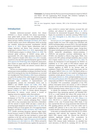
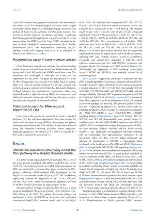
FIGURE 2
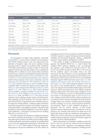
JNK-IN-5A derivatives demonstrated comparable therapeutic efficacy to resmetirom in MASLD cell models. (A) TAG contents assay, and TAG
contents normalized by protein quantification. Accumulated TAG on 1-week DNL steatosis with resmetirom 10 µM, 5 µM, and 5 µM compound-treated
cells was investigated. Assay was performed as triplicated. (B) Comparison of resmetirom and JNK-IN-5A derivatives for the DNL pathway, involving
transcription factor and enzyme protein expression levels via Western blot analysis. Band intensity is measured using ImageJ. (C) 5 µM resmetirom
and JNK-IN-5A derivatives lipid droplet and cytoskeleton fluorescence staining. Green: neutral lipids, Red: Cytoskeleton (F-actin), Scale bar, 100 µm.
5 random ROI of GFP intensities are measured with Image J. Data are represented as mean±SD. Significance was tested using t-test (*P < 0.05).
Frontiers in Pharmacology frontiersin.org
07
Kim et al. 10.3389/fphar.2026.1823203
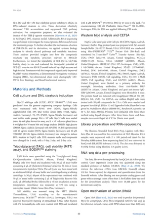
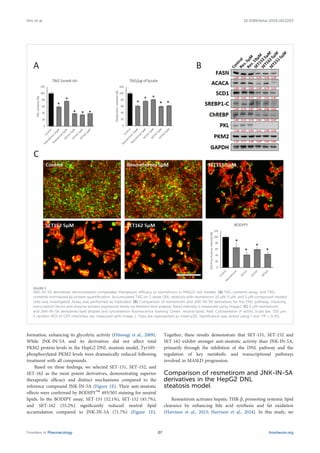
FIGURE 3
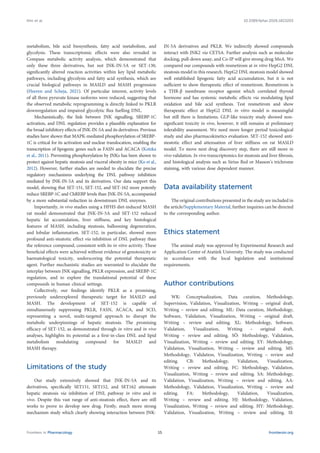
JNK-IN-5A derivatives exhibited distinct transcriptional profiles compared to the reference compound. (A) PCA plot showing sample distribution of SET151,
SET152, SET162, SET130, JNK-IN-5A, and Control groups (both n = 4) based on principal component analysis of RNA-seq gene expression profile. (B) Bar plot
showing the numbers of differentially expressed genes (DEGs) in each treatment group. (C) Heatmaps of the Jaccard index showing the similarity of up- and
downregulated genes across different treatment groups. (D) Heatmap showing the differential expression (presented as log2 fold change) of 40 genes
derived from the MASLD-associated module in different treatment groups. p Represent significantly differentially expressed genes (DEGs) in each group (cut-off
p values for DEGs (p < 0.05). (E) Dot plot showing the GSOA results conducted based on the biological process category from the Gene Ontology (GO) dataset.
The size of the dot indicates the gene ratio, i.e., the DEGs assigned to the corresponding pathway relative to the total analysed DEGs., and the dot’s colour
indicates the adjusted p value. (F) Bar plot showing the normalized enrichment score (NES) of metabolic dysfunction–associated steatotic liver disease (MASLD)
and metabolic dysfunction–associated steatohepatitis (MASH) related genes. RNA sequencing samples were prepared as quadruple.
Frontiers in Pharmacology frontiersin.org
08
Kim et al. 10.3389/fphar.2026.1823203
compared the therapeutic effects of resmetirom and JNK-IN-5A
derivatives in the HepG2 DNL steatosis model (Figure 2).
Resmetirom treatment resulted in a dose-dependent reduction in
intracellular TAG levels (Figure 2A). Specifically, 10 µM (62.4%) and
5 µM (78.2%) of resmetirom reduced the TAG content. SET-151
(42.8%), SET-152 (38.3%), and SET-162 (43.6%), also significantly
reduced TAG content at 5 μM. When normalized to total protein
content, 10 µM (61.2%) and 5 µM (75.7%) of resmetirom, 5 µM
(80.5%) of SET151, 5 µM (59.9%) of SET152 and 5 µM (61.8%) of
SET162 reduced the TAG levels compared to the untreated
control group.
Western blot analysis of the DNL pathway proteins further
demonstrated the superior inhibitory effects of JNK-IN-5A
derivatives compared to resmetirom (Figure 2B). FASN (0.66 &
0.71), ACACA (0.84 & 0.80) and SCD1 (0.63 & 0.73) protein levels
were decreased in the 5µM and 10µ resmetirom treated group. In
contrast, treatment with 5 μM of SET-151, SET-152, and SET-162
resulted in more pronounced reductions: FASN (0.40, 0.38, 0.45),
ACACA (0.22, 0.38, 0.79), and SCD1 (0.36, 0.42, 0.51), respectively.
Moreover, the JNK-IN-5A derivatives showed more potent
suppression of the key DNL transcription factors SREBP1-c and
ChREBP compared to resmetirom. SREBP1-c expression was
reduced by resmetirom at 5 μM (0.74) and 10 μM (0.87),
whereas SET-151, SET-152, and SET-162 further decreased
SREBP1-c expression (0.49, 0.68, and 0.86). ChREBP expression
(0.29, 0.42, and 0.42) was also markedly reduced by JNK-IN-5A
derivatives for SET-151, SET-152, and SET-162, compared to
reductions observed with resmetirom at 5 μM (0.72) and 10 μM
(0.75). Importantly, JNK-IN-5A derivatives almost completely
suppressed PKL expression, reducing it to undetectable levels. In
contrast, resmetirom-treated cells maintained 87% and 93% of PKL
expression at 5 μM and 10 μM, respectively. PKM2 expression was
moderately reduced across all treatment groups: resmetirom 5 μM
(0.71), resmetirom 10 μM (0.85), SET-151 (0.86), SET-152 (0.80),
and SET-162 (0.80).
Lipid staining further confirmed these findings. BODIPY™493/
503 staining demonstrated that 5 μM resmetirom reduced neutral
lipid accumulation to 65.9% of the control level, whereas SET-151,
SET-152, and SET-162 showed more pronounced reductions to
41.8%, 59.5%, and 43.6%, respectively (Figure 2C). Collectively,
these results indicate that JNK-IN-5A derivatives exhibit stronger
inhibitory effects on the DNL steatosis pathway and intracellular
lipid accumulation compared to resmetirom at HepG2 DNL in vitro
model. It highlighting JNK-IN-5A derivatives as a DNL pathway-
targeting therapeutics for MASLD.
JNK-IN-5A derivatives exhibit distinct
transcriptional profiles
To investigate the transcriptional differences induced by JNK-
IN-5A and its novel derivatives, we performed global RNA
sequencing (RNA-seq) analysis on treated HepG2 cells. Principal
Component Analysis (PCA) revealed three distinct gene
expression clusters corresponding to the treatment groups
(Figure 3A). SET-130 clustered closely with JNK-IN-5A, whereas
SET-151, SET-152, and SET-162 formed a separate, tightly grouped
cluster. All treatment groups were distinct from the untreated
control group.
In terms of global transcriptional impact, SET-151, SET-152,
and SET-162 induced substantially higher numbers of differentially
expressed genes (DEGs) compared to SET-130 and JNK-IN-5A
(Figure 3B). Jaccard index analysis of the DEGs further
confirmed that SET-151, SET-152, and SET-162 shared a high
degree of similarity in both upregulated and downregulated gene
sets (Figure 3C), suggesting a consistent and distinct mechanism of
action among these three derivatives. To assess the functional
consequences of these transcriptional changes, we first examined
the expression of a previously defined MASLD-associated gene
module (n = 40) (Yang et al., 2021). All compounds, including
JNK-IN-5A and its derivatives, significantly downregulated the
expression of genes within this MASLD module (Figure 3D).
Notably, SET-151, SET-152, and SET-162 produced a more
pronounced downregulation effect, with a marked reduction in
PKLR expression.
Gene Set Overrepresentation Analysis (GSOA) revealed that the
downregulated genes in all treatment groups were enriched in
pathways related to lipid metabolism and glycolysis. Importantly,
SET-151, SET-152, and SET-162 uniquely suppressed genes
associated with fatty acid metabolism and primary bile acid
biosynthesis, pathways highly relevant to MASLD pathogenesis
(Figure 3E). To further explore the clinical relevance of these
findings, we retrieved MASLD- and MASH-associated genes
from the DisGeNET database (773 and 316 genes, respectively),
(Piñero et al., 2015), and performed Gene Set Enrichment Analysis
(GSEA). The results demonstrated that all treatment groups reduced
the enrichment scores of MASLD-associated genes; however, SET-
151, SET-152, and SET-162 were significantly more effective in this
regard (Figure 3F). Interestingly, a reduction in the enrichment
scores of MASH-related genes was observed exclusively in cells
treated with SET-151, SET-152, and SET-162, but not in cells treated
with JNK-IN-5A or SET-130 (Figure 3F). Collectively, these
transcriptomic analyses demonstrate that SET-151, SET-152, and
SET-162 exhibit distinct and stronger transcriptional modulation
compared to the parental compound JNK-IN-5A, supporting their
superior therapeutic potential for MASLD and MASH.
JNK-IN-5A derivatives exhibit distinct
metabolic profiles
To comprehensively characterize the metabolic alterations
induced by JNK-IN-5A and its derivatives, we performed
Compass metabolic activity analysis (Wagner et al., 2021) based
on global transcriptomic profiling. The analysis was conducted
using the RECON3D human metabolic network model, which
encompasses 10,600 reactions, 5,835 metabolites and 2,248 genes.
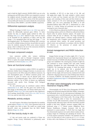
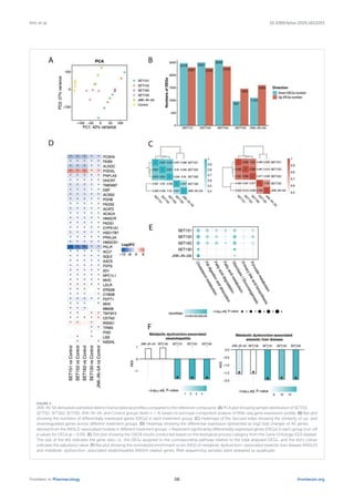
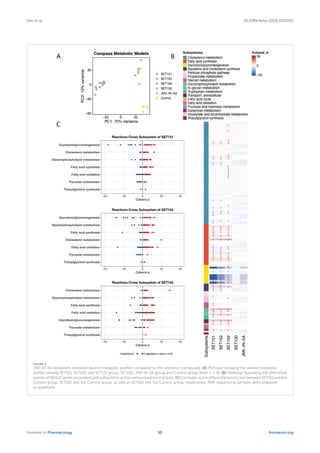
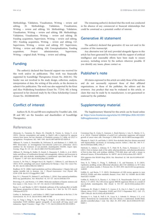
PCA of Compass output revealed three distinct metabolic clusters
corresponding to the treatment groups: (1) SET-151, SET-152, and
SET-162; (2) SET-130 and JNK-IN-5A; and (3) the untreated control
group (Figure 4A). These results highlight the distinct metabolic
states induced by the three most potent JNK-IN-5A derivatives
compared to both the parental compound and the control.
We next performed differential reaction activity analysis to
quantify metabolic alterations between the treatment and control
groups. Cohen’s d effect sizes were calculated for each reaction, and
significance was determined using Benjamini–Hochberg adjusted
Wilcoxon rank-sum tests (p < 0.05), excluding reactions classified
Frontiers in Pharmacology frontiersin.org
09
Kim et al. 10.3389/fphar.2026.1823203
FIGURE 4
JNK-IN-5A derivatives exhibited distinct metabolic profiles compared to the reference compound. (A) PCA plot showing the distinct metabolic
profiles among SET151, SET152, and SET172 group, SET130, JNK-IN-5A group and Control group (both n = 4). (B) Heatmap illustrating the differential
activity of MASLD genes associated with subsystems across various treatment groups. (C) Compass-score differential activity test between SET151 and the
Control group, SET152 and the Control group, as well as SET162 and the Control group, respectively. RNA sequencing samples were prepared
as quadruple.
Frontiers in Pharmacology frontiersin.org
10
Kim et al. 10.3389/fphar.2026.1823203
under the “Miscellaneous” subsystem. The SET-151, SET-152, and
SET-162 groups exhibited 9,214, 9,580, and 9,945 significantly
altered reactions, respectively. In contrast, no significantly
changed reactions were detected in the SET-130 and JNK-IN-5A
groups. As shown in Figure 4B, reactions related to lipid
metabolism, including glycolysis, cholesterol metabolism, fatty
acid synthesis, and pyruvate metabolism, were significantly
altered in response to SET-151, SET-152, and SET-162 treatment,
compared to the control group. Notably, SET-130 and JNK-IN-5A
treatments did not induce significant changes in these key metabolic
pathways (Supplementary Figure S1).
To further assess disease-relevant metabolic changes, we
extracted 115 MASLD-associated reactions from the Compass
results. The majority of these reactions were involved in
cholesterol metabolism, fatty acid synthesis, and glycolysis/
gluconeogenesis pathways (Figure 4C). Remarkably, activity
levels of the three pyruvate kinase isoforms encoded by the
PKLR gene were significantly reduced following SET-151, SET-
152, and SET-162 treatment. This finding suggests that these
JNK-IN-5A derivatives effectively disrupt pyruvate kinase-
regulated glycolysis, contributing to their therapeutic effects
in MASLD. Collectively, these metabolic analyses demonstrate
that SET-151, SET-152, and SET-162 induce distinct and robust
metabolic remodelling compared to JNK-IN-5A, with
pronounced inhibition of lipids and pyruvate metabolism,
supporting their superior potential as therapeutic candidates
for MASLD. Among these hit compounds, SET-152 exhibited
the strongest downregulation of glycolysis and cholesterol
metabolism pathways. This suggests that SET-152 selected for
the further in vivo stud.
GLP (good laboratory Practice)-like toxicity
assessment of SET-152 in rats demonstrates
its safety
After testing the efficacy of SET-152 in an in vitro model of
steatosis and revealing its MoA based on global transcriptomics and
systems biology approach, we conducted a GLP-like 7-day oral
tolerability study in Wistar rats to evaluate its safety at three dose
levels (30, 100, and 300 mg/kg). The study included a vehicle control
group (4 males and four females) and three dose groups, each
consisting of three male and three female rats. The oral gavage
dosing was administered once daily in the morning for seven
consecutive days.
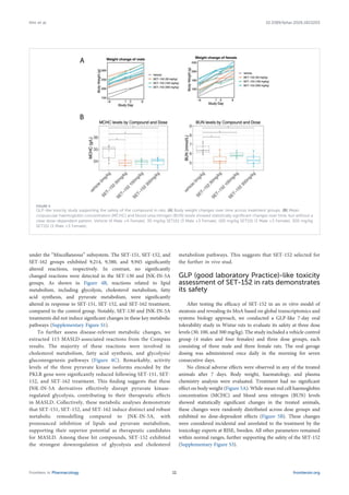
No clinical adverse effects were observed in any of the treated
animals after 7 days. Body weight, haematology, and plasma
chemistry analysis were evaluated. Treatment had no significant
effect on body weight (Figure 5A). While mean red cell haemoglobin
concentration (MCHC) and blood urea nitrogen (BUN) levels
showed statistically significant changes in the treated animals,
these changes were randomly distributed across dose groups and
exhibited no dose-dependent effects (Figure 5B). These changes
were considered incidental and unrelated to the treatment by the
toxicology experts at RISE, Sweden. All other parameters remained
within normal ranges, further supporting the safety of the SET-152
(Supplementary Figure S3).
FIGURE 5
GLP-like toxicity study supporting the safety of the compound in rats. (A) Body weight changes over time across treatment groups. (B) Mean
corpuscular haemoglobin concentration (MCHC) and blood urea nitrogen (BUN) levels showed statistically significant changes over time, but without a
clear dose-dependent pattern. Vehicle (4 Male +4 Female), 30 mg/kg SET151 (3 Male +3 Female), 100 mg/kg SET151 (3 Male +3 Female), 300 mg/kg
SET151 (3 Male +3 Female).
Frontiers in Pharmacology frontiersin.org
11
Kim et al. 10.3389/fphar.2026.1823203
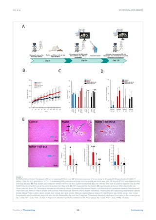
FIGURE 6
SET152 exhibited distinct therapeutic efficacy in reversing MASH in vivo. (A) Schematic overview of in vivo study. 6–8 weeks 29 SD rats (Control 8, MASH 7,
MASH + JNK-IN-5A 7, and MASH + SET152 7) underwent MASH induction via a high-sucrose and fat diet for 84 days. JNK-5A-IN and SET152 were treated for the
following 42 days. (B) Body weight was measured weekly over the 126 days experimental period. (C) Liver stiffness (kPa) was recorded at baseline (Day 0), after
MASH induction (Day 84), and at the end of drug treatment (Day 126). (D) MRI measured liver fat content. (E) Haematoxylin and eosin (H&E) staining for liver
tissue collected at day 126. Histological features are indicated as follows: portal area (blue arrow), hepatic cord (blue bracket), panlobular steatosis (black bracket),
macrovesicular steatosis-macro droplet (black arrow), macrovesicular steatosis-micro droplet (red arrow), hepatocytes with ballooning degeneration (yellow
arrow), lobular inflammation (green arrow), and central vein (gray arrow). Data are represented as mean±SD. Significance was tested using t-test. Group
comparisons were conducted using one-way ANOVA followed by the post hoc Tukey test. * Represent statistical significance compared to the control group
(*p < 0.05, **p < 0.01, ***p < 0.001). # Represent statistical significance relative to the MASH group (#p < 0.05, ##p < 0.01, ###p < 0.001).
Frontiers in Pharmacology frontiersin.org
12
Kim et al. 10.3389/fphar.2026.1823203
Evaluation of JNK-IN-5A and SET-152 in a
high-fat, high-sucrose diet-induced in vivo
MASH model
To further assess the therapeutic potential of JNK-IN-5A and
SET-152 in rats, we developed a high-fat, high-sucrose (HFHS) diet-
induced MASH animal model (Figure 6A). Rats were fed the HFHS
diet for 84 days (12 weeks) to induce MASH, which was confirmed
using non-invasive methods, including two-dimensional (2D) shear
wave elastography and magnetic resonance imaging (MRI).
Following MASH induction, rats were treated with either JNK-
IN-5A or SET-152 at a dose level of 30 mg/kg via oral gavage for
42 days (6 weeks). Body weight was monitored weekly throughout
the study (Figure 6B). By 126 days, the MASH group exhibited a
significant increase in body weight (343 ± 50 g) compared to the
control group (300 ± 25.6 g). Notably, the JNK-IN-5A (304 ± 23.9 g)
and SET-152 (298 ± 24.3 g) treatment groups maintained their body
weights comparable to those of healthy controls.
Liver stiffness, a marker of fibrosis, was evaluated using 2D shear
wave elastography on day 126 (Figure 6C). The MASH group exhibited
significantly increased liver stiffness (36.7 ± 3.5 kPa) compared to
controls (25.2 ± 4.3 kPa). Treatment with JNK-IN-5A (29.2 ± 0.4 kPa)
and SET-152 (29.1 ± 2.9 kPa) significantly reduced liver stiffness
compared to the untreated MASH group. Hepatic fat content was
assessed by MRI at day 84 (post-MASH induction) and day 126
(following treatment) (Figure 6D). 42 days of treatment with JNK-
IN-5A (16.8% ± 10.3%) and SET-152 (15.0% ± 8.5%) significantly
reduced hepatic fat accumulation compared to the untreated MASH
group (24.5% ± 3.4%). Histopathological analyses were performed on
liver tissues using haematoxylin and eosin (H&E) staining (Figure 6E).
Steatosis, ballooning degeneration, and lobular inflammation were
scored according to standard criteria: steatosis (0–3), ballooning
(0–2), and inflammation (0–3). The MASH group displayed
significant pathological features, with elevated steatosis (1.43 ± 0.98),
ballooning degeneration (1.29 ± 0.49), and lobular inflammation (0.71 ±
0.49) scores. JNK-IN-5A treatment reduced steatosis (0.86 ± 0.90),
ballooning degeneration (0.29 ± 0.76), and eliminated lobular
inflammation. Remarkably, SET-152 treatment led to a 90%
reduction in steatosis (0.14 ± 0.38), an 89% reduction in ballooning
degeneration (0.14 ± 0.38), and an 80% reduction in lobular
inflammation (0.14 ± 0.38).
Blood biochemistry analyses were conducted at the end of the
126 days study to evaluate liver function and metabolic health
(Table 1). The MASH group exhibited significant elevations in
serum alanine aminotransferase (ALT) and TAG levels, indicative
of liver injury and dyslipidaemia. Treatment with both JNK-IN-5A and
SET-152 significantly reduced ALT and TAG levels, indicating an
improvement in liver damage and metabolic dysfunction. To evaluate
the safety of the compound in the MASH model, we performed a
micronucleus (MN) assay to assess genotoxicity and conducted
haematological analyses (Table S1). No significant differences in
MN frequency were observed among the control, MASH, MASH +
JNK-IN-5A, and MASH + SET-152 groups, indicating no detectable
genotoxicity. Similarly, haematological parameters remained within
normal ranges across all groups, suggesting that neither compound
induced haematological toxicity (Table 2). Collectively, these findings
demonstrate that both JNK-IN-5A and its derivative SET-152
effectively attenuate MASH-related pathological features, including
hepatic fat accumulation, liver stiffness, inflammation, and
biochemical markers, without inducing observable toxicity in an in
vivo MASH model. Notably, SET-152 exhibited promising therapeutic
efficacy, supporting its potential candidate for MASH treatment.
TABLE 1 Blood biochemistry of liver injury and MASLD progression.
Groups Control MASH MASH + JNK-IN-5A MASH + SET152
ALP (U/L) 158.71 ± 26.71 104.29 ± 27,35** 93.29 ± 27,67*** 118.57 ± 21.97
AST (U/L) 236.11 ± 24.65 262.86 ± 41.5 225.39 ± 90.87 216.6 ± 40.84
ALT (U/L) 52.01 ± 17.5 118.63 ± 32,39*** 32.68 ± 6,31###
38.37 ± 15,65###
LDH (U/L) 1951.71 ± 485.58 2523.29 ± 515.64 1623.67 ± 541.00 2212.83 ± 389.82
Triglyceride (mg/dL) 81.71 ± 18.22 184.43 ± 53,89*** 108.43 ± 31,66##
96.14 ± 37,73###
Total cholesterol (mg/dL) 61.57 ± 9.64 76.86 ± 4.91 78.86 ± 19.80 54.86 ± 8,30#
HDL cholesterol (mg/dL) 41.00 ± 6.51 38.86 ± 6.31 42.14 ± 13.12 36.71 ± 8.18
LDL cholesterol (mg/dL) 10.57 ± 2.51 12.86 ± 3.18 11.29 ± 2.21 8.43 ± 2.44
Total protein (g/dL) 7.21 ± 0.50 7.56 ± 0.38 8.16 ± 0,99* 7.54 ± 0.54
Albumin (g/dL) 3.20 ± 0.21 3.63 ± 0.22 3.81 ± 0,45* 3.49 ± 0.30
Glucose (mg/dL) 145.57 ± 31.65 205.00 ± 88.24 208.43 ± 64.02 228.57 ± 165.46
Total bilirubin (mg/dL) 0.09 ± 0.05 0.11 ± 0.07 0.10 ± 0.08 0.10 ± 0.05
Creatine (mg/dL) 0.44 ± 0.03 0.53 ± 0.14 0.53 ± 0.12 0.43 ± 0.16
Uric acid (mg/dL) 0.98 ± 0.35 1.33 ± 0.24 1.61 ± 0.93 1.35 ± 0.45
Serum biochemical markers were measured to evaluate liver function and metabolic alterations associated with MASH induction and treatment.
Data are represented as mean±SD. Significance was tested using t-test. Group comparisons were conducted using one-way ANOVA followed by the post hoc Tukey test. *p value < 0.05, #p
value < 0.05 to MASH group.
Frontiers in Pharmacology frontiersin.org
13
Kim et al. 10.3389/fphar.2026.1823203
Discussion
The dysregulation of hepatic lipid metabolism, particularly
increased DNL, is a central feature of MASLD and its progressive
form, MASH. Multiple enzymes in the DNL pathway have been
identified as therapeutic targets, with several candidates showing
promising results in clinical and preclinical studies. Denifanstat
(TVB-2640), a FASN inhibitor currently in a Phase 2 b clinical trial,
suppresses FASN activity and reduces hepatic TAG accumulation by
inhibiting DNL. In addition to its anti-steatotic effects, Denifanstat
has been shown to attenuate steatohepatitis through the deactivation
of hepatic stellate cells (Loomba et al., 2021a; Syed-Abdul et al., 2020;
O’Farrell et al., 2022) and has demonstrated anti-cancer activity in
models of lung carcinoma, breast cancer, astrocytoma, and colon
cancer (Fries et al., 2023; Kelly et al., 2023; Falchook et al., 2021; Cao
et al., 2022). Similarly, Firsocostat, an ACACA inhibitor that has
completed a Phase 2 clinical trial, effectively reduces hepatic DNL
(Kirby et al., 2021) and improves liver fibrosis (Loomba et al., 2021b;
Alkhouri et al., 2022; Alkhouri et al., 2020). Several preclinical
ACACA inhibitors, including CP-640186 (Corominas-Faja et al.,
2014; Hess et al., 2010), Soraphen A (Corominas-Faja et al., 2014;
Schreurs et al., 2009), and TOFA (Li et al., 2013), have also been
reported to reduce weight gain, hepatic steatosis, and inflammation,
while exhibiting anti-cancer properties. The anti-cancer effects of
the FASN and ACACA-targeting molecules are attributed mainly to
the suppression of lipid synthesis, a metabolic pathway essential for
both energy storage and the production of key cellular components,
including membranes and signalling molecules. Importantly, lipid
biosynthesis is often markedly upregulated in cancer cells to support
rapid proliferation, a phenomenon associated with the Warburg
effect (Burns and Manda, 2017).
Our study expands on this therapeutic paradigm by identifying
PKL isoform, encoded by the PKLR gene, as a novel, highly disease-
associated target for MASLD and MASH. Systems biology analysis,
combined with global transcriptomic profiling, has identified PKLR
as one of the most significantly associated genes with MASLD and
HCC (Lee et al., 2017). PKLR catalyses the final step of glycolysis,
linking glucose and pyruvate metabolism to the DNL pathway by
providing precursors for fatty acid synthesis. Notably, the inhibition
of PKLR is expected to suppress both glycolysis-derived substrate
availability and downstream lipogenesis, offering a multifaceted
approach to target metabolic dysregulation in MASLD.
In this context, we applied a computational drug repurposing
approach and identified JNK-IN-5A as a small molecule that
modulates PKLR expression. Building on this, we synthesized
and evaluated a series of JNK-IN-5A derivatives (SET-151, SET-
152, SET-162, and SET-130), three of which (SET-151, SET-152, and
SET-162) exhibited superior anti-steatotic efficacy. Our data
demonstrate that these derivatives not only downregulate PKLR
expression but also suppress the expression of critical enzymes in the
DNL pathway (FASN, ACACA) and SCD. SCD is a well-established
therapeutic target for MASLD and certain cancers (Sun et al., 2024;
Jeyakumar and Vajreswari, 2022; Min and Kim, 2023; Sen et al.,
2023; Katoh et al., 2022). Pharmacological inhibition of SCD has
been shown to exert beneficial effects in metabolic diseases and
cancer. For example, the SCD inhibitor E6446 suppresses both SCD
expression and the transcription factor ATF3, leading to impaired
adipogenic differentiation and reduced hepatic lipogenesis (Wang
et al., 2023). Furthermore, Ascenzi et al. demonstrated the pivotal
role of SCD as a key therapeutic target in cancer (Ascenzi et al.,
2021). Inhibition of SCD reduces MUFA synthesis, resulting in the
accumulation of SFAs. Elevated intracellular SFA levels are known
to trigger cellular stress responses, including enhanced autophagy.
Excessive autophagy can, in turn, promote apoptosis, lipotoxicity,
and ferroptosis. Based on these established mechanisms, we
hypothesize that the marked reduction of SCD protein
expression observed following treatment with JNK-IN-5A and its
derivatives may contribute to the reduced cell viability seen in the
HepG2 DNL steatosis model. The simultaneous downregulation of
PKLR, FASN, ACACA, and SCD by our compounds offers a unique
multiple-targeting strategy that disrupts both substrate supply and
enzymatic execution of lipid biosynthesis.
Further transcriptomic and metabolic modelling revealed that
SET-151, SET-152, and SET-162 induced broad transcriptional
changes beyond PKLR suppression, downregulating pathways
essential for MASLD and MASH progression, including pyruvate
TABLE 2 Blood haematology following MASH induction and treatment.
Groups Control MASH MASH + JNK-IN-5A MASH + SET152
PCT (%) 0.68 ± 0.12 0.71 ± 0.10 0.77 ± 0.14 0.72 ± 0.16
PLT (10
^
3/μL) 845.57 ± 158.62 871.29 ± 128.53 947.71 ± 123.89 904.86 ± 140.16
MCHC (g/dL) 28.24 ± 0.96 28.97 ± 0.69 29.27 ± 1.14 29.71 ± 1.11
MCH (fL) 17.41 ± 0.60 17.84 ± 0.59 18.04 ± 0.48 17.81 ± 0.60
MCV (fL) 61.67 ± 2.07 61.51 ± 1.66 61.69 ± 2.87 60.11 ± 2.25
HCT (%) 54.40 ± 2.67 55.51 ± 2.31 55.41 ± 5.42 51.99 ± 3.93
HGB (g/dL) 15.19 ± 1.27 16.09 ± 0.76 16.20 ± 1.51 15.40 ± 1.27
RBC (10
^
6/μL) 8.83 ± 0.60 9.04 ± 0.52 8.98 ± 0.65 8.66 ± 0.73
WBC (10
^
3/μL) 5.81 ± 2.27 4.70 ± 1.50 4.27 ± 1.67 5.75 ± 3.07
Haematological analysis was performed to evaluate systemic effects of MASH, and subsequent treatment with JNK-IN-5A, or SET152. Parameters include platelet count (PLT), procalcitonin
(PCT), mean corpuscular haemoglobin concentration (MCHC), mean corpuscular haemoglobin (MCH), mean corpuscular volume (MCV), haematocrit (HCT), haemoglobin (HGB), red blood
cell count (RBC), and white blood cell count (WBC).
Data are represented as mean±SD., Group comparisons were conducted using one-way ANOVA, followed by the post hoc Tukey test.
Frontiers in Pharmacology frontiersin.org
14
Kim et al. 10.3389/fphar.2026.1823203
metabolism, bile acid biosynthesis, fatty acid metabolism, and
glycolysis. These transcriptomic effects were also revealed in
Compass metabolic activity analysis, which demonstrated that
only these three derivatives, but not JNK-IN-5A or SET-130,
significantly altered reaction activities within key lipid metabolic
pathways, including glycolysis and fatty acid synthesis, which are
crucial biological pathways in MASLD and MASH progression
(Heeren and Scheja, 2021). Of particular interest, activity levels
of all three pyruvate kinase isoforms were reduced, suggesting that
the observed metabolic reprogramming is directly linked to PKLR
downregulation and impaired glycolytic flux fuelling DNL.
Mechanistically, the link between JNK signalling, SREBP-1C
activation, and DNL regulation provides a plausible explanation for
the broad inhibitory effects of JNK-IN-5A and its derivatives. Previous
studies have shown that MAPK-mediated phosphorylation of SREBP-
1C is critical for its activation and nuclear translocation, enabling the
transcription of lipogenic genes such as FASN and ACACA (Kotzka
et al., 2011). Preventing phosphorylation by JNKs has been shown to
protect against hepatic steatosis and visceral obesity in mice (Ko et al.,
2012). However, further studies are needed to elucidate the precise
regulatory mechanisms underlying the DNL pathway inhibition
mediated by JNK-IN-5A and its derivatives. Our data support this
model, showing that SET-151, SET-152, and SET-162 more potently
reduce SREBP-1C and ChREBP levels than JNK-IN-5A, accompanied
by a more substantial reduction in downstream DNL enzymes.
Importantly, in vivo studies using a HFHS diet-induced MASH
rat model demonstrated that JNK-IN-5A and SET-152 reduced
hepatic fat accumulation, liver stiffness, and key histological
features of MASH, including steatosis, ballooning degeneration,
and lobular inflammation. SET-152, in particular, showed more
profound anti-steatotic effect via inhibition of DNL pathway than
the reference compound, consistent with its in vitro activity. These
beneficial effects were achieved without evidence of genotoxicity or
haematological toxicity, underscoring the potential therapeutic
agent. Further mechanistic studies are warranted to elucidate the
interplay between JNK signalling, PKLR expression, and SREBP-1C
regulation, and to explore the translational potential of these
compounds in human clinical settings.
Collectively, our findings identify PKLR as a promising,
previously underexplored therapeutic target for MASLD and
MASH. The development of SET-152 is capable of
simultaneously suppressing PKLR, FASN, ACACA, and SCD,
representing a novel, multi-targeted approach to disrupt the
metabolic underpinnings of hepatic steatosis. The promising
efficacy of SET-152, as demonstrated through in vitro and in vivo
analyses, highlights its potential as a first-in-class DNL and lipid
metabolism modulating compound for MASLD and
MASH therapy.
Limitations of the study
Our study extensively showed that JNK-IN-5A and its
derivatives, specifically SET151, SET152, and SET162 attenuate
hepatic steatosis via inhibition of DNL pathway in vitro and in
vivo. Despite this vast range of anti-steatosis effect, there are still
works to prove to develop new drug. Firstly, much more strong
mechanism study which clearly showing interaction between JNK-
IN-5A derivatives and PKLR. We indirectly showed compounds
interact with JNK2 via CETSA. Further analysis such as molecular
docking, pull-down assay, and Co-IP will give strong drug MoA. We
compared our compounds with resmetirom at in vitro HepG2 DNL
steatosis model in this research. HepG2 DNL steatosis model showed
well established lipogenic fatty acid accumulation, but it is not
sufficient to show therapeutic effect of resmetirom. Resmetirom is
a THR-β membrane receptor agonist which correlated thyroid
hormone and has systemic metabolic effects via modulating lipid
oxidation and bile acid synthesis. Test resmetirom and show
therapeutic effect at HepG2 DNL in vitro model is meaningful
but still there is limitations. GLP-like toxicity study showed non-
significant toxicity in vivo, however, it still remains at preliminary
tolerability assessment. We need more longer period toxicological
study and also pharmacokinetics evaluation. SET-152 showed anti-
steatotic effect and attenuation of liver stiffness on rat MASLD
model. To move next drug discovery step, there are still more in
vivo validation. In vivo transcriptomics for steatosis and liver fibrosis,
and histological analysis such as Sirius Red or Masson’s trichrome
staining, with various dose dependent manner.
Data availability statement
The original contributions presented in the study are included in
the article/Supplementary Material, further inquiries can be directed
to the corresponding author.
Ethics statement
The animal study was approved by Experimental Research and
Application Center of Atatürk University. The study was conducted
in accordance with the local legislation and institutional
requirements.
Author contributions
WK: Conceptualization, Data curation, Methodology,
Supervision, Validation, Visualization, Writing – original draft,
Writing – review and editing. ML: Data curation, Methodology,
Software, Validation, Visualization, Writing – original draft,
Writing – review and editing. XL: Methodology, Software,
Validation, Visualization, Writing – original draft,
Writing – review and editing. SÖ: Methodology, Validation,
Visualization, Writing – review and editing. EY: Methodology,
Validation, Visualization, Writing – review and editing. MS:
Methodology, Validation, Visualization, Writing – review and
editing. CB: Methodology, Validation, Visualization,
Writing – review and editing. FC: Methodology, Validation,
Visualization, Writing – review and editing. SA: Methodology,
Validation, Visualization, Writing – review and editing. AA:
Methodology, Validation, Visualization, Writing – review and
editing. FA: Methodology, Validation, Visualization,
Writing – review and editing. HJ: Methodology, Validation,
Visualization, Writing – review and editing. HY: Methodology,
Validation, Visualization, Writing – review and editing. SI:
Frontiers in Pharmacology frontiersin.org
15
Kim et al. 10.3389/fphar.2026.1823203
Methodology, Validation, Visualization, Writing – review and
editing. JS: Methodology, Validation, Visualization,
Writing – review and editing. SA: Methodology, Validation,
Visualization, Writing – review and editing. BB: Methodology,
Validation, Visualization, Writing – review and editing. JB:
Funding acquisition, Supervision, Writing – review and editing.
MU: Funding acquisition, Writing – review and editing. CZ:
Supervision, Writing – review and editing. HT: Supervision,
Writing – review and editing. AM: Conceptualization, Funding
acquisition, Project administration, Supervision,
Writing – original draft, Writing – review and editing.
Funding
The author(s) declared that financial support was received for
this work and/or its publication. This work was financially
supported by ScandiEdge Therapeutics (Grant No. 2026-01). The
funder was not involved in the study design, collection, analysis,
interpretation of data, the writing of this article, or the decision to
submit it for publication. The work was also supported by the Knut
and Alice Wallenberg Foundation (Grant No. 77254). ML is being
sponsored in her doctoral study by the China Scholarship Council
(Grant No. 202208440189).
Conflict of interest
Authors SI, JS, SA and BB were employed by Trustlife Labs. AM,
JB and MU are the founders and shareholders of ScandiEdge
Therapeutics.
The remaining author(s) declared that this work was conducted
in the absence of any commercial or financial relationships that
could be construed as a potential conflict of interest.
Generative AI statement
The author(s) declared that generative AI was not used in the
creation of this manuscript.
Any alternative text (alt text) provided alongside figures in this
article has been generated by Frontiers with the support of artificial
intelligence and reasonable efforts have been made to ensure
accuracy, including review by the authors wherever possible. If
you identify any issues, please contact us.
Publisher’s note
All claims expressed in this article are solely those of the authors
and do not necessarily represent those of their affiliated
organizations, or those of the publisher, the editors and the
reviewers. Any product that may be evaluated in this article, or
claim that may be made by its manufacturer, is not guaranteed or
endorsed by the publisher.
Supplementary material
The Supplementary Material for this article can be found online
at: https://www.frontiersin.org/articles/10.3389/fphar.2026.1823203/
full#supplementary-material
References
Abruscato, G., Tarantino, R., Mauro, M., Chiarelli, R., Vizzini, A., Arizza, V., et al.
(2025). Glucose consumption and uptake in HepG2 cells is improved by aqueous
extracts from leaves, but not rhizomes, of Posidonia oceanica (L.) delile via GLUT-4
upregulation. Protoplasma 262, 1483–1493. doi:10.1007/s00709-025-02076-8
Alkhouri, N., Lawitz, E., Noureddin, M., DeFronzo, R., and Shulman, G. I. (2020). GS-
0976 (firsocostat): an investigational liver-directed acetyl-CoA carboxylase (ACC)
inhibitor for the treatment of non-alcoholic steatohepatitis (NASH). Expert Opin.
Investig. Drugs 29, 135–141. doi:10.1080/13543784.2020.1668374
Alkhouri, N., Herring, R., Kabler, H., Kayali, Z., Hassanein, T., Kohli, A., et al. (2022).
Safety and efficacy of combination therapy with semaglutide, cilofexor and firsocostat in
patients with non-alcoholic steatohepatitis: a randomised, open-label phase II trial.
J. Hepatol. 77, 607–618. doi:10.1016/j.jhep.2022.04.003
Ascenzi, F., De Vitis, C., Maugeri-Saccà, M., Napoli, C., Ciliberto, G., and Mancini, R.
(2021). SCD1, autophagy and cancer: implications for therapy. J. Exp. Clin. Cancer Res.
40, 265. doi:10.1186/s13046-021-02067-6
Bradbury, M. W. (2006). Lipid metabolism and liver inflammation. I. Hepatic fatty acid
uptake: possible role in steatosis. Am. J. Physiol.-Gastrointest. Liver Physiol. 290,
G194–G198. doi:10.1152/ajpgi.00413.2005
Bray, N. L., Pimentel, H., Melsted, P., and Pachter, L. (2016). Near-optimal probabilistic
RNA-seq quantification. Nat. Biotechnol. 34, 525–527. doi:10.1038/nbt.3519
Browning, J. D., and Horton, J. D. (2004). Molecular mediators of hepatic steatosis and
liver injury. J. Clin. Invest. 114, 147–152. doi:10.1172/JCI200422422
Burns, J. S., and Manda, G. (2017). Metabolic pathways of the warburg effect in health
and disease: perspectives of choice, chain or chance. Int. J. Mol. Sci. 18, 2755. doi:10.
3390/ijms18122755
Canbay, A., Bechmann, L., and Gerken, G. (2007). Lipid metabolism in the liver. Z. Für
Gastroenterol. 45, 35–41. doi:10.1055/s-2006-927368
Cao, D., Yang, J., Deng, Y., Su, M., Wang, Y., Feng, X., et al. (2022). Discovery of a
mammalian FASN inhibitor against xenografts of non-small cell lung cancer and
melanoma. Signal Transduct. Target. Ther. 7, 273. doi:10.1038/s41392-022-01099-4
Corominas-Faja, B., Cuyàs, E., Gumuzio, J., Bosch-Barrera, J., Leis, O., Martin, Á. G.,
et al. (2014). Chemical inhibition of acetyl-CoA carboxylase suppresses self-renewal
growth of cancer stem cells. Oncotarget 5, 8306–8316. doi:10.18632/oncotarget.2059
Dhamija, E., Paul, S. B., and Kedia, S. (2019). Non-alcoholic fatty liver disease associated
with hepatocellular carcinoma: an increasing concern. Indian J. Med. Res. 149, 9–17.
doi:10.4103/ijmr.IJMR_1456_17
Falchook, G., Infante, J., Arkenau, H.-T., Patel, M. R., Dean, E., Borazanci, E., et al.
(2021). First-in-human study of the safety, pharmacokinetics, and pharmacodynamics
of first-in-class fatty acid synthase inhibitor TVB-2640 alone and with a taxane in
advanced tumors. EClinicalMedicine 34, 100797. doi:10.1016/j.eclinm.2021.100797
FDA approves first MASH drug (2024). Nat. Biotechnol. 42, 540. doi:10.1038/s41587-
024-02220-4
Fries, B. D., Tobias, F., Wang, Y., Holbrook, J. H., and Hummon, A. B. (2023).
Lipidomics profiling reveals differential alterations after FAS inhibition in 3D Colon
cancer cell culture models. J. Proteome Res. 23, 2919–2933. doi:10.1021/acs.jproteome.
3c00593
Griffett, K., and Burris, T. P. (2023). Development of LXR inverse agonists to treat
MAFLD, NASH, and other metabolic diseases. Front. Med. 10, 1102469. doi:10.3389/
fmed.2023.1102469
Griffett, K., Welch, R. D., Flaveny, C. A., Kolar, G. R., Neuschwander-Tetri, B. A., and
Burris, T. P. (2015). The LXR inverse agonist SR9238 suppresses fibrosis in a model of
non-alcoholic steatohepatitis. Mol. Metab. 4, 353–357. doi:10.1016/j.molmet.2015.
01.009
Grohmann, M., Wiede, F., Dodd, G. T., Gurzov, E. N., Ooi, G. J., Butt, T., et al. (2018).
Obesity drives STAT-1-Dependent NASH and STAT-3-Dependent HCC. Cell. 175,
1289–1306.e20. doi:10.1016/j.cell.2018.09.053
Harrison, S. A., Taub, R., Neff, G. W., Lucas, K. J., Labriola, D., Moussa, S. E., et al.
(2023). Resmetirom for nonalcoholic fatty liver disease: a randomized, double-blind,
placebo-controlled phase 3 trial. Nat. Med. 29, 2919–2928. doi:10.1038/s41591-023-
02603-1
Frontiers in Pharmacology frontiersin.org
16
Kim et al. 10.3389/fphar.2026.1823203
Harrison, S. A., Bedossa, P., Guy, C. D., Schattenberg, J. M., Loomba, R., Taub, R., et al.
(2024). A phase 3, randomized, controlled trial of resmetirom in NASH with liver
fibrosis. N. Engl. J. Med. 390, 497–509. doi:10.1056/NEJMoa2309000
Hatziagelaki, E., Paschou, S. A., Schön, M., Psaltopoulou, T., and Roden, M. (2022).
NAFLD and thyroid function: pathophysiological and therapeutic considerations.
Trends Endocrinol. Metab. 33, 755–768. doi:10.1016/j.tem.2022.08.001
Heeren, J., and Scheja, L. (2021). Metabolic-associated fatty liver disease and lipoprotein
metabolism. Mol. Metab. 50, 101238. doi:10.1016/j.molmet.2021.101238
Hess, D., Chisholm, J. W., and Igal, R. A. (2010). Inhibition of stearoylCoA desaturase
activity blocks cell cycle progression and induces programmed cell death in lung cancer
cells. PloS One 5, e11394. doi:10.1371/journal.pone.0011394
Hitosugi, T., Kang, S., Vander Heiden, M. G., Chung, T.-W., Elf, S., Lythgoe, K., et al.
(2009). Tyrosine phosphorylation inhibits PKM2 to promote the warburg effect and
tumor growth. Sci. Signal. 2, ra73. doi:10.1126/scisignal.2000431
Iqbal, S., Sebhaoui, J., Ashraf, S., Ozcan, M., Kim, W., Belmen, B., et al. (2024). Design
and synthesis of novel JNK inhibitors targeting liver pyruvate kinase for the treatment of
non-alcoholic fatty liver disease and hepatocellular carcinoma. Bioorg. Chem. 147,
107425. doi:10.1016/j.bioorg.2024.107425
Ito, M., Nagasawa, M., Omae, N., Tsunoda, M., Ishiyama, J., Ide, T., et al. (2013). A novel
JNK2/SREBP-1c pathway involved in insulin-induced fatty acid synthesis in human
adipocytes. J. Lipid Res. 54, 1531–1540. doi:10.1194/jlr.M031591
Jeyakumar, S. M., and Vajreswari, A. (2022). Stearoyl-CoA desaturase 1: a potential
target for non-alcoholic fatty liver disease? perspective on emerging experimental
evidence. World J. Hepatol. 14, 168–179. doi:10.4254/wjh.v14.i1.168
Kannt, A., Wohlfart, P., Madsen, A. N., Veidal, S. S., Feigh, M., and Schmoll, D. (2021).
Activation of thyroid hormone receptor-β improved disease activity and metabolism
independent of body weight in a mouse model of non-alcoholic steatohepatitis and
fibrosis. Br. J. Pharmacol. 178, 2412–2423. doi:10.1111/bph.15427
Karim, G., and Bansal, M. B., (2023). Resmetirom: an orally administered, small-
molecule, liver-directed, β-selective THR agonist for the treatment of non-alcoholic fatty
liver disease and non-alcoholic steatohepatitis. Eur. Endocrinol. 19, 60. doi:10.17925/EE.
2023.19.1.60
Katoh, Y., Yaguchi, T., Kubo, A., Iwata, T., Morii, K., Kato, D., et al. (2022). Inhibition of
stearoyl-CoA desaturase 1 (SCD1) enhances the antitumor T cell response through
regulating β-catenin signaling in cancer cells and ER stress in T cells and synergizes with
anti-PD-1 antibody. J. Immunother. Cancer 10, e004616. doi:10.1136/jitc-2022-004616
Kelly, M. J., Pietranico-Cole, S., Larigan, J. D., Haynes, N.-E., Reynolds, C. H., Scott, N.,
et al. (2014). Discovery of 2-[3,5-dichloro-4-(5-isopropyl-6-oxo-1,6-dihydropyridazin-
3-yloxy)phenyl]-3,5-dioxo-2,3,4,5-tetrahydro[1,2,4]triazine-6-carbonitrile (MGL-
3196), a highly selective thyroid hormone receptor β agonist in clinical trials for the
treatment of dyslipidemia. J. Med. Chem. 57, 3912–3923. doi:10.1021/jm4019299
Kelly, W., Diaz Duque, A. E., Michalek, J., Konkel, B., Caflisch, L., Chen, Y., et al. (2023).
Phase II investigation of TVB-2640 (denifanstat) with bevacizumab in patients with first
relapse high-grade astrocytoma. Clin. Cancer Res. Off. J. Am. Assoc. Cancer Res. 29,
2419–2425. doi:10.1158/1078-0432.CCR-22-2807
Kim, W., Li, M., Jin, H., Yang, H., Türkez, H., Uhlén, M., et al. (2023). Characterization
of an in vitro steatosis model simulating activated de novo lipogenesis in MAFLD
patients. iScience 26, 107727. doi:10.1016/j.isci.2023.107727
King, Z. A., Lu, J., Dräger, A., Miller, P., Federowicz, S., Lerman, J. A., et al. (2016). BiGG
models: a platform for integrating, standardizing and sharing genome-scale models.
Nucleic Acids Res. 44, D515–D522. doi:10.1093/nar/gkv1049
Kirby, B. J., Lutz, J. D., Yue, M. S., Garrison, K. L., Qin, A. R., Ampaw, L., et al. (2021).
Organic anion transporting polypeptide inhibition dramatically increases plasma
exposure but not pharmacodynamic effect nor inferred hepatic intracellular
exposure of firsocostat. Clin. Pharmacol. Ther. 109, 1334–1341. doi:10.1002/cpt.2105
Kleiner, D. E., Brunt, E. M., Van Natta, M., Behling, C., Contos, M. J., Cummings, O. W.,
et al. (2005). Design and validation of a histological scoring system for nonalcoholic fatty
liver disease. Hepatol. Balt. Md 41, 1313–1321. doi:10.1002/hep.20701
Kotzka, J., Knebel, B., Haas, J., Kremer, L., Jacob, S., Hartwig, S., et al. (2012). Preventing
phosphorylation of sterol regulatory element-binding protein 1a by MAP-kinases
protects mice from fatty liver and visceral obesity. PloS One 7, e32609. doi:10.1371/
journal.pone.0032609
Kotzka, J., Knebel, B., Janssen, O. E., Schaefer, J. R., Soufi, M., Jacob, S., et al. (2011).
Identification of a gene variant in the master regulator of lipid metabolism SREBP-1 in a
family with a novel form of severe combined hypolipidemia. Atherosclerosis 218,
134–143. doi:10.1016/j.atherosclerosis.2011.05.008
Lee, S., Zhang, C., Liu, Z., Klevstig, M., Mukhopadhyay, B., Bergentall, M., et al. (2017).
Network analyses identify liver-specific targets for treating liver diseases. Mol. Syst. Biol.
13, 938. doi:10.15252/msb.20177703
Li, S., Qiu,L., Wu, B.,Shen, H., Zhu, J., Zhou, L., et al. (2013). TOFA suppresses ovarian cancer
cell growth in vitro and in vivo. Mol. Med. Rep. 8, 373–378. doi:10.3892/mmr.2013.1505
Liao, X., Ozcan, M., Shi, M., Kim, W., Jin, H., Li, X., et al. (2023). Open MoA: revealing
the mechanism of action (MoA) based on network topology and hierarchy.
Bioinformatics 39, btad666. doi:10.1093/bioinformatics/btad666
Lin, K.-X., Wu, Z.-Y., Qin, M.-L., and Zeng, H.-C. (2025). Bisphenol S induces lipid
metabolism disorders in HepG2 and SK-Hep-1 cells via oxidative stress. Toxics 13, 44.
doi:10.3390/toxics13010044
Liu, Z., Zhang, C., Lee, S., Kim, W., Klevstig, M., Harzandi, A. M., et al. (2019). Pyruvate
kinase L/R is a regulator of lipid metabolism and mitochondrial function. Metab. Eng.
52, 263–272. doi:10.1016/j.ymben.2019.01.001
Loomba, R., Mohseni, R., Lucas, K. J., Gutierrez, J. A., Perry, R. G., Trotter, J. F., et al.
(2021a). TVB-2640 (FASN inhibitor) for the treatment of nonalcoholic steatohepatitis:
FASCINATE-1, a randomized, placebo-controlled phase 2a trial. Gastroenterology 161,
1475–1486. doi:10.1053/j.gastro.2021.07.025
Loomba, R., Noureddin, M., Kowdley, K. V., Kohli, A., Sheikh, A., Neff, G., et al.
(2021b). Combination therapies including cilofexor and firsocostat for bridging fibrosis
and cirrhosis attributable to NASH. Hepatol. Baltim. Md 73, 625–643. doi:10.1002/hep.
31622
Love, M. I., Huber, W., and Anders, S. (2014). Moderated estimation of fold change and
dispersion for RNA-seq data with DESeq2. Genome Biol. 15, 550. doi:10.1186/s13059-
014-0550-8
Lu, Q., Tian, X., Wu, H., Huang, J., Li, M., Mei, Z., et al. (2021). Metabolic changes of
hepatocytes in NAFLD. Front. Physiol. 12, 710420. doi:10.3389/fphys.2021.710420
Mardinoglu, A., and Palsson, B. Ø. (2025). Genome-scale models in human
metabologenomics. Nat. Rev. Genet. 26, 123–140. doi:10.1038/s41576-024-00768-0
Min, J.-Y., and Kim, D.-H. (2023). Stearoyl-CoA desaturase 1 as a therapeutic
biomarker: focusing on cancer stem cells. Int. J. Mol. Sci. 24, 8951. doi:10.3390/
ijms24108951
O’Farrell, M., Duke, G., Crowley, R., Buckley, D., Martins, E. B., Bhattacharya, D., et al.
(2022). FASN inhibition targets multiple drivers of NASH by reducing steatosis,
inflammation and fibrosis in preclinical models. Sci. Rep. 12, 15661. doi:10.1038/
s41598-022-19459-z
Piñero, J., Queralt-Rosinach, N., Bravo, À., Deu-Pons, J., Bauer-Mehren, A., Baron, M.,
et al. (2015). DisGeNET: a discovery platform for the dynamical exploration of human
diseases and their genes. Database J. Biol. Databases Curation 2015, bav028. doi:10.1093/
database/bav028
Pittala, S., Haspula, D., Cui, Y., Yang, W.-M., Kim, Y.-B., Davis, R. J., et al. (2024). G12/
13-mediated signaling stimulates hepatic glucose production and has a major impact on
whole body glucose homeostasis. Nat. Commun. 15, 9996. doi:10.1038/s41467-024-
54299-7
Riazi, K., Azhari, H., Charette, J. H., Underwood, F. E., King, J. A., Afshar, E. E., et al.
(2022). The prevalence and incidence of NAFLD worldwide: a systematic review and
meta-analysis. Lancet Gastroenterol. Hepatol. 7, 851–861. doi:10.1016/S2468-1253(22)
00165-0
Rida, R., and Kreydiyyeh, S. (2023). Effect of FTY720P on lipid accumulation in
HEPG2 cells. Sci. Rep. 13, 19716. doi:10.1038/s41598-023-46011-4
Sanders, F. W. B., and Griffin, J. L. (2016). De novo lipogenesis in the liver in health and
disease: more than just a shunting yard for glucose. Biol. Rev. 91, 452–468. doi:10.1111/
brv.12178
Schreurs, M., van Dijk, T. H., Gerding, A., Havinga, R., Reijngoud, D.-J., and Kuipers, F.
(2009). Soraphen, an inhibitor of the acetyl-CoA carboxylase system, improves
peripheral insulin sensitivity in mice fed a high-fat diet. Diabetes Obes. Metab. 11,
987–991. doi:10.1111/j.1463-1326.2009.01078.x
Sefried, S., Häring, H.-U., Weigert, C., and Eckstein, S. S. (2018). Suitability of
hepatocyte cell lines HepG2, AML12 and THLE-2 for investigation of insulin
signalling and hepatokine gene expression. Open Biol. 8, 180147. doi:10.1098/rsob.
180147
Sen, U., Coleman, C., and Sen, T. (2023). Stearoyl coenzyme A desaturase-1: multitasker
in cancer, metabolism, and ferroptosis. Trends Cancer 9, 480–489. doi:10.1016/j.trecan.
2023.03.003
Sheka, A. C., Adeyi, O., Thompson, J., Hameed, B., Crawford, P. A., and Ikramuddin, S.
(2020). Nonalcoholic steatohepatitis: a review. JAMA 323, 1175–1183. doi:10.1001/jama.
2020.2298
Shen, Q., Bai, Y., Chang, K. C. N., Wang, Y., Burris, T. P., Freedman, L. P., et al. (2011).
Liver X receptor-retinoid X receptor (LXR-RXR) heterodimer cistrome reveals
coordination of LXR and AP1 signaling in keratinocytes. J. Biol. Chem. 286,
14554–14563. doi:10.1074/jbc.M110.165704
Sinton, M. C., Meseguer-Ripolles, J., Lucendo-Villarin, B., Wernig-Zorc, S., Thomson,
J. P., Carter, R. N., et al. (2021). A human pluripotent stem cell model for the analysis of
metabolic dysfunction in hepatic steatosis. iScience 24, 101931. doi:10.1016/j.isci.2020.
101931
Stacklies, W., Redestig, H., Scholz, M., Walther, D., and Selbig, J. (2007). pcaMethods--a
bioconductor package providing PCA methods for incomplete data. Bioinforma. Oxf.
Engl. 23, 1164–1167. doi:10.1093/bioinformatics/btm069
Subramanian, A., Tamayo, P., Mootha, V. K., Mukherjee, S., Ebert, B. L., Gillette, M. A.,
et al. (2005). Gene set enrichment analysis: a knowledge-based approach for interpreting
genome-wide expression profiles. Proc. Natl. Acad. Sci. 102, 15545–15550. doi:10.1073/
pnas.0506580102
Frontiers in Pharmacology frontiersin.org
17
Kim et al. 10.3389/fphar.2026.1823203
Sun, Q., Xing, X., Wang, H., Wan, K., Fan, R., Liu, C., et al. (2024). SCD1 is the critical
signaling hub to mediate metabolic diseases: mechanism and the development of its
inhibitors. Biomed. Pharmacother. 170, 115586. doi:10.1016/j.biopha.2023.115586
Syed-Abdul, M. M., Parks, E. J., Gaballah, A. H., Bingham, K., Hammoud, G. M.,
Kemble, G., et al. (2020). Fatty acid synthase inhibitor TVB-2640 reduces hepatic
de novo lipogenesis in males with metabolic abnormalities. Hepatol. Balt. Md 72,
103–118. doi:10.1002/hep.31000
Wagner, A., Wang, C., Fessler, J., DeTomaso, D., Avila-Pacheco, J., Kaminski, J., et al.
(2021). Metabolic modeling of single Th17 cells reveals regulators of autoimmunity. Cell.
184, 4168–4185.e21. doi:10.1016/j.cell.2021.05.045
Wang, W., Kong, Y., Wang, X., Wang, Z., Tang, C., Li, J., et al. (2023).
Identification of novel SCD1 inhibitor alleviates nonalcoholic fatty liver
disease: critical role of liver-adipose axis. Cell. Commun. Signal. 21, 268.
doi:10.1186/s12964-023-01297-9
Wu, T., Hu, E., Xu, S., Chen, M., Guo, P., Dai, Z., et al. (2021). clusterProfiler 4.0: a
universal enrichment tool for interpreting omics data. Innov. Camb. Mass 2, 100141.
doi:10.1016/j.xinn.2021.100141
Yang, W., and Lu, Z. (2013). Nuclear PKM2 regulates the warburg effect. Cell. Cycle 12,
3343–3347. doi:10.4161/cc.26182
Yang, H., Arif, M., Yuan, M., Li, X., Shong, K., Türkez, H., et al. (2021). A network-based
approach reveals the dysregulated transcriptional regulation in non-alcoholic fatty liver
disease. iScience 24, 103222. doi:10.1016/j.isci.2021.103222
Yu, G., Wang, L.-G., Han, Y., and He, Q.-Y. (2012). clusterProfiler: an R package for
comparing biological themes among gene clusters. Omics J. Integr. Biol. 16, 284–287.
doi:10.1089/omi.2011.0118
Yuan, M., Shi, M., Yang, H., Ashraf, S., Iqbal, S., Turkez, H., et al. (2025). Targeting
PKLR in liver diseases. Trends Endocrinol. Metab. 36, S1043276025000542–1110. doi:10.
1016/j.tem.2025.03.009
Zhang, C., Shi, M., Kim, W., Arif, M., Klevstig, M., Li, X., et al. (2022). Discovery of
therapeutic agents targeting PKLR for NAFLD using drug repositioning. eBioMedicine
83, 104214. doi:10.1016/j.ebiom.2022.104214
Zhou, J., Tripathi, M., Ho, J. P., Widjaja, A. A., Shekeran, S. G., Camat, M. D., et al.
(2022). Thyroid hormone decreases hepatic steatosis, inflammation, and fibrosis in a
dietary mouse model of nonalcoholic steatohepatitis. Thyroid. Off. J. Am. Thyroid. Assoc.
32, 725–738. doi:10.1089/thy.2021.0621
Zhu, A., Ibrahim, J. G., and Love, M. I. (2019). Heavy-tailed prior distributions for
sequence count data: removing the noise and preserving large differences.
Bioinformatics 35, 2084–2092. doi:10.1093/bioinformatics/bty895
Frontiers in Pharmacology frontiersin.org
18
Kim et al. 10.3389/fphar.2026.1823203