SPIN-SPIN COUPLING
IN
NMR-SPECTROSCOPY
AKSHAY KUMAR.U
1st M-Pharm
Dept of pharmaceutical chemistry
National college of pharmacy
CONTENTS
• INTRODUCTION
• THEORY
• NUCLEAR MAGNETIC DOUBLE RESONANCE(NMDR)
• REFERENCES
INTRODUCTION
• Many proton spectra have resonance band splits into doublets, triplets, quartets etc. this
phenomenon is known as spin-spin coupling or spin-spin splitting.
• The interaction of the spin of a proton with that of another proton or protons attached to an
adjacent carbon is the main cause of spin-spin coupling.
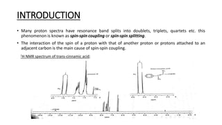
1H NMR spectrum of trans-cinnamic acid;
• The aromatic protons; five in number give rise to the peaks at δ 7.4 and δ 7.55 and the carboxyl
proton is at δ 12.5.
• Proton HA appears as two lines on the spectrum (centered on δ 6.45) and proton HX appears as
two lines (centered on δ 7.8).
• Each signal is split into a doublet, the separation between the two HA lines is the same as the
separation between the two HX lines.
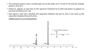
1H NMR spectra of 1,1,2-trichloroethane;
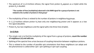
• The spectrum of 1,1,2-trichloro ethane; the signal from proton HA appears as a triplet while the
proton HX is doublet.
• The multiplicity of lines is related to the number of protons in neighbouring groups.
• In 1,1,2-trichloro ethane proton HX have only one neighbouring proton and it appears as a two
line signal (doublet).
• Proton HA has two neighbours and the signal splits into three lines (triplet).
• The simple rule is to find the multiplicity of the signal from a group of protons, count the number
of neighbours (n) and add one.
• Splitting of the spectral lines arises because of coupling interaction between neighbour protons.
• This is related to the number of possible spin orientations that these neighbours can adopt and
the phenomenon is called either spin- spin splitting or spin-spin coupling.
“The number of lines (multiplicity) observed in the NMR signal for a group of protons is not
related to the number of protons in that group”
(n+1) Rule
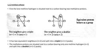
• First the lone methine hydrogen is situated next to a carbon bearing two methylene protons.
• It has two equivalent neighbours (n=2) and split into a triplet (n+1=3 peaks).
• The methylene protons are situated next to a carbon bearing only one methine hydrogen (n=1)
and split into a doublet (n+1=2 peaks).
1,1,2-trichloro ethane
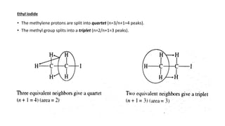
• The methylene protons are split into quartet (n=3/n+1=4 peaks).
• The methyl group splits into a triplet (n=2/n+1=3 peaks).
Ethyl iodide
THEORY
• Consider two vicinal protons similar to the alkene protons in cinnamic acid (HA & HX).
• These protons having different magnetic environments, come to resonance at different positions
in NMR spectrum.
• They do not give rise to single peaks (singlets) but doublets.
• The separation between the lines of each doublet is equal and this spacing is known as the
coupling constant (J).
Explanation for the occurrence of doublet and triplet
Doublet(trans cinnamic acid)
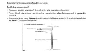
• Resonance position for proton A depends on its total magnetic environment.
• Proton X itself magnetic and have its nuclear magnet either aligned with proton A or opposed to
proton A.
• Thus proton X can either increase the net magnetic field experienced by A (X-aligned/parallel) or
decrease it (X-opposed/antiparallel).
• This means they comes to resonance twice, hence doublet.
• The mutual magnetic influence between proton A & proton X is not transmitted through the
space but via the electrons in the intervening bonds.
• Nuclear spin of proton A couples with electron spin of C HA, C C, C HX binding electrons.
• Thus A and X protons of cinnamic acid give rise to characteristic pair of doublets caused by two
protons undergoing spin coupling.
• The two spin orientation of proton A and proton X arising is equal in molecules throughout the
sample, the two lines in each doublet are of equal intensity.
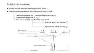
Triplet(1,1,2-trichloro ethane)
• Proton A have two neighbouring protons X and X’.
• They have three different possible combination of spin:
1. The nuclear spins X and X’ can both be parallel to A ( ).
2. Both can be antiparallel to A ( ).
3. One can be parallel and the other antiparallel.
X parallel with X’ antiparallel ( ).
X antiparallel with X’ parallel ( ).
• These three distinct energy situation give rise to triplet.
• The relative intensities predicted by the above spin coupling mechanism are:
• First order prediction always depart from real spectra since the doublet and triplet spectra are
slightly distorted.
• The inner lines being little more intense than the outer line that is humping towards the centre.
• This only shows the direction to proton which is coupled and there is no change in their intensity.
1:1 in doublets
1:2:1 in triplets
NUCLEAR MAGNETIC DOUBLE RESONANCE
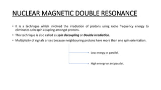
• It is a technique which involved the irradiation of protons using radio frequency energy to
eliminates spin-spin coupling amongst protons.
• This technique is also called as spin decoupling or Double irradiation.
• Multiplicity of signals arises because neighbouring protons have more than one spin orientation.
Low energy or parallel.
High energy or antiparallel.
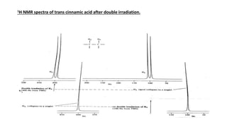
1H NMR spectra of trans cinnamic acid after double irradiation.
• Proton A appears as a doublet because of the two spin of proton X.
• Irradiate X with correct radio frequency energy we can stimulate rapid transition between the two
spin states.
• This makes the life time of a nucleus in any one spin state is too short to resolve the coupling with
proton A.
• This makes proton A resonance only once not twice, so appears only as a singlet.
• To perform this operation, in addition to the basic NMR instrument, a second tunable radio
frequency source is required for the irradiation proton X at a necessary frequency.
• So the simultaneous use of two radio frequency sources, this technique is called as double
irradiation.
• The nuclear spins during the process are less coupled than before, hence it is called as spin
decoupling
REFERENCES
• William kemp, Organic spectroscopy, third edition; 135-141.
• Pavia ,Lampman ,Kriz ,Vyvyan. Introduction to spectroscopy ,Fourth
edition;131-132.
• https://en.wikipedia.org/wiki/Nuclear magnetic resonance
spectroscopy