SCREENING METHODS OF ANTIULCER
AGENTS
1
Peptic ulcer is a break in the inner lining of the stomach, first part of the small
intestine or sometimes the lower esophagus. An ulcer in the stomach is known as
a gastric ulcer while that in the first part of the intestines is known as a duodenal ulcer.
Causes:
 Acid, pepsin, bile
 Helicobacter pylori
 NSAIDs & other drugs
 Smoking, Alcohol, stress
 Free radicals
 Oily, spicy, irregular dietary habit
 Hereditary factors
2Peptic ulcer
INVITRO METHODS
 Gastrin Binding Assay
 Iotidine Binding Assay
 H+ / K+-ATPase Inhibition Assay
3
Gastrin Binding Assay
Principle:
Major stimuli for gastric acid secretion.
Gastrin ( G cells of gastric antrum)
Bind to CCK2 receptors on parietal cells --> release HCl
Bind to CCK2 receptors on ECL cells -->Histamine --> act on H2
receptors of parietal cells --> release HCl
Compounds with gastrin receptor antagonistic activity --> can be
potential antiulcer agents
4
Procedure:
 Fundic gland suspension (Guinea pig stomach)
 Incubated with 50μl [125I] Gastrin -->
1) In buffer alone (for total binding)
2)In presence of unlabeled gastrin (for non-specific binding)
3) In presence of test compound (for competition assay)
 For 90 minutes at 37°C
 Ice cold buffer, in Microcentrifuge tubes, is layered with incubated mixture.
 Centrifuged for 5 minutes at 10,000 g
 Radioactivity is quantified in pellet after discarding the supernatant.
5
Evaluation:
 Total binding, non-specific binding and specific binding are determined.
 Percentage of specifically bound Gastrin displaced by a given concentration of
the test compound calculated.
 The higher the displaced Gastrin , more is the gastrin antagonistic activity of test
compound.
6
H+ / K+-ATPase Inhibition Assay
H+/K+ - ATPase or proton pump --> final step in the synthesis of acid by parietal
cells
Procedure:
 Homogenate of 80 ng Microsomal gastric H+/K+ - ATPase (pig gastric
mucosa) incubated with 100μl buffer, 1mM ATP and Test compound in
microtiter plate for 30 mins at 37°
 Reaction is stopped by adding Malachite green (colorimetric agent)
 After 10 seconds, 15% sodium citrate is added for 45 minutes
 Release of orthophosphate from ATP quantified by colorimeter at 570 nm
7
Evaluation:
 Percentage inhibition of H+/K+ - ATPase is calculated.
 Lesser the orthophosphate released, more is the
 inhibition of H+/K+ - ATPase by test compound.
%Inhibition =
8
[activity (control)- activity(test)]
Activity (control)
* 100
INVIVO METHODS
 Pylorus Ligation in Rats
 Stress Ulcer Model
 Histamine-Induced Gastric Ulcer
 Ethanol-Induced Mucosal Damage
 Acetic Acid-Induced Gastric Ulcer
 Indomethacin induced gastric ulcers
 Cysteamine-Induced Duodenal Ulcer
 Dimaprit-Induced Duodenal Ulcer
 Mepirizole -Induced Duodenal Ulcers
 Reserpine-Induced Chronic Ulcers
9
Pylorus Ligation in Rats
This procedure was described by Shay et al.
Principle:
 Pylorus is ligated over a certain period of time.
 Accumulation of gastric acid causes ulceration.
Procedure:
 Wistar rats weighing 150-200 grams
 Fasting : 48 hours ; water ad libitum.
 Housed singly in cages with raised bottoms of wide wire mesh to avoid
coprophagy.
 Under anaesthesia, a one-inch midline abdominal incision is given below
the xiphoid process.
10
 Under anaesthesia, a one-inch midline abdominal incision is given
below the xiphoid process.
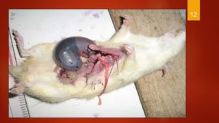
 Pylorus is ligated without damaging its blood supply.
 Stomach is replaced and abdominal wall closed with
sutures.
 Test compounds are given either orally or injected s.c.
 About 17-19 hours after pyloric ligation, rats are
sacrificed and stomachs are dissected out.
 Contents of the stomach are drained into a graduated centrifuge tube
and acidity determined by titration with 0.1 N NaOH.
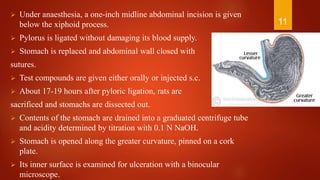
 Stomach is opened along the greater curvature, pinned on a cork
plate.
 Its inner surface is examined for ulceration with a binocular
microscope.
11
12
 The Ulcer Index is calculated and the Ulcer Severity graded.
Ulcer severity is graded as:
0 - No ulcer
1 - Superficial ulcer
2 - Deep ulcer
3 - Perforation
Ulcer index:
UI = UN+ US + UP * 10-1
Where, UN = Avg. of number of animals
US = Avg. of severity scores
UP = Percentage of animals with ulcer
13
Stress Ulcer Model
Selye (1936) , for the first time , described the use of
restraints for production of Gastric ulcers
Uses:
 Technically simple
 Do not require anesthesia or surgery
 Lesions located in glandular region of stomach
 As psychogenic factors are involved in the
pathogenesis of gastric ulcers, psychotropic drugs
could be evaluated.
14
Restraint- induced ulcers
Principle:
Stress plays a significant role in the pathogenesis
of gastric ulcers.
Procedure:
 Albino rats weighing 150-200 g are used
 Fasted for 36 hours before experiment
 Drug was administered orally or subcutaneously
 30 min later animals were subjected to restraint
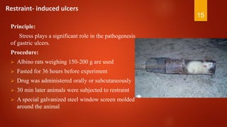
 A special galvanized steel window screen molded
around the animal
15
 The limbs of the animal are tied ,so that it cannot move
 Rats were kept under restraint for 24 hours
 Animals are sacrificed and their stomach is dissected& opened along greater
curvature. UI & ulcer severity are determined
16
Cold water immersion induced ulcers
Principle:
Exposure of cold conditions to restrained animals accelerates the occurrence of
gastric ulcers.
Procedure:
 Wistar rats weighing 150-200 grams are used.
 After fasting the animals for 16 hours, the test compound is administered orally
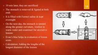
 Rats are then placed individually in restraint cages vertically, and then
 immersed in water upto abdominal, at 22°C for 1 hour.
 Then rats are removed from the cages, dried
 Evan’s blue (30mg/kg) injected i.v. via the tail vein
17
 10 min later, they are sacrificed
 The stomach is removed & ligated at both
ends
 It is filled with Formol saline & kept
overnight
 On the next day, the stomach is opened
along the greater curvature, washed in
warm water and examined for ulcerative
lesions
 Evan’s blue helps in evaluation of lesion
score.
 Calculation: Adding the lengths of the
longest diameter of the lesions
18
Swimming stress ulcer
Procedure :
 Albino rats ; either sex
 Fasted for 24 hrs; free access to water
 Rats forced to swim in deep concrete
tube filled with water at 230C for 5 hrs
 Animals removed and Sacrificed &
stomach removed ; opened along
greater curvature
 Severity grading done ; Ulcer Index
calculated
19
Histamine induced gastric stress
Principle:
Gastric acid secretion is increased when histamine is administered
intraperitoneally
Procedure:
 Guinea pig weighing 300-400 grams are taken
 Fasted for 36 hours before experiment; water ad libitum
 Histamine acid sulphate (50 mg ) was administered i.p.
 Promethazine hydrochloride 5 mg is injected i.p. 15 min before and 15 min
after histamine to protect the animals against histamine toxicity.
 The test drugs is administered before 30-45 mins before the histamine
injection.
20
 Four hours after histamine injection, guinea pigs are sacrificed and
stomach dissected out.
 The animals are sacrificed and their stomachs dissected out.
The degree of ulceration grading is done as follows
 Type 0 : No visible ulcers
 Type 1 : 10 or less small ulcers, 1-3 mm in diameter
 Type 2 : 11 or more ulcers, 1-3 mm in diameter
 Type 3 : 1 or more ulcers, 4-6 mm in diameter
 Type 4 : 1 or bigger ulcers, 7 mm or more in diameter
 Type 5 : Gastric or esophageal wall perforation
21
Ethanol-Induced Mucosal Damage
Principle:
Using transmission densitometer it is possible to quantify the extent of gastric lesion
induced by ethanol, by measuring the optical density of photographic negatives of
gastric mucosa.
Procedure:
 Wistar rats weighing 150-200 grams are taken
 Fasted for 18 hours before experiment; water ad libitum.
 Rats are given test drugs or standard drug orally.
 30 mins later 1 ml/200gm of 99.80% alcohol is administered orally.
 After 1 hour, Rats are sacrificed and stomachs dissected out.
 Severity score and ulcer index are calculated
22
 Using a transmission densitometer,the photo graph of the tissue were taken.
Damaged areas have lower optical density values.
EVALUATION:
 The significance of differences in optical density between control and
ethanol-treated tissue is evaluated.
23
2424Acetic acid induced gastric ulcers
Principle:
 Acetic acid enhances the ulceration in stomach by increasing the acidity of
stomach contents.
 This model produces chronic ulcers that resembles human ulcers and it is used to
screen drugs for gastroprotective effect.
Procedure:
 Albino rats used
 0.05 ml of Acetic acid (1-30%) injected in the submucosal layer of the stomach
 Penetrating peptic ulcers : adhered to Liver
 Chronic ulcers with repeated healing and re-aggravation.
 Effect of test drug given twice daily for 10-15 days is noted.
Indomethacin -induced chronic ulcers
Principle:
 NSAID induced gastric damage ; by blocking COX enzyme, cytoprotective
prostaglandin production inhibited.
Procedure:
 Oral administration of Indomethacin (25 mg/kg) after 24h fasting male albino rats.
 Indomethacin is dissolved in sodium bicarbonate to form a clear solution.
 Gastric ulcer can be examined after 4h and opening the stomach along the greater
curvature & washed in normal saline.
 Wounds are located in the glandular region under a simple microscope.
25
Cysteamine-Induced Duodenal Ulcer
Selye and Szabo (1973)
Cysteamine HCl (β-mercaptoethylamine HCl)
Principle:
Pathogenesis :
 Inhibition of alkaline mucus production
 Increased gastric acid secretion
 Increased serum gastrin levels
 Delayed gastric emptying.
26
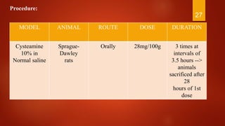
MODEL ANIMAL ROUTE DOSE DURATION
Cysteamine
10% in
Normal saline
Sprague-
Dawley
rats
Orally 28mg/100g 3 times at
intervals of
3.5 hours -->
animals
sacrificed after
28
hours of 1st
dose
27
Procedure:
 Perforating duodenal ulcer are produced that are located 2-4mm from the
pylorus, mainly on the anterior wall of the duodenum.
 The ulcer and its features in test group are compared to those in control group.
28
Dimaprit-Induced Duodenal Ulcer
Principle:
 H2 receptor agonist
 Induced gastric erosion in rats after single i.v. dose.
 Duodenal ulcer in guinea pigs after repeated s.c. dose.
Procedure:
 Female Sprague rats (150-200 g)or female guinea pigs (250-300 g) are taken
 Fasted for 24 hours before experiment; free access to water
 In rats ,Dimaprit is given in a dose of 100mg/kg intravenously, single dose
 The animal is sacrificed one hour later & stomach dissected out for evaluation
of gastric erosions
 The test drug or vehicle is given orally 60 min before injecting dimaprit
29
Mepirizole -Induced Duodenal Ulcers
 Okabe et al(1982)- described the production of Duodenal Ulcers in rats by
mepirizole , a NSAIDS
 This is used in screening of antacids and H2 receptors antagonist
Procedure:
 Male Sprague – Dawley rats (200-220g)
 Mepirizole(200mg/kg) in 1% carboxymethyl cellulose solution via gastric
intubation
 Subsequently, rats kept in cages with raised mesh bottom anddeprived of food and
water for 24 hrs
 Leads to, ulceration in proximal duodenum anderosions in antrum.
 Anti ulcer therapy started 24 hrs afterMepirizole administration
30
 11th day, Animal sacrificed
 Duodenum and Stomach evaluated for ulcer area under microscope
 Ulcer or erosion indices are calculated from the sum of area of ulcers &
erosions respectively
31
Reserpine-Induced Chronic Ulcers
 The mechanism of ulcer formation is by histamine liberation from the mast cells
in stomach wall which increase in gastric acid secretion
Procedure:
 Female Sprague – Dawley rats; 130-180g
 Fasted for 48 hrs, Free access to 0.8% sucrose in 0.2% NaCl w/v
 Liquid diet withdrawn 1hr before starting
 Animals injected with Test drug i.p. ½ hr later, Reserpine(5mg/kg) or Vehicle
injected i.p.
 4 hrs later, Animal sacrificed, Stomach removed, Examined for mucosal lesions
32
33