Protein - Unit 3 - Biochemistry - B.sc Nursing 2nd Sem Notes
Detailed exploration of protein digestion, amino acid classification, metabolism, urea cycle, related disorders, and inborn errors of aromatic amino acid metabolism accordidng to syllabus of B.Sc. Nursing 2nd Sem by INC.
Protein - Unit 3 - Biochemistry - B.sc Nursing 2nd Sem Notes
1.
Biochemistry
Unit – 3
Proteins
:-Mr. Priyanshu Anand
%%%%%%%%%%%%%%%%%%%%%%%%%%%%%%%%%%%%%%%%%%%%%%%%%%%%%%%%%%
Contents :-
Classification of amino acids based on nutrition, metabolic rate with examples
Digestion, absorption & metabolism of protein & related disorders
Biologically important compounds synthesized from various amino acids (only names)
In born errors of amino acid metabolism – only aromatic amino acids (in brief)
Plasma protein – types, function & normal values
Causes of proteinuria, hypoproteinemia, hyper-gamma globinemia
Principle of electrophoresis, normal & abnormal electrophoretic patterns (in brief).
%%%%%%%%%%%%%%%%%%%%%%%%%%%%%%%%%%%%%%%%%%%%%%%%%%%%%%%%%%
PROTEIN
Proteins are among the most abundant organic molecules in living systems and are
way more diverse in structure and function than other classes of macromolecules.
A single cell can contain thousands of proteins, each with a unique function.
Although their structures, like their functions, vary greatly, all proteins are made
up of one or more chains of amino acids.
Amino acids as building blocks of peptides and proteins.
Classification of Amino Acids
a) Nutritional Classification
Amino acids are divided into essential and non-essential groups based on whether
the body can synthesize them.
2.
Essential aminoacids: Cannot be synthesized in sufficient amounts, must be
supplied through diet. Examples: Arginine, Valine, Histidine, Isoleucine,
Leucine, Lysine, Methionine, Phenylalanine, Threonine, Tryptophan.
Non-essential amino acids: Synthesized in the body, not required in diet.
Examples: Glycine, Alanine, Serine, Cysteine, Aspartate, Asparagine,
Glutamate, Glutamine, Tyrosine, Proline.
b) Metabolic Fate Classification
Amino acids are grouped based on what their carbon skeletons yield after
catabolism:
Glucogenic amino acids → precursors for glucose/glycogen. Examples:
Alanine, Aspartate, Glycine, Methionine.
Ketogenic amino acids → precursors for ketone bodies/fat. Examples:
Leucine, Lysine.
Both glucogenic & ketogenic → yield glucose and ketone bodies. Examples:
Isoleucine, Phenylalanine, Tryptophan, Tyrosine.
Digestion, Absorption & Metabolism of Proteins
Digestion
Stomach:
o HCl denatures proteins, exposing peptide bonds.
o Pepsinogen → pepsin (active enzyme) hydrolyzes proteins into
peptides.
Small intestine:
o Pancreatic enzymes: trypsin, chymotrypsin, carboxypeptidase.
o Brush border peptidases break peptides into amino acids.
End products: Amino acids, dipeptides, tripeptides.
Absorption
Occurs mainly in jejunum and ileum.
Amino acids absorbed via active transport (Na⁺ dependent carriers).
Dipeptides/tripeptides absorbed by H⁺-linked transporters, hydrolyzed inside
enterocytes.
3.
Metabolism
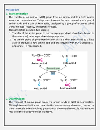
1. Transamination
The transferof an amino ( NH2) group from an amino acid to a keto acid is
known as transamination. This process involves the interconversion of a pair of
amino acids and a pair of keto acids, catalysed by a group of enzymes called
transaminases (recently, aminotransferases).
Transamination occurs in two stages :
1 Transfer of the amino group to the coenzyme pyridoxal phosphate (bound to
the coenzyme) to form pyridoxamine phosphate.
2 The amino group of pyridoxamine phosphate is then transferred to a keto
acid to produce a new amino acid and the enzyme with PLP (Pyridoxal 5′-
phosphate) is regenerated.
2. Deamination
The removal of amino group from the amino acids as NH3 is deamination.
Although transamination and deamination are separately discussed, they occur
simultaneously, often involving glutamate as the central molecule. Deamination
may be either oxidative or non-oxidative.
4.
Oxidative deamination
It isthe liberation of free ammonia from the amino group of amino acids
coupled with oxidation. This takes place mostly in liver and kidney. The purpose
of oxidative deamination is to provide NH3 for urea synthesis and -keto acids
for a variety of reactions, including energy generation.
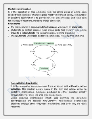
Key Enzyme
The main enzyme is glutamate dehydrogenase, which acts on glutamate.
Glutamate is central because most amino acids first transfer their amino
group to α-ketoglutarate (via transamination), forming glutamate.
Then glutamate undergoes oxidative deamination, releasing free ammonia.
Non-oxidative deamination
It is the removal of an amino group from an amino acid without involving
oxidation. This reaction occurs mainly in the liver and kidney, similar to
oxidative deamination. Ammonia produced is either excreted directly
through kidney or eners the urea cycle (inside liver).
Unlike oxidative deamination (which uses enzymes like glutamate
dehydrogenase and requires NAD⁺/NADP⁺), non-oxidative deamination
proceeds through other enzymatic mechanisms that don’t rely on redox
reactions.
5.
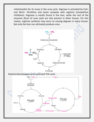
3. Urea cycle
Detoxifiesammonia and converts it to urea in liver. “Urea is the end product of
protein metabolism… known as Krebs-Henseleit Urea cycle.”
Urea has two amino ( NH2) groups, one derived from NH3 and the other from
aspartate. Carbon atom is supplied by CO2. Urea synthesis is a five-step cyclic
process, with five distinct enzymes. The first two enzymes are present in
mitochondria while the rest are localized in cytosol. The details of urea cycle are
described :
1. Synthesis of carbamoyl phosphate : Carbamoyl phosphate synthase I (CPS
I) of mitochondria catalyses the condensation of NH4+
ions with CO2 to
form carbamoyl phosphate. This step consumes two ATP and is
irreversible, and rate-limiting. CPS I requires N acetylglutamate for its
activity. Another enzyme, carbamoyl phosphate synthase II (CPS II)—
involved in pyrimidine synthesis—is present in cytosol. It accepts amino
group from glutamine and does not require N-acetylglutamate for its
activity.
2. Formation of citrulline : Citrulline is synthesized from carbamoyl
phosphate and ornithine by ornithine transcarbamoylase. Ornithine is
regenerated and used in urea cycle. Therefore, its role is comparable to
that of oxaloacetate in citric acid cycle. Ornithine and citrulline are basic
amino acids. (They are never found in protein structure due to lack of
codons). Citrulline produced in this reaction is transported to cytosol by a
transporter system.
3. Synthesis of arginosuccinate : Argino succinate synthase condenses
citrulline with aspartate to produce arginosuccinate. The second amino
group of urea is incorporated in this reaction. This step requires ATP which
is cleaved to AMP and pyrophosphate (PPi). The latter is immediately
broken down to inorganic phosphate (Pi).
4. Cleavage of arginosuccinate : Argino succinase cleaves arginosuccinate to
give arginine and fumarate. Arginine is the immediate precursor for urea.
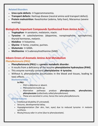
Fumarate liberated here provides a connecting link with TCA cycle,
gluconeogenesis etc.
5. Formation of urea : Arginase is the fifth and final enzyme that cleaves
arginine to yield urea and ornithine. Ornithine, so regenerated, enters
6.
mitochondria for itsreuse in the urea cycle. Arginase is activated by Co2+
and Mn2+. Ornithine and lysine compete with arginine (competitive
inhibition). Arginase is mostly found in the liver, while the rest of the
enzymes (four) of urea cycle are also present in other tissues. For this
reason, arginine synthesis may occur to varying degrees in many tissues.
But only the liver can ultimately produce urea.
Relationship between urea cycle and TCA cycle :
7.
Related Disorders
Ureacycle defects → hyperammonemia.
Transport defects: Hartnup disease (neutral amino acid transport defect).
Protein malnutrition: Kwashiorkor (edema, fatty liver), Marasmus (severe
wasting).
Biologically Important Compounds Synthesized from Amino Acids
Tryptophan → serotonin, melatonin, niacin.
Tyrosine → catecholamines (dopamine, norepinephrine, epinephrine),
thyroid hormones, melanin.
Histidine → histamine.
Glycine → heme, creatine, purines.
Glutamate → GABA.
Methionine → S-adenosylmethionine (SAM).
Inborn Errors of Aromatic Amino Acid Metabolism
Phenylketonuria (PKU):
Phenylketonuria (PKU) is a genetic metabolic disorder.
It results from a deficiency of the enzyme phenylalanine hydroxylase (PAH).
This enzyme normally converts phenylalanine → tyrosine.
Without it, phenylalanine accumulates in the blood and tissues, leading to
toxic effects.
Biochemical Basis
In PKU:
o PAH is defective or absent.
o Phenylalanine builds up.
o Alternative pathways produce phenylpyruvate, phenyllactate,
phenylacetate (collectively called phenylketones).
o These are excreted in urine, hence the name phenylketonuria.
Clinical Features
Intellectual disability (if untreated).
Seizures, developmental delay.
Hypopigmentation (fair skin, hair, eyes) due to reduced tyrosine → melanin
synthesis.
Musty/mousy odor in urine (due to phenylacetate).
8.
Diagnosis
Newborn screening:Guthrie bacterial inhibition test or tandem mass
spectrometry.
Elevated blood phenylalanine levels.
Management
Dietary restriction of phenylalanine (low-protein diet).
Supplement with tyrosine (becomes essential in PKU).
Avoid aspartame (contains phenylalanine).
In some cases, BH4 supplementation helps if the defect is in cofactor metabolism.
Alkaptonuria:
Alkaptonuria is a rare autosomal recessive genetic disorder.
It results from a deficiency of the enzyme homogentisate oxidase.
This enzyme normally breaks down homogentisic acid in the
tyrosine/phenylalanine catabolic pathway.
Without it, homogentisic acid accumulates in the body.
Biochemical Basis
Normal pathway: Phenylalanine → Tyrosine → Homogentisic acid →
Maleylacetoacetate (via homogentisate oxidase).
In Alkaptonuria: Homogentisic acid cannot be metabolized further → builds up in
tissues and is excreted in urine.
Clinical Features
Dark urine: Urine turns black on standing due to oxidation of homogentisic acid.
Ochronosis: Bluish-black pigmentation in connective tissues (cartilage, sclera, skin).
Arthropathy: Degenerative joint disease, especially in spine and large joints, due to
pigment deposition.
Possible cardiac valve involvement and kidney stones.
Diagnosis
Observation of black urine.
Detection of homogentisic acid in urine (chromatography or spectrophotometry).
Genetic testing for mutations in the HGD gene (homogentisate dioxygenase).
Management
No complete cure.
Dietary restriction of phenylalanine and tyrosine may help reduce homogentisic acid
levels.
Vitamin C supplementation may slow ochronosis by reducing oxidation.
Symptomatic treatment for arthritis and joint pain.
9.
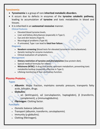
Tyrosinemia:
Tyrosinemia isa group of rare inherited metabolic disorders.
It occurs due to defects in enzymes of the tyrosine catabolic pathway,
leading to accumulation of tyrosine and toxic metabolites in blood and
tissues.
It is inherited in an autosomal recessive manner.
Clinical Features
Elevated blood tyrosine levels.
Liver and kidney disturbances (especially in Type I).
Eye and skin lesions (Type II).
Neurological problems (Type III).
If untreated, Type I can lead to fatal liver failure.
Diagnosis
Newborn screening (blood tests for elevated tyrosine or succinylacetone).
Genetic testing for enzyme mutations.
Clinical evaluation of symptoms.
Management
Dietary restriction of tyrosine and phenylalanine (low-protein diet).
Special medical formulas for infants.
Nitisinone (NTBC): A drug that blocks upstream metabolism, preventing toxic
metabolite buildup (especially effective in Type I).
Lifelong monitoring of liver and kidney function.
Plasma Proteins
Types
Albumin: Major fraction, maintains osmotic pressure, transports fatty
acids, bilirubin, drugs.
Globulins:
o α1 (antitrypsin), α2 (ceruloplasmin, haptoglobin), β (transferrin,
complement), γ (immunoglobulins).
Fibrinogen: Clotting factor.
Functions
Osmotic balance (albumin).
Transport (albumin, transferrin, ceruloplasmin).
Immunity (γ-globulins).
Clotting (fibrinogen).
10.
Normal Values
Totalplasma protein: 6–8 g/dL.
Albumin: 3.5–5 g/dL.
Globulins: 2–3 g/dL.
Protein Disorders
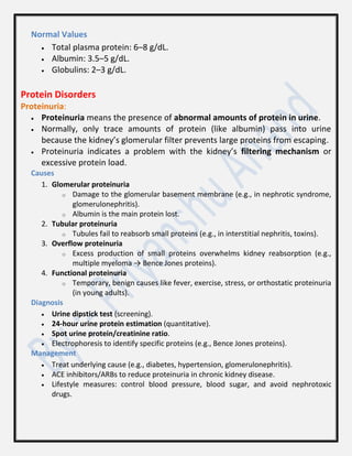
Proteinuria:
Proteinuria means the presence of abnormal amounts of protein in urine.
Normally, only trace amounts of protein (like albumin) pass into urine
because the kidney’s glomerular filter prevents large proteins from escaping.
Proteinuria indicates a problem with the kidney’s filtering mechanism or
excessive protein load.
Causes
1. Glomerular proteinuria
o Damage to the glomerular basement membrane (e.g., in nephrotic syndrome,
glomerulonephritis).
o Albumin is the main protein lost.
2. Tubular proteinuria
o Tubules fail to reabsorb small proteins (e.g., in interstitial nephritis, toxins).
3. Overflow proteinuria
o Excess production of small proteins overwhelms kidney reabsorption (e.g.,
multiple myeloma → Bence Jones proteins).
4. Functional proteinuria
o Temporary, benign causes like fever, exercise, stress, or orthostatic proteinuria
(in young adults).
Diagnosis
Urine dipstick test (screening).
24-hour urine protein estimation (quantitative).
Spot urine protein/creatinine ratio.
Electrophoresis to identify specific proteins (e.g., Bence Jones proteins).
Management
Treat underlying cause (e.g., diabetes, hypertension, glomerulonephritis).
ACE inhibitors/ARBs to reduce proteinuria in chronic kidney disease.
Lifestyle measures: control blood pressure, blood sugar, and avoid nephrotoxic
drugs.
11.
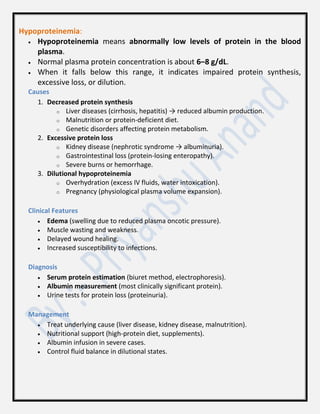
Hypoproteinemia:
Hypoproteinemia meansabnormally low levels of protein in the blood
plasma.
Normal plasma protein concentration is about 6–8 g/dL.
When it falls below this range, it indicates impaired protein synthesis,
excessive loss, or dilution.
Causes
1. Decreased protein synthesis
o Liver diseases (cirrhosis, hepatitis) → reduced albumin production.
o Malnutrition or protein-deficient diet.
o Genetic disorders affecting protein metabolism.
2. Excessive protein loss
o Kidney disease (nephrotic syndrome → albuminuria).
o Gastrointestinal loss (protein-losing enteropathy).
o Severe burns or hemorrhage.
3. Dilutional hypoproteinemia
o Overhydration (excess IV fluids, water intoxication).
o Pregnancy (physiological plasma volume expansion).
Clinical Features
Edema (swelling due to reduced plasma oncotic pressure).
Muscle wasting and weakness.
Delayed wound healing.
Increased susceptibility to infections.
Diagnosis
Serum protein estimation (biuret method, electrophoresis).
Albumin measurement (most clinically significant protein).
Urine tests for protein loss (proteinuria).
Management
Treat underlying cause (liver disease, kidney disease, malnutrition).
Nutritional support (high-protein diet, supplements).
Albumin infusion in severe cases.
Control fluid balance in dilutional states.
12.
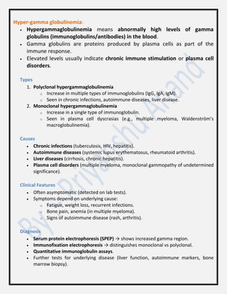
Hyper-gamma globulinemia:
Hypergammaglobulinemiameans abnormally high levels of gamma
globulins (immunoglobulins/antibodies) in the blood.
Gamma globulins are proteins produced by plasma cells as part of the
immune response.
Elevated levels usually indicate chronic immune stimulation or plasma cell
disorders.
Types
1. Polyclonal hypergammaglobulinemia
o Increase in multiple types of immunoglobulins (IgG, IgA, IgM).
o Seen in chronic infections, autoimmune diseases, liver disease.
2. Monoclonal hypergammaglobulinemia
o Increase in a single type of immunoglobulin.
o Seen in plasma cell dyscrasias (e.g., multiple myeloma, Waldenström’s
macroglobulinemia).
Causes
Chronic infections (tuberculosis, HIV, hepatitis).
Autoimmune diseases (systemic lupus erythematosus, rheumatoid arthritis).
Liver diseases (cirrhosis, chronic hepatitis).
Plasma cell disorders (multiple myeloma, monoclonal gammopathy of undetermined
significance).
Clinical Features
Often asymptomatic (detected on lab tests).
Symptoms depend on underlying cause:
o Fatigue, weight loss, recurrent infections.
o Bone pain, anemia (in multiple myeloma).
o Signs of autoimmune disease (rash, arthritis).
Diagnosis
Serum protein electrophoresis (SPEP) → shows increased gamma region.
Immunofixation electrophoresis → distinguishes monoclonal vs polyclonal.
Quantitative immunoglobulin assays.
Further tests for underlying disease (liver function, autoimmune markers, bone
marrow biopsy).
13.
Management
Treat theunderlying cause (infection, autoimmune disease, liver disorder, plasma
cell malignancy).
Supportive care (manage anemia, infections).
Electrophoresis
Electrophoresis is a biochemical technique used to separate charged
molecules—like proteins, nucleic acids, or amino acids—based on their
movement in an electric field.
Principle
Separation of charged molecules in an electric field.
Proteins migrate based on charge and size. Document line: “The
movement of charged particles in an electric field… known as
electrophoresis.”
Normal Pattern
Albumin (largest peak).
α1, α2, β, γ globulins.
Abnormal Patterns
Hypoalbuminemia: Liver disease, nephrotic syndrome.
Monoclonal spike (M-protein): Multiple myeloma.
Polyclonal increase in γ-globulins: Chronic infection, autoimmune disease.