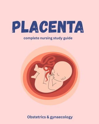
PLACENTA
complete nursing study guide
Obstetrics & gynaecology
Anatomy & Structure
Development & Formation
Functions
Placental Hormones
Placental Circulation
Placental Abnormalities
Exam Points & MCQs
Includes:
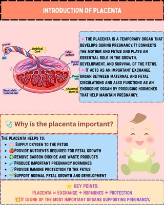
Introduction of placenta
🌸The placenta is a temporary organ that
develops during pregnancy. It connects
the mother and fetus and plays an
essential role in the growth,
development, and survival of the fetus.
🌸It acts as an important exchange
organ between maternal and fetal
circulations and also functions as an
endocrine organ by producing hormones
that help maintain pregnancy.
⭐key points:
Placenta = Exchange + Hormones + Protection
💥It is one of the most important organs supporting pregnancy.
key points:
key points:
Placenta = Exchange + Hormones + Protection
It is one of the most important organs supporting pregnancy
🩺Why is the placenta important?
The placenta helps to:
🫁Supply oxygen to the fetus
🍎Provide nutrients required for fetal growth
♻️Remove carbon dioxide and waste products
🧪Produce important pregnancy hormones
🛡️Provide immune protection to the fetus
🌱Support normal fetal growth and development
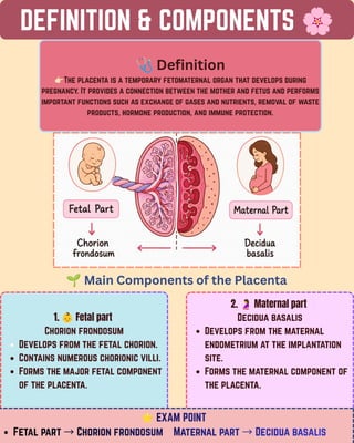
DEFINITION & COMPONENTS 🌸
🩺Definition
👉🏻The placenta is a temporary fetomaternal organ that develops during
pregnancy. It provides a connection between the mother and fetus and performs
important functions such as exchange of gases and nutrients, removal of waste
products, hormone production, and immune protection.
1. 👶Fetal part
Chorion frondosum
Develops from the fetal chorion.
Contains numerous chorionic villi.
Forms the major fetal component
of the placenta.
🌱Main Components of the Placenta
2. 🤰Maternal part
Decidua basalis
Develops from the maternal
endometrium at the implantation
site.
Forms the maternal component of
the placenta.
⭐EXAM POINT
Fetal part →Chorion frondosum Maternal part →Decidua basalis
EXAM POINT
EXAM POINT
Fetal part →Chorion frondosum Maternal part →Decidua basalis
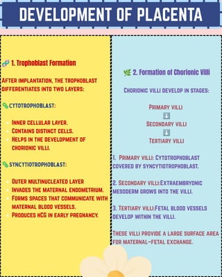
DEVELOPMENT OF PLACENTA
🧬1. Trophoblast Formation
After implantation, the trophoblast
differentiates into two layers:
🦠Cytotrophoblast:
Inner cellular layer.
Contains distinct cells.
Helps in the development of
chorionic villi.
🦠Syncytiotrophoblast:
Outer multinucleated layer
Invades the maternal endometrium.
Forms spaces that communicate with
maternal blood vessels.
Produces hCG in early pregnancy.
🌿2. Formation of Chorionic Villi
Chorionic villi develop in stages:
Primary villi
⬇️
Secondary villi
⬇️
Tertiary villi
1. Primary villi: Cytotrophoblast
covered by syncytiotrophoblast.
2. Secondary villi:Extraembryonic
mesoderm grows into the villi.
3. Tertiary villi:Fetal blood vessels
develop within the villi.
These villi provide a large surface area
for maternal–fetal exchange.
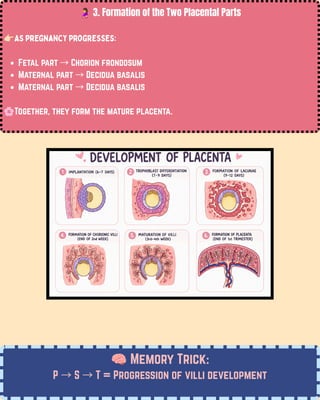
🤰3. Formation of the Two Placental Parts
👉🏻As pregnancy progresses:
Fetal part →Chorion frondosum
Maternal part →Decidua basalis
🌸Together, they form the mature placenta.
🧠Memory Trick:
P →S →T = Progression of villi development
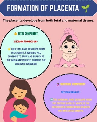
FORMATION OF PLACENTA 🌱
👶FETAL COMPONENT:
Chorion frondosum:-
⭐The fetal part develops from
the chorion. Chorionic villi
continue to grow and branch at
the implantation site, forming the
chorion frondosum.
🤰MATERNAL COMPONENT:
Decidua basalis:-
➡️The maternal part develops from
the decidua basalis, which is the
specialized endometrium beneath
the implanted embryo.
The placenta develops from both fetal and maternal tissues.
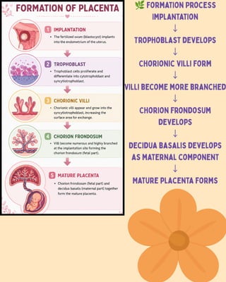
🌿Formation Process
Implantation
↓
Trophoblast develops
↓
Chorionic villi form
↓
Villi become more branched
↓
Chorion frondosum
develops
↓
Decidua basalis develops
as maternal component
↓
Mature placenta forms
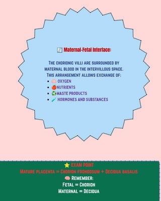
🔄Maternal–Fetal Interface:
The chorionic villi are surrounded by
maternal blood in the intervillous space.
This arrangement allows exchange of:
🫁OXYGEN
🍎NUTRIentS
♻️waste products
🧪hormones and substances
⭐EXAM POINT
Mature placenta = Chorion frondosum + Decidua basalis
🧠Remember:
Fetal = Chorion
Maternal = Decidua
LOCATION & ATTACHMENT OF
PLACENTA 🤰
📍Normal Location:
The placenta usually implants in the upper part of
the uterus, most commonly on the:
Anterior wall
Posterior wall
Fundal region
Sometimes the lateral wall
The exact location can vary from one pregnancy to
another.
🔗How is the Placenta Attached?
👉🏻The placenta is attached to the uterine wall at the
site where implantation occurs.
👉🏻The fetal side of the placenta is connected to the
fetus through the umbilical cord.
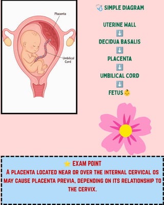
⭐EXAM POINT
A placenta located near or over the internal cervical os
may cause placenta previa, depending on its relationship to
the cervix.
🩺Simple Diagram
UTERINE WALL
⬇️
Decidua basalis
⬇️
Placenta
⬇️
Umbilical cord
⬇️
fetus👶
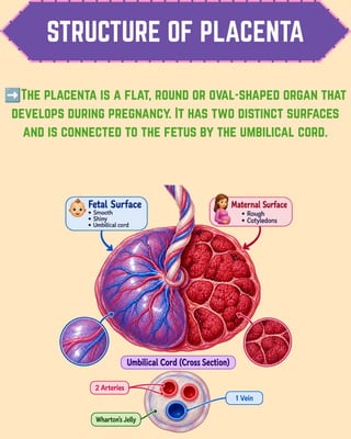
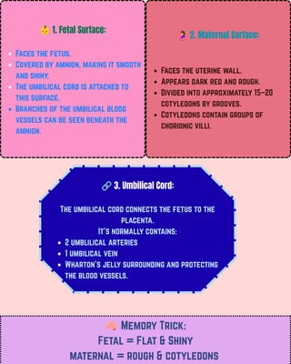
STRUCTURE OF PLACENTA
➡️The placenta is a flat, round or oval-shaped organ that
develops during pregnancy. It has two distinct surfaces
and is connected to the fetus by the umbilical cord.
👶1. Fetal Surface:
Faces the fetus.
Covered by amnion, making it smooth
and shiny.
The umbilical cord is attached to
this surface.
Branches of the umbilical blood
vessels can be seen beneath the
amnion.
🤰2. Maternal Surface:
Faces the uterine wall.
Appears dark red and rough.
Divided into approximately 15–20
cotyledons by grooves.
Cotyledons contain groups of
chorionic villi.
🔗3. Umbilical Cord:
The umbilical cord connects the fetus to the
placenta.
It's normally contains:
2 umblilical arteries
1 umbilical vein
Wharton's jelly surrounding and protecting
the blood vessels.
🧠Memory Trick:
Fetal = Flat & Shiny
maternal = rough & cotyledons
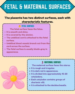
FETAL & MATERNAL SURFACES
👶FETAL SURFACE:
The fetal surface faces the fetus.
It is smooth and shiny.
It is covered by the amnion.
The umbilical cord is attached to the fetal
surface.
Umbilical blood vessels branch out from the
cord across the surface.
The fetal surface is usually bluish-gray in
appearance.
🤰MATERNAL SURFACE:
The maternal surface faces the uterus.
It is rough and irregular.
It is dark red in appearance.
It is divided into approximately 15–20
cotyledons.
Each cotyledon contains groups of
chorionic villi.
It is attached to the decidua basalis.
The placenta has two distinct surfaces, each with
characteristic features.
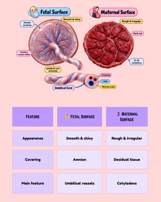
Feature 👶Fetal Surface
🤰Maternal
Surface
Appearance Smooth & shiny Rough & irregular
Covering Amnion Decidual tissue
Main feature Umbilical vessels Cotyledons
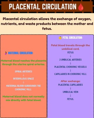
PLACENTAL CIRCULATION🩸
Placental circulation allows the exchange of oxygen,
nutrients, and waste products between the mother and
fetus.
🤰MATERNAL CIRCULATION:
Maternal blood reaches the placenta
through the uterine spiral arteries.
Spiral arteries
↓
Intervillous space
↓
Maternal blood surrounds the
chorionic villi.
Maternal blood does not normally
mix directly with fetal blood.
👶FETAL CIRCULATION:
Fetal blood travels through the
umbilical cord.
Fetus
↓
2 Umbilical arteries
↓
Placental chorionic vessels
↓
Capillaries in chorionic villi.
After exchange:
Placental capillaries
↓
Umbilical vein
↓
Fetus.
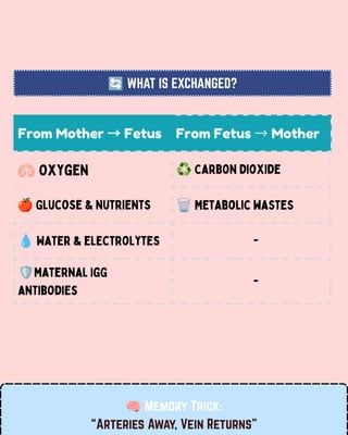
From Mother →Fetus From Fetus →Mother
🫁Oxygen ♻️Carbon dioxide
🍎Glucose & nutrients 🗑️Metabolic wastes
💧Water & electrolytes -
🛡️Maternal IgG
antibodies
-
🔄WHAT IS EXCHANGED?
🧠Memory Trick:
“Arteries Away, Vein Returns”
PLACENTAL BARRIER 🛡️
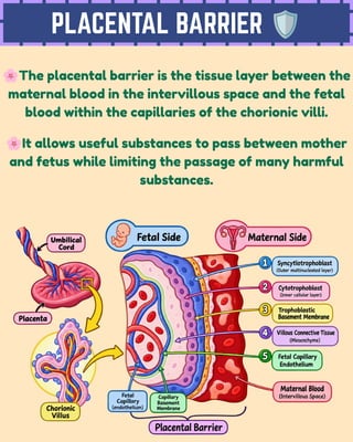
🌸The placental barrier is the tissue layer between the
maternal blood in the intervillous space and the fetal
blood within the capillaries of the chorionic villi.
🌸It allows useful substances to pass between mother
and fetus while limiting the passage of many harmful
substances.
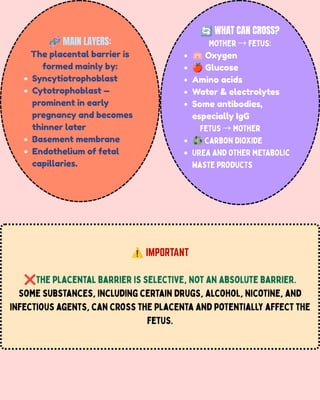
🧬MAIN LAYERS:
The placental barrier is
formed mainly by:
Syncytiotrophoblast
Cytotrophoblast —
prominent in early
pregnancy and becomes
thinner later
Basement membrane
Endothelium of fetal
capillaries.
🔄WHAT CAN CROSS?
Mother →Fetus:
🫁Oxygen
🍎Glucose
Amino acids
Water & electrolytes
Some antibodies,
especially IgG
Fetus →Mother
♻️Carbon dioxide
Urea and other metabolic
waste products
⚠️IMPORTANT
❌The placental barrier is selective, not an absolute barrier.
Some substances, including certain drugs, alcohol, nicotine, and
infectious agents, can cross the placenta and potentially affect the
fetus.
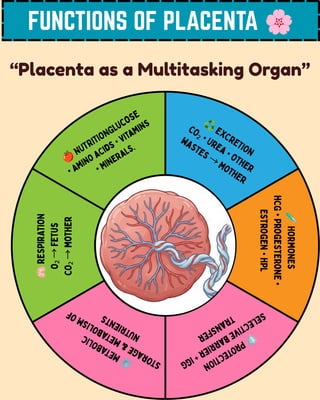
FUNCTIONS OF PLACENTA 🌸
“Placenta as a Multitasking Organ”
🍎NUTRITIONGlucose
• Amino acids • Vitamins
• Minerals.
♻️EXCRETION
CO₂ • Urea • Other
wastes →Mother
🧪
HORMONES
hCG
•
Progesterone
•
Estrogen
•
hPL
🫁
RESPIRATION
O₂
→
Fetus
CO₂
→
Mother
🛡️
P
R
O
T
E
C
T
I
O
N
S
e
l
e
c
t
i
v
e
b
a
r
r
i
e
r
+
I
g
G
t
r
a
n
s
f
e
r
⚙️
M
E
T
A
B
O
L
I
C
S
t
o
r
a
g
e
&
m
e
t
a
b
o
l
i
s
m
o
f
n
u
t
r
i
e
n
t
s
PLACENTAL HORMONES 🧪🌸
PLACENTAL HORMONES
PLACENTAL HORMONES
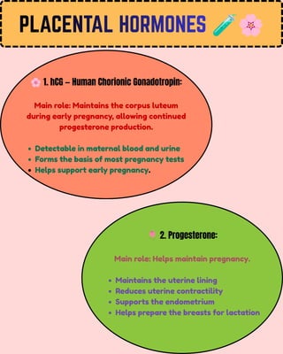
🌸1. hCG — Human Chorionic Gonadotropin:
Main role: Maintains the corpus luteum
during early pregnancy, allowing continued
progesterone production.
Detectable in maternal blood and urine
Forms the basis of most pregnancy tests
Helps support early pregnancy.
🌷2. Progesterone:
Main role: Helps maintain pregnancy.
Maintains the uterine lining
Reduces uterine contractility
Supports the endometrium
Helps prepare the breasts for lactation
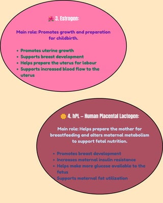
🌺3. Estrogen:
Main role: Promotes growth and preparation
for childbirth.
Promotes uterine growth
Supports breast development
Helps prepare the uterus for labour
Supports increased blood flow to the
uterus
🌼4. hPL — Human Placental Lactogen:
Main role: Helps prepare the mother for
breastfeeding and alters maternal metabolism
to support fetal nutrition.
Promotes breast development
Increases maternal insulin resistance
Helps make more glucose available to the
fetus
Supports maternal fat utilization
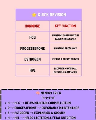
Hormone Key Function
hCG
Maintains corpus luteum
early in pregnancy
Progesterone Maintains pregnancy
Estrogen Uterine & breast growth
hPL
Lactation + maternal
metabolic adaptation
⭐QUICK REVISION
🧠MEMORY TRICK
“H-P-E-H”
H →hCG →Helps maintain corpus luteum
P →Progesterone →Pregnancy maintenance
E →Estrogen →Expansion & growth
H →hPL →Helps lactation & fetal nutrition
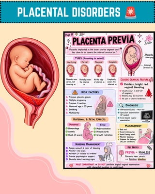
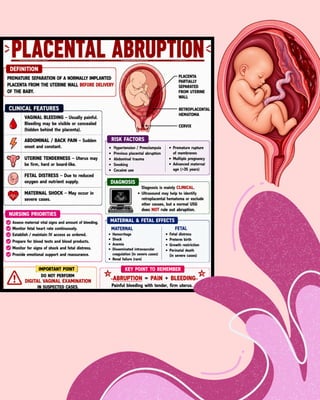
PLACENTAL DISORDERS 🚨
THANK YOU FOR
READING! 💌
✨CREATED BY
AQSHA
Keep Learning • Keep Growing • Keep Caring