Article
iScience
Phospho-JNK agonists show promising effects for
the treatment of hepatocellular carcinoma
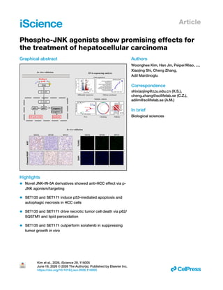
Graphical abstract
Highlights
• Novel JNK-IN-5A derivatives showed anti-HCC effect via p-
JNK agonism/targeting
• SET135 and SET171 induce p53-mediated apoptosis and
autophagic necrosis in HCC cells
• SET135 and SET171 drive necrotic tumor cell death via p62/
SQSTM1 and lipid peroxidation
• SET135 and SET171 outperform sorafenib in suppressing
tumor growth in vivo
Authors
Woonghee Kim, Han Jin, Peipei Miao, ...,
Xiaojing Shi, Cheng Zhang,
Adil Mardinoglu
Correspondence
shixiaojing@zzu.edu.cn (X.S.),
cheng.zhang@scilifelab.se (C.Z.),
adilm@scilifelab.se (A.M.)
In brief
Biological sciences
Kim et al., 2026, iScience 29, 116005
June 19, 2026 © 2026 The Author(s). Published by Elsevier Inc.
https://doi.org/10.1016/j.isci.2026.116005 ll
iScience
Article
Phospho-JNK agonists show promising effects
for the treatment of hepatocellular carcinoma
Woonghee Kim,1,11 Han Jin,1,2,11 Peipei Miao,3 Mehmet Ozcan,4 Xinmeng Liao,1 Mengzhen Li,1 Shazia Iqbal,5
Jihad Sebhaoui,5,6 Sajda Ashraf,5 Burcu Belmen,5 Hasan Turkez,7 Jan Boren,8 Mathias Uhlen,1 Xiaojing Shi,3,*
Cheng Zhang,1,9,* and Adil Mardinoglu1,10,12,*
1Science for Life Laboratory, KTH – Royal Institute of Technology, 17165 Stockholm, Sweden
2Central Laboratory, Tianjin Medical University General Hospital, Tianjin 300052, P.R. China
3Tianjian Laboratory of Advanced Biomedical Sciences, Academy of Medical Sciences, Laboratory Animal Center, State Key Laboratory of
Esophageal Cancer Prevention & Treatment, Zhengzhou University, Zhengzhou 450052, P.R. China
4Faculty of Medicine, Zonguldak Bulent Ecevit University, Zonguldak 67600, Turkiye
5Trustlife Labs Drug Research & Development Center, Istanbul 34774, Turkiye
6Life and Health Sciences Laboratory, FMP, Abdelmalek Essaadi University, Tetouan 93000, Morocco
7Department of Medical Biology, Faculty of Medicine, Atatürk University, Erzurum 25030, Turkey
8Department of Molecular and Clinical Medicine, University of Gothenburg, Sahlgrenska University Hospital, 41390 Gothenburg, Sweden
9Institute of Liver Studies, Faculty of Life Science & Medicine, King’s College London, London SE5 9RS, UK
10Centre for Host-Microbiome Interactions, Faculty of Dentistry, Oral & Craniofacial Sciences, King’s College London, London SE1 9RT, UK
11These authors contributed equally
12Lead contact
*Correspondence: shixiaojing@zzu.edu.cn (X.S.), cheng.zhang@scilifelab.se (C.Z.), adilm@scilifelab.se (A.M.)
https://doi.org/10.1016/j.isci.2026.116005
SUMMARY
Hepatocellular carcinoma (HCC) remains difficult to treat due to its limited targets. Hence, we introduced
phosphorylated c-Jun N-terminal kinase (p-JNK) as an anti-HCC target protein and investigated JNK-IN-
5A and six derivatives (SET135, SET156, SET158, SET159, SET171, and SET172) which stabilize p-JNK.
In vitro, these compounds outperformed sorafenib and regorafenib, inducing stronger p53-mediated
cell-cycle arrest, autophagy, apoptosis, and reduced invasiveness via JNK/c-Jun pathways. RNA-seq
profiling revealed distinct mechanisms: SET135 triggered autophagic necrosis via p62/SQSTM1, while
SET171 induced reactive oxygen species (ROS)-driven necrosis. Systems biology analysis confirmed their
enhanced efficacy. A 7-day GLP-like rat toxicity study showed SET135 and SET171 were well-tolerated.
In vivo study performed with 21-day treatment of SET135 or SET171 showed superior anti-tumor effects
compared to sorafenib via apoptotic mechanisms in HCC-transplanted mice. These findings highlight
JNK-IN-5A derivatives as promising HCC therapeutic candidates capable of inducing both apoptotic and
necrotic cell death.
INTRODUCTION
Primary liver cancer ranks as the sixth most prevalent cancer
globally and holds the third position in terms of mortality. Hepa­
tocellular carcinoma (HCC) represents the predominant form of
primary liver cancer.1
The significant risk factors for developing
HCC include chronic infection with hepatitis B virus (HBV) or
hepatitis C virus (HCV), heavy alcohol consumption, metabolic
dysfunction-associated steatohepatitis (MASH), and exposure
to aflatoxins or pyrrolizidine alkaloids. These risk factors under­
score the complex etiology of HCC, highlighting the importance
of understanding and mitigating these risks in its prevention and
management.2–4
Patients diagnosed with early-stage HCC are typically
advised to undergo surgical liver resection or liver transplanta­
tion. Those in the intermediate stage or unable to undergo sur­
gery may be considered for chemotherapy.5
However, the
application of chemotherapy in treating liver cancer faces sig­
nificant challenges, including a high rate of chemoresistance.6
This resistance is partly due to the liver’s fundamental role in
filtering foreign substances from the bloodstream, which can
limit the effectiveness of systemic chemotherapy. To enhance
the efficacy of chemotherapy, medical professionals often
employ regional chemotherapy techniques such as hepatic ar­
tery infusion (HAI) or transarterial chemoembolization (TACE).7
These methods deliver chemotherapy directly to the liver, aim­
ing to increase drug concentration at the tumor site while mini­
mizing systemic exposure and side effects. Multi-target kinase
inhibitors, like sorafenib and regorafenib, have become widely
used in the treatment of liver cancer.8,9
These drugs reduce
angiogenesis, the process of new blood vessel formation, by in­
hibiting key receptors such as the vascular endothelial growth
factor receptor-2 (VEGFR-2) and the platelet-derived growth
factor receptor (PDGFR), which are found in endothelial cells.
iScience 29, 116005, June 19, 2026 © 2026 The Author(s). Published by Elsevier Inc. 1
This is an open access article under the CC BY license (http://creativecommons.org/licenses/by/4.0/).
ll
OPEN ACCESS
Additionally, they target cancer cell growth by inhibiting spe­
cific kinases, such as Raf-1 and c-Kit.10,11
This multifaceted
approach inhibits tumor growth and metastasis by simulta­
neously disrupting angiogenesis and cellular proliferation
pathways.
Despite the integration of multi-target kinase inhibitors with
chemoembolization techniques in the treatment of HCC, the pri­
mary objectives of chemotherapy remain tumor size reduction
and anti-angiogenesis.12
To develop more effective anti-HCC
agents, researchers are focusing on designing new drug candi­
dates that target specific proteins.13
In our previous research,
we employed systems biology methodologies, along with co-
expression network analysis, to identify pyruvate kinase L/R
(PKLR) as a potential therapeutic target for HCC patients.13–15
Following this discovery, we conducted a computational drug re­
positioning study to identify small molecules that could inhibit or
modulate PKLR gene expression. We identified JNK-IN-5A as a
potent inhibitor of PKLR expression, exhibiting cytotoxic effects
on the HepG2 human liver cancer cell line using in vitro assays.16
JNK-IN-5A is a small-molecule inhibitor originally developed to
target c-Jun N-terminal kinases (JNKs). Targeting JNKs may
present a promising strategy for HCC patients with sorafenib
resistance. The lack of response to sorafenib in HCC has been
associated with the activation of JNKs, which is linked to the
increased expression of the cancer stem cell marker CD133.10
This finding suggests that inhibitors like JNK-IN-5A could offer
a new therapeutic target for HCC patients, particularly those
who exhibit resistance to current frontline treatments such as
sorafenib.
In this study, we selected six JNK-IN-5A derivatives that
showed the highest toxicity in our previous analysis.17
We first
performed in vitro experiments to test the anti-HCC efficacy of
selected derivatives, including SET135, SET156, SET158,
SET159, SET171, and SET172. We observed that these com­
pounds demonstrated significant activation of the p53 tumor
suppressor pathway, leading to cell-cycle arrest, enhanced
autophagy, and mitochondrial apoptosis. Notably, the most
potent compounds, SET135 and SET171, induced necrotic
cell death alongside apoptosis. SET135 was found to trigger
p62-involved autophagic necrotic cell death, whereas
SET171 induced extreme levels of reactive oxygen species
(ROS), leading to lipid peroxidation and subsequent necrotic
cell death. Second, we revealed the MoA of JNK-IN-5A
derivatives by conducting global transcriptomics analysis
and employing a systems biology-based approach. We
performed bioinformatics analysis to elucidate the distinct
pathways that are affected by these compounds. Further­
more, we conducted a 7-day GLP-like toxicity study in rats
to assess the safety of these compounds. Finally, a 21-day
mouse study suggested that SET135 and SET171 potently
induce apoptotic tumor death in the transplanted Huh7 cells,
showing a superior therapeutic effect and stability compared
to sorafenib. This comprehensive approach underscores the
unique MoA of each derivative, highlighting their potential as
targeted therapies for HCC. Our findings highlight the thera­
peutic potential of these compounds in treating HCC, particu­
larly by targeting specific cellular pathways to induce cancer
cell death.
RESULTS
JNK-IN-5A derivatives induce apoptotic cell death, cell-
cycle arrest, and autophagy via activation of phospho-
JNKs pathway
A structure-guided design strategy was employed to develop
next-generation JNK-IN-5A derivatives. The chemical reactivity
and modifiable regions of the parent scaffold were analyzed,
identifying three primary sites for optimization: the naphthalene
moiety, cyclohexane ring, and amide linker. Each site was sys­
tematically substituted with alternative functional groups to
explore structure-activity relationships (SAR) and improve target
engagement with PKLR. Molecular docking studies were per­
formed using the crystal structure of JNK in complex with
compound JNK-5e to guide structural refinement.18
Designed
molecules were ranked according to docking S-scores, root-
mean-square deviation (RMSD) values, and the retention of
key hydrogen-bonding and hydrophobic interactions observed
in the JNK-5e complex.17
Based on computational prioritization,
37 derivatives were synthesized via systematic chemical deriva­
tization of JNK-IN-5A, utilizing amide coupling, heteroaryl substi­
tution, and selective alkylation reactions under optimized condi­
tions. Thirty-seven JNK-IN-5A derivatives were screened for
their anti-HCC effect on HepG2 cells via the cell viability
assay17,19
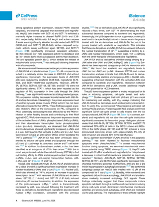
(Figure S1A). We selected six derivatives based on
their potent toxicity against HepG2 human liver cancer cell lines,
including the top four most toxic compounds, SET159, SET158,
SET171, and SET156, along with two additional drugs, SET135
and SET172, which demonstrated high toxicity after 2 and
4 days of treatment. The structures of these six compounds
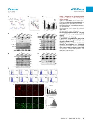
are presented in Figure 1A.
We reassessed the anti-HCC effects of JNK-IN-5A and the six
derivatives alongside the FDA-approved liver cancer drugs,
including sorafenib and regorafenib, using the HepG2 cells
(Figure 1). Initially, we tested the cell viability using the MTT assay
at a concentration of 10 μM for up to 4 days (Figure 1B). The com­
pounds significantly reduced cellular viability: sorafenib (51% ±
3.6), regorafenib (41.1% ± 2.4), JNK-IN-5A (59.7% ± 2.3),
SET135 (43.1% ± 2.7), SET156 (53.7% ± 4.3), SET158
(51.9% ± 0.8), SET159 (61.6% ± 2.9), SET171 (67.4% ± 5.7),
and SET172 (62.7% ± 4.0) at day 2 (Figure 1C). Sorafenib
(19.2% ± 3.4) and regorafenib (13.4% ± 2.3) demonstrated a
time-dependent decrease in cell viability at day 4. Notably,
SET171 showed the lowest cell viability at 1.1% ± 1.1 on day
4, while SET135 also exhibited a strong cell viability inhibition ef­
fect at 15.9% ± 1.4 (Figures 1B and 1C). Both SET135 and
SET171 significantly induced necrotic cell death, as evidenced
by propidium iodide (PI) staining (Figure S1D). The IC50 values
of the compounds were observed (Figure S1E). Based on IC50
values, all compounds were treated at 10 μM to observe fully
affected HepG2 cells by compounds. These data suggest that
JNK-IN-5A and six derivatives may operate through a different
MoA compared to sorafenib and regorafenib, providing a prom­
ising avenue for future drug development for the treatment of
HCC. We examined the expression of proteins associated with
the apoptosis pathway using western blot analysis. This was
conducted on HepG2 cells treated for 2 days with 10 μM com­
pounds (Figure 1D). JNK-IN-5A and six derivatives showed
2 iScience 29, 116005, June 19, 2026
iScience
Article
ll
OPEN ACCESS
Figure 1. Six JNK-IN-5A derivatives induce
apoptosis, cell-cycle arrest, and autophagy
via p53 pathway
(A) Structure of the JNK-IN-5A and six derivatives.
(B and C) Time-dependent cell viability assay (MTT)
for day 1 to day 4 at 10 μM drug concentration.
(D) Apoptosis pathway proteins western blot anal­
ysis of whole lysate.
(E) p-JNK pathway proteins western blot analysis of
whole lysate.
(F) Nuclear protein western blot analysis.
(G) Cellular thermal shift assay (CETSA). Cells were
treated at 20 μM for 2 h and had heat shock at 55◦
C
for 3 min.
(H) FACS analysis with PI staining.
(I) Mitochondrial membrane potential assay. 2 μM
TMRE staining on 10 μM drug-treated HepG2 cells
for 1 day. Scale bars, 100 μm.
(J) Autophagy assay. Autophagic vacuoles were
stained with GFP fluorescence. GFP fluorescence
signals were measured by region of interest (ROI)
using ImageJ program. Scale bars, 100 μm. Data
are represented as mean ± SD. Significance was
tested using t test (*p < 0.05).
iScience 29, 116005, June 19, 2026 3
iScience
Article
ll
OPEN ACCESS
strong apoptosis protein expression, cleaved PARP, cleaved
caspase3, and cleaved caspase9 than sorafenib and regorafe­
nib. HepG2 cells treated with SET135 and SET171 exhibited a
markedly higher increase in cleaved PARP (5.45- and 5.39-
fold, respectively). Additionally, full length and active caspase
3 levels were significantly elevated in cells treated with SET135
(38.90-fold) and SET171 (39.36-fold). Active caspase3 enzy­
matic activity assay confirmed again SET135 and SET171
(Figure S1B) significantly increased caspase3 activation.
SET135 showed 0.71-fold caspase3 activity to docetaxel and
SET171 shoed same level as caspase3 activity than docetaxel.
The anti-apoptotic protein Bcl-2, which inhibits the release of
mitochondrial cytochrome,20
was reduced following treatment
with all compounds.
We also evaluated these suspected target proteins expression
of JNK-IN-5A and six derivatives (Figure 1E). All compounds re­
sulted in a relatively similar decrease in JNK1/2/3 p54 without
significance. Conversely, the expression levels of JNK1/2/3
p46 were reduced by sorafenib (0.68-fold), regorafenib (0.79-
fold), and SET171(0.89-fold) significantly. However, JNK-IN-
5A, SET135, SET156, SET158, SET159, and SET172 were not
significantly altered. STAT1, which has been reported as the
regulator of PKL expression in liver cells through the JNKs
pathway,21
was significantly reduced in all drug-treated groups.
PKL expression notably decreased in cells treated with JNK-IN-
5A and six derivatives. Interestingly, the protein expression level
of another pyruvate kinase muscle (PKM) isoform has not been
affected compared to that of PKL. These findings suggest a spe­
cific inhibitory effect of the compounds on PKL compared to
PKM, potentially decreasing the side effects led by the PKM in­
hibition on other healthy tissues and contributing to their MoA
against HCC. We further measured the protein expression levels
of the activated form of JNKs, phosphorylated JNKs (p-JNKs),
and their downstream transcription factor phosphorylated
c-Jun (p-c-Jun). Interestingly, we observed that JNK-IN-5A
and its derivatives showed significantly increased p-JNKs and
p-c-Jun. Compounds that activate p-JNKs and p-c-Jun have
been shown to have an anti-cancer effect, which facilitates the
TNF-α pathway.22,23
JNK-IN-5A has been previously docu­
mented to induce cell-cycle arrest and apoptosis through the
p53 and p21 pathways in pancreatic cancer and T cell leuke­
mia.24,25
In addition, its downstream protein, c-Jun, has been
identified as an antagonist of p53 in liver cancer.26
With this in
mind, we performed cellular thermal shift assay (CETSA) to find
out binding affinity to JNKs and observed nuclear translocated
p-JNKs, c-Jun, and anti-cancer transcription factors, p53,
c-Myc, and p21 (Figures 1F and 1G).
HepG2 cells treated with 10 μM of JNK-IN-5A and derivatives
for 2 days showed a significant protein expression increase in
nuclear-localized p-JNKs and c-Jun (Figure 1F). Nuclear p53,
which also showed as TNF-α, induced an increase in apoptotic
transcription factor23
with treatment of JNK-IN-5a and six deriv­
atives. SET135 (11.14-fold) and SET171 (7.87-fold) induced
distinctive p53 nuclear localization. In addition, the expression
of the proto-oncogene c-Myc,27
which is transcriptionally
repressed by p53, was reduced following the treatment with
these six derivatives. Sorafenib and regorafenib also decreased
nuclear c-Myc expression, consistent with the previous
studies.28,29
The six derivatives and JNK-IN-5A also significantly
reduced c-Myc levels, with SET171 demonstrating the most
substantial decrease compared to sorafenib and regorafenib.
Interestingly, the p53-dependent tumor suppressor protein p21
was found to be specifically localized in the nucleus in the groups
treated with the six derivatives and JNK-IN-5A, but not in the
groups treated with sorafenib or regorafenib. This indicates
that these six derivatives and JNK-IN-5A may uniquely influence
the nuclear translocation of p21,30
suggesting a binding to p-
JNKs and nuclear translocation, which leads TNF-α pathway
from the other therapies with sorafenib and regorafenib.
JNK-IN-5A and six derivatives showed strong binding to p-
JNK rather than JNK1 and JNK2 in HepG2 cells (Figure 1G). Sor­
afenib has been reported to regulate p-JNK via interaction with
JNKs.31
We found that sorafenib and regorafenib binds to
JNK1 but not JNK2 and p-JNK. However, CETSA and nuclear
translocation analyses indicate that JNK-IN-5A and its deriva­
tives preferentially stabilize and engage p-JNK in HepG2 cells,
suggesting enhanced interaction with the activated JNK pool
compared to sorafenib and regorafenib. Interaction between
JNK-IN-5A derivatives and p-JNK provide additional insight
into their potential for HCC treatment.
The p53 tumor suppressor protein is widely recognized for its
roles in inducing apoptosis, cell-cycle arrest, and auto­
phagy.32–35
Studies suggest that both p53 and p21 are crucial
for maintaining cell viability, with IC50 assays indicating that
JNK-IN-5A and six derivatives exert a robust cell-cycle arrest ef­
fect. To verify this, we conducted PI fluorescence-activated cell
sorting (FACS) analysis. PI staining and FACS analysis confirmed
significant G2/M cell-cycle arrest in cells treated with the six
compounds and JNK-IN-5A (Figure 1H; Table 1). In contrast, sor­
afenib and regorafenib did not alter the cell-cycle distribution
significantly compared to the control group. Histogram plots re­
vealed that JNK-IN-5A, SET156, SET158, SET159, and SET172
maintained 20%–30% of cells in the G0/G1 phase and 60%–
70% in the G2/M phase. SET135 and SET171 induced a more
pronounced cell-cycle arrest, with approximately 5%–6% of
cells in G0/G1 and around 89% in the G2/M phase.
Bcl-2, a protein downstream of JNKs involved in the apoptotic
pathway,36
has been reported to facilitate mitochondrial
apoptosis when phosphorylated.24
To assess mitochondrial
function during apoptosis, we examined mitochondrial mem­
brane potential using TMRE staining (Figure 1I). HepG2 cells
treated with 10 μM of sorafenib and regorafenib for 1 day showed
a decrease in mitochondrial membrane potential, as previously
reported.37
Similarly, JNK-IN-5A, SET135, SET156, SET158,
SET159, SET171, and SET172 also significantly decreased mito­
chondrial membrane potential. Furthermore, JNK and Bcl-2 are
known to participate in autophagic cell death.38,39
We performed
autophagy assays on HepG2 cells treated with 10 μM of the
compounds for 1 day (Figure 1J). Notably, while sorafenib and
regorafenib did not induce autophagy, JNK-IN-5A and six deriv­
atives markedly increased autophagy GFP signals. These
findings collectively suggest that these compounds induce
apoptotic cell death through a multifaceted mechanism involving
strong cell-cycle arrest, diminished mitochondrial membrane
potential, and pronounced autophagy, all of which are mediated
via the p53 pathway. The implications of these effects highlight
4 iScience 29, 116005, June 19, 2026
iScience
Article
ll
OPEN ACCESS
the potential of these compounds as multifunctional agents in
HCC therapy.
We have shown that JNK-IN-5A and six derivatives induce
apoptotic cell death, cell-cycle arrest, and autophagy through
the activation of the p53 tumor suppressor pathway via p-
JNKs activation. This mechanism highlights the compounds
that engage a critical regulatory pathway in cell biology, exploit­
ing the role of the p53 protein in maintaining cellular integrity by
triggering responses to DNA damage, stress signals, and aber­
rant growth signals. By activating this pathway, the JNK-IN-5A
and six derivatives effectively promote the removal of damaged
or cancerous cells, providing a targeted approach to HCC ther­
apy that harnesses the body’s natural defences against tumor
progression.
SET135 and SET171 reprogram the necrotic cell death
In our detailed investigation of the effects of the six derivatives on
cell death mechanisms, we monitored the progression of
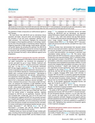
necrotic cell death for up to 4 days. Notably, treatment with
10 μM SET135 and SET171 induced pronounced cytotoxicity
from day 1 to day 4 (Figure 2A). As previously mentioned,
SET135 also triggers apoptosis (Figure 1D). The concurrent in­
duction of apoptosis and necrosis observed in this study is remi­
niscent of the effects reported for Tanshinone IIA treatment on
HepG2 cells, a process termed necroptosis.40
Necroptosis is
characterized as programmed cell necrosis, distinguishing it
from apoptosis in several key ways.41,42
Unlike apoptosis, during
necroptosis, the protein p62/SQSTM1 recruits autophagic pro­
teins to form a necrosome, leading to necrotic cell death.43
To
further understand this process in the context of SET135 treat­
ment, we analyzed the expression of p62 (SQSTM1) and its
phosphorylated form, phospho-p62 (S349)—an activated state
of p62—using western blot analysis (Figure 2B). Treatment of
HepG2 cells with 10 μM SET135 for 2 days resulted in signifi­
cantly elevated levels of both p62 and p-S349 p62. We found
LC3, the central protein in autophagy pathway, relatively
increased by SET135 but not significant. However, SET135
only increased p62, p-S349 p62, and LC3 at the same time.
This observation suggests that SET135 may activate necropto­
sis in HepG2 cells through mechanisms involving both the upre­
gulation of p62, p-S349 p62, and LC3 contributing to the forma­
tion of the necrosome complex and showing necrotic cell death.
For further confirmation of necroptosis induced necrosis, we co-
treated Necrostatin-144
with SET135 (Figure 2C). 10 μM
Necrostatin-1 co-treated with SET135 showed significant
reduced necrotic cell death (52.9%–24.6%).
Oxidative stress can induce cell death through multiple path­
ways, including mitochondrial damage via caspase-3 activation,
DNA damage through cleaved PARP, and the initiation of auto­
phagy.45–47
To understand the mechanism behind cell death
induced by JNK-IN-5A and six derivatives, we assessed
cellular ROS levels in HepG2 cells treated with 10 μM of the com­
pounds for 1 day. Cells were stained with the ROS detector
2′
,7′
-dichlorodihydrofluorescein diacetate (DCFDA). The fluores­
cence image analysis showed that SET171 significantly
increased cellular ROS (Figure 2D). Notably, SET171 was asso­
ciated with pronounced late necrotic cell death from days 3 and
4 (Figure 2A).
Previous studies have demonstrated that elevated cellular
ROS can induce lipid peroxidation, leading to membrane rupture
and necrotic cell death, a process referred to as ferroptosis.48,49
To confirm lipid peroxidation, we employed a ratiometric lipid
peroxidation sensor staining (Figure 2E). This sensor stains
cellular lipids with RFP fluorescence, which is replaced by GFP
fluorescence upon lipid peroxidation. SET171 exhibited a statis­
tically significant increase in the GFP/RFP ratio, indicating lipid
peroxidation, unlike other compounds tested. We also measured
the expression level of glutathione peroxidase 4 (GPX4), a crucial
enzyme that mitigates lipid peroxidation and plays a key role in
preventing ferroptosis.50
HepG2 cells treated with sorafenib
and regorafenib showed significantly higher GPX4 expression
compared to the control (Figure 2B), while treatments with
JNK-IN-5A, SET156, SET158, SET159, and SET172 resulted in
similar or slightly increased GPX4 levels (1.00–1.29-fold). In
contrast, SET135 and SET171 showed a significantly decrease
in GPX4 expression levels (0.70-fold and 0.63-fold, respectively).
We also tested ferroptosis inhibitor Ebselen51
with SET171.
Not as much as necrosis inhibition effect of Necrostatin-1 with
SET135, 10 μM ebselen significantly decreased necrotic cell
death, 83.7%–60.5%, by SET171 (Figure 2C).
These results suggest that SET171 induces ferroptosis
through mechanisms involving increased cellular ROS, lipid per­
oxidation, and reduced GPX4 expression.
The ability of SET135 and SET171 to simultaneously induce
apoptosis and necrosis. SET135 and SET171 showed necropto­
sis and ferroptosis features. However, it is not clear that how
much necrotic cell death affect to anti-HCC effect and how
related to apoptotic cell death. Despite unclear specific pathway
between apoptosis and necrosis, SET135 and SET171 represent
a promising approach to overcome resistance to apoptosis in
cancer cells, highlighting their potential as therapeutic agents
in HCC treatment strategies.52
JNK-IN-5A derivatives inhibit cancer invasion and
mobility in HepG2 cells
We assessed the impact of the compounds on the invasiveness
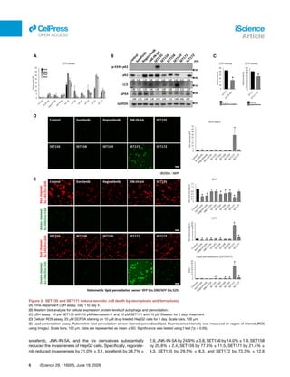
and motility of HepG2 cells (Figure 3). Invasion assay conducted
with a concentration of 1 μM revealed that regorafenib,
Table 1. Cell population on PI FACs analysis
Control Sorafenib Regorafe-nib JNK-IN-5A SET135 SET156 SET158 SET159 SET171 SET172
G0/G1 62.4 62.2 70.5 22.7 5.5 30.1 31.6 30.5 6.6 16.7
S 15.4 10.1 9.7 4.4 2.0 2.3 3.5 3.3 3.6 2.7
G2/M 16.2 19.5 14.9 67.3 79.9 60.8 60.3 62.4 79.1 73.2
JNK-IN-5A and six derivatives induce G2/M cell-cycle arrest. SET135 and SET171 demonstrated the most significant cell cycle arrest potency, nearly
80% G2/M phase accumulation, which represents stronger effect than other compounds and FDA-approved anti-HCC drugs.
iScience 29, 116005, June 19, 2026 5
iScience
Article
ll
OPEN ACCESS
sorafenib, JNK-IN-5A, and the six derivatives substantially
reduced the invasiveness of HepG2 cells. Specifically, regorafe­
nib reduced invasiveness by 21.0% ± 3.1, sorafenib by 28.7% ±
2.9, JNK-IN-5A by 24.9% ± 3.8, SET156 by 14.0% ± 1.9, SET158
by 20.8% ± 2.4, SET159 by 77.8% ± 11.5, SET171 by 21.4% ±
4.3, SET135 by 29.5% ± 8.3, and SET172 by 72.3% ± 12.6
Figure 2. SET135 and SET171 induce necrotic cell death by necroptosis and ferroptosis
(A) Time-dependent LDH assay. Day 1 to day 4.
(B) Western blot analysis for cellular expression protein levels of autophage and peroxidation.
(C) LDH assay. 10 μM SET135 with 10 μM Necrostatin-1 and 10 μM SET171 with 10 μM Ebselen for 2 days treatment.
(D) Cellular ROS assay. 20 μM DCFDA staining on 10 μM drug-treated HepG2 cells for 1 day. Scale bars, 100 μm.
(E) Lipid peroxidation assay. Ratiometric lipid peroxidation sensor-stained peroxidised lipid. Fluorescence intensity was measured on region of interest (ROI)
using ImageJ. Scale bars, 100 μm. Data are represented as mean ± SD. Significance was tested using t test (*p < 0.05).
6 iScience 29, 116005, June 19, 2026
iScience
Article
ll
OPEN ACCESS
(Figure 3A). Western blot analysis revealed that sorafenib, regor­
afenib, JNK-IN-5A, and the six derivatives led to a decrease in
MMP9 levels (Figure 3B), a matrix metalloproteinase implicated
in cancer invasion and metastasis. Furthermore, the expression
levels of proteins involved in cancer cell migration and motility,
such as α-tubulin53–55
and β-actin,56,57
were significantly
reduced.
Additionally, a wound-healing assay was performed to eval­
uate the effects of the drugs on cancer cell motility. After
achieving a confluent state, a wound was created in the
HepG2 cell layer using a pipette tip, and the wound width was
measured through image analysis until the control HepG2 cells
closed the wounded area by day 4 (Figure 3C). The results
showed that all tested compounds delayed wound healing
compared to untreated HepG2 cells, which achieved 100%
wound closure by day 4. Specifically, regorafenib delayed
wound healing by 44.8% ± 17.4, sorafenib by 35.0% ± 9.9,
JNK-IN-5A by 32.7% ± 10.4, SET156 by 26.2% ± 15.3,
SET158 by 39.5% ± 6.1, SET159 by 35.5% ± 2.6, SET171 by
36.4% ± 14.0, SET135 by 31.2% ± 9.4, and SET172 by
60.7% ± 5.0. These findings suggest that the tested drugs effec­
tively impair the invasiveness and motility of HepG2 cells, high­
lighting their potential as therapeutic agents in limiting HCC pro­
gression and metastasis.
JNK-IN-5A derivatives show unique transcriptional
profiles in HepG2 cells
To elucidate the changes in gene expression and biological path­
ways induced by drug treatment, we conducted RNA sequencing
(RNA-seq) analysis on HepG2 cells treated with JNK-IN-5A, six
derivatives, sorafenib and regorafenib. The analysis revealed
distinct transcriptional profiles for each treatment group
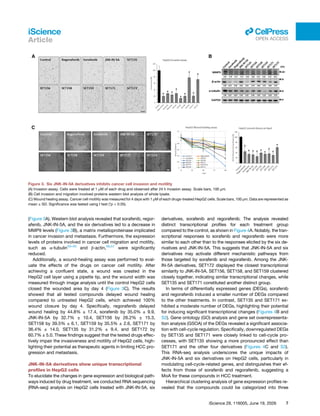
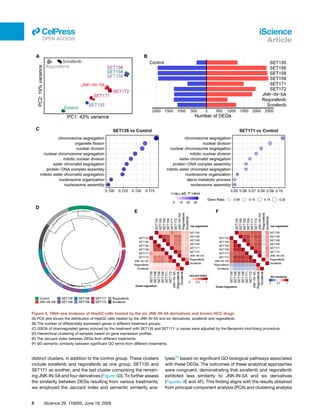
compared to the control, as shown in Figure 4A. Notably, the tran­
scriptional responses to sorafenib and regorafenib were more
similar to each other than to the responses elicited by the six de­
rivatives and JNK-IN-5A. This suggests that JNK-IN-5A and six
derivatives may activate different mechanistic pathways from
those targeted by sorafenib and regorafenib. Among the JNK-
IN-5A derivatives, SET172 displayed the closest transcriptional
similarity to JNK-IN-5A. SET156, SET158, and SET159 clustered
closely together, indicating similar transcriptional changes, while
SET135 and SET171 constituted another distinct group.
In terms of differentially expressed genes (DEGs), sorafenib
and regorafenib induced a smaller number of DEGs compared
to the other treatments. In contrast, SET135 and SET171 ex­
hibited a moderate number of DEGs, highlighting their potential
for inducing significant transcriptional changes (Figures 4B and
S2). Gene ontology (GO) analysis and gene set overrepresenta­
tion analysis (GSOA) of the DEGs revealed a significant associa­
tion with cell-cycle regulation. Specifically, downregulated DEGs
by SET135 and SET171 were closely linked to cell-cycle pro­
cesses, with SET135 showing a more pronounced effect than
SET171 and the other four derivatives (Figures 4C and S3).
This RNA-seq analysis underscores the unique impacts of
JNK-IN-5A and six derivatives on HepG2 cells, particularly in
modulating cell-cycle-related genes, and distinguishes their ef­
fects from those of sorafenib and regorafenib, suggesting a
MoA for these compounds in HCC treatment.
Hierarchical clustering analysis of gene expression profiles re­
vealed that the compounds could be categorized into three
Figure 3. Six JNK-IN-5A derivatives inhibits cancer cell invasion and motility
(A) Invasion assay. Cells were treated at 1 μM of each drug and observed after 24 h invasion assay. Scale bars, 100 μm.
(B) Cell invasion and migration involved proteins western blot analysis of whole lysate.
(C) Wound healing assay. Cancer cell motility was measured for 4 days with 1 μM of each drugs-treated HepG2 cells. Scale bars, 100 μm. Data are represented as
mean ± SD. Significance was tested using t test (*p < 0.05).
iScience 29, 116005, June 19, 2026 7
iScience
Article
ll
OPEN ACCESS
distinct clusters, in addition to the control group. These clusters
include sorafenib and regorafenib as one group, SET135 and
SET171 as another, and the last cluster comprising the remain­
ing JNK-IN-5A and four derivatives (Figure 4D). To further assess
the similarity between DEGs resulting from various treatments,
we employed the Jaccard index and semantic similarity ana­
lyses58
based on significant GO biological pathways associated
with these DEGs. The outcomes of these analytical approaches
were congruent, demonstrating that sorafenib and regorafenib
exhibited less similarity to JNK-IN-5A and six derivatives
(Figures 4E and 4F). This finding aligns with the results obtained
from principal component analysis (PCA) and clustering analysis
Figure 4. RNA-seq analyses of HepG2 cells treated by the six JNK-IN-5A derivatives and known HCC drugs
(A) PCA plot shows the distribution of HepG2 cells treated by the JNK-IN-5A and six derivatives, sorafenib and regorafenib.
(B) The number of differentially expressed genes in different treatment groups.
(C) GSOA of downregulated genes induced by the treatment with SET135 and SET171. p values were adjusted by the Benjamini-Hochberg procedure.
(D) Hierarchical clustering of samples based on gene expression profiles.
(E) The Jaccard index between DEGs from different treatments.
(F) GO semantic similarity between significant GO terms from different treatments.
8 iScience 29, 116005, June 19, 2026
iScience
Article
ll
OPEN ACCESS
(Figures 4A–4E), reinforcing the notion that JNK-IN-5A and six
derivatives potentially engage distinct biological mechanisms
compared to sorafenib and regorafenib. Within the group of
JNK-IN-5A and three derivatives, including SET156, SET158,
and SET159, there was a close relationship, indicating a high de­
gree of similarity among their effects. These compounds also
showed a notable resemblance to JNK-IN-5A, suggesting
shared pathways or targets that might underlie their therapeutic
actions.
To thoroughly assess the pathways disrupted by the treat­
ments, gene set enrichment analysis (GSEA) was conducted us­
ing hallmark gene sets from the Molecular Signatures Database
(MSigDB)59
and the Kyoto Encyclopedia of Genes and Genomes
(KEGG) pathway.60
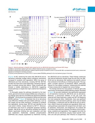
Figure 5A illustrates that all six derivatives
more effectively inhibited cell-cycle-related pathways, such as
E2F targets and the G2M checkpoint, compared to sorafenib
and regorafenib. Among these, SET135 was identified as the
most potent inhibitor, closely followed by SET171. Moreover,
all treatments were found to upregulate apoptosis signaling.
Notably, JNK-IN-5A and six derivatives exhibited liver-specific
effects, specifically inhibiting bile acid metabolism and fatty
acid metabolism. Additionally, oxidative phosphorylation
(OXPHOS) was uniquely suppressed by treatments involving
the JNK-IN-5A and six derivatives. These findings underscore
that while all treatments robustly hinder the cell cycle, the JNK-
IN-5A and six derivatives offer additional liver-specific actions
that contribute to their efficacy in inhibiting cell growth. This sug­
gests a broader spectrum of therapeutic effects, particularly
relevant for liver-related conditions, underscoring the potential
of these compounds for targeted liver cancer therapy.
The findings derived from the analysis of hallmark gene sets
were further corroborated by KEGG pathway analysis. Illustrated
in Figure 5B, pyruvate metabolism was significantly suppressed
in all treatments involving the JNK-IN-5A and six derivatives.
Moreover, a pronounced inhibition of the cell cycle was
observed with these treatments, notably with SET135 displaying
the most substantial effect. Additionally, there was significant
suppression of the peroxisome pathway, which may be related
to autophagy, in treatments with the JNK-IN-5A and six deriva­
tives, unlike in those with sorafenib and regorafenib. When eval­
uating pathways associated with programmed cell death, such
as apoptosis, endocytosis, and phagosome, the analysis did
not reveal a clear distinction between the effects of the six deriv­
atives, sorafenib and regorafenib. This suggests that while the
JNK-IN-5A and six derivatives uniquely impact specific meta­
bolic and cell-cycle pathways, their influence on programmed
Figure 5. Altered pathways in HepG2 cells treated by the six JNK-IN-5A derivatives and known HCC drugs
(A) GSEA of representative hallmark gene sets in different treatment groups compared to control.
(B) GSEA of representative KEGG pathways in different treatment groups compared to control. For (A) and (B), NES: normalized enrichment score; p values were
adjusted by the Benjamini-Hochberg procedure.
(C) Activity scores (presented as Z score) of the 14 cancer-related PROGENy pathways for the nine treatment groups in this study.
iScience 29, 116005, June 19, 2026 9
iScience
Article
ll
OPEN ACCESS
cell death pathways may share similarities with that of sorafenib
and regorafenib. These insights suggest that the unique thera­
peutic potential of the JNK-IN-5A and six derivatives could be
attributed to their specific mechanisms of action on metabolic
regulation and cell-cycle control, rather than a fundamentally
different approach to inducing programmed cell death.
Using PROGENy, a computational algorithm designed to
infer the activity of 14 cancer-related signaling pathways from
gene expression data,61
we observed that SET135 and
SET171 significantly activated the p53 signaling pathways,
confirming the findings shown in Figure 1. Furthermore, while
sorafenib and regorafenib showed a high level of activation of
the TGFβ pathway, such activation was not observed in treat­
ments with the six derivatives. This suggests a different func­
tional profile of these compounds compared to the two FDA-
approved cancer drugs.
Our comprehensive analyses using HepG2 cells indicate that
although all treatments are capable of inhibiting cancer cell
growth, with SET135 and SET171 being the most effective of
the six derivatives, JNK-IN-5A likely possesses unique and
liver-specific effects that distinguish it from sorafenib and regor­
afenib. This distinction suggests that JNK-IN-5A and six deriva­
tives might offer alternative therapeutic mechanisms, potentially
providing new avenues for the treatment of HCC, particularly by
targeting pathways such as p53, which are crucial in the regula­
tion of cancer cell survival and death.
Normal liver cell line and GLP-like toxicity study in rats
supporting the safety of the compounds
We have checked toxicity to normal cells on the THLE-2 non-
tumorigenic human liver epithelial cell line in vitro (Figure S1C).
All compounds were treated at 5 μM concentration to 5,000 cells
for 2 days. JNK-IN-5A and derivatives showed significantly
reduced toxicity at THLE-2 cells as sorafenib and regorafenib.
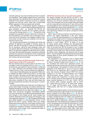
Seven-day oral tolerability study was conducted in Wistar rats
to evaluate the safety of SET135 and SET171 at three dose
levels: 30 mg/kg, 100 mg/kg, and 300 mg/kg body weight. The
study included a vehicle control group (four males and four fe­
males) and three dose groups for each compound, with each
dose group consisting of three male and three female rats.
Dosing was performed once daily in the morning for 7 consecu­
tive days.
No clinical adverse effects were observed in the treated animal
groups. Body weight, hematology, and plasma chemistry were
assessed throughout the study. We observed that the treatment
with these compounds did not affect body weight (Figure 6A).
While mean corpuscular hemoglobin concentration (MCHC)
and mean corpuscular volume (MCV) showed statistically signif­
icant changes in some treated animals, these changes were
randomly distributed across dose groups and showed no clear
dose-dependent response (Figure 6B). Phosphate (PHOS) levels
were slightly elevated in a dose-dependent manner in both
SET135 and SET171 groups, whereas blood urea nitrogen
(BUN) levels remained within the normal range, indicating no kid­
ney toxicity (Figure 6C). These minor variations were considered
incidental and unrelated to treatment. All other parameters re­
mained within normal ranges, further supporting the safety of
both compounds (Figure S6).
JNK-IN-5A derivatives show in vivo anti-tumor activity
Our analysis indicated that both SET135 and SET171 have
potent inhibitory effects on HCC cell models. Hence, we evalu­
ated the in vivo anti-tumor activities of these two compounds
in Huh7 HCC xenograft tumor animal models. Mice were treated
with the compounds every other day for 21 days. We found that
the body weights of mice in all treatment groups were not signif­
icantly different from those of the control group (Figure 7A). We
observed that JNK-IN-5A, SET135, SET171, and sorafenib treat­
ment significantly inhibited tumor growth compared to vehicle
control (Figure 7B).
After 21 days of compound treatment, we performed visual in­
spection and weight measurement of the tumor tissues
(Figures 7C and 7D). Daily administration of 10 mg/kg sorafenib
decreased tumor weight to 52.27% compared to vehicle control.
JNK-IN-5A at 10 mg/kg in the treated group showed a 58.6%
reduction in tumor weight. SET171 at doses of 10 and
20 mg/kg treated groups markedly and dose-dependently
decreased the tumor weight to 73.87% and 50.05%, respec­
tively. On the other hand, SET135 at doses of 5 and 10 mg/kg
in the treated groups showed a markedly and dose-dependently
decrease in tumor weight to 67.19% and 42.65% compared to
the vehicle control, respectively. We observed that 20 mg/kg
SET171- and 10 mg/kg SET135-treated groups showed stronger
inhibitory effects on tumor growth compared to the 10 mg/kg
sorafenib-treated group.
We investigated in situ apoptosis using a cleaved PARP
IHC, TUNEL assay and cell-cycle arrest using ki67 IHC for
21 days of compound-treated tumor tissues (Figures 7E and
7F). Consistent with overall tumor suppressive activity, we
detected significantly induced cell-cycle arrest and apoptosis
in all treated groups. Cell proliferation marker protein ki67 was
decreased at 20 mg/kg SET171 up to 21% and 10 mg/kg
SET135 up to 5.7%, respectively. SET171 and SET135 treated
group also showed increased cleaved PARP IHC staining
intensity to 3- to 4-fold. 10 mg/kg sorafenib- and 10 mg/kg
JNK-IN-5A-treated groups induced 14.27% and 11.54%
TUNEL positive cells, respectively. 10 mg/kg and 20 mg/kg
SET171-treated groups showed significant and dose-depen­
dently induced apoptosis at 4.71% and 14.84%. SET135
also showed highly increased apoptotic cells by 5.64%
at 5 mg/kg dose and 25.4% at 10 mg/kg dose. Notably,
the 10 mg/kg SET135-treated group showed stronger
TUNEL-positive cells than the 10 mg/kg sorafenib-treated
group.
Throughout the treatment period, we did not observe obvious
toxicities including body weight change of mice in all treatment
groups compared to the vehicle group (Figure 7A). We per­
formed pathological and hematological examinations for six ma­
jor organs, including the heart, liver, spleen, lung, and kidney tis­
sues, based on H&E staining (Figures 7G and S7). We found that
there are no significant differences in lesions or toxic-damage
sites between all 21-day treated groups compared to the vehicle
control. Hematological and biochemical blood tests for 21 days
of compound-treated mice also showed that JNK-IN-5A,
SET135, and SET171 have fewer side effects than sorafenib
(Figure S7). Hematological test for WBC, RBC, HGB, MCH,
MCHC, MCV, PLT, HCT ,and serum biochemical test for ALT,
10 iScience 29, 116005, June 19, 2026
iScience
Article
ll
OPEN ACCESS
AST, γ-GT, TBIL, DBIL, ALP, ALB, BUN, UA, and CREA showed
no significant changes in the hematological and serum biochem­
ical parameters. These in vivo analyses indicated that deriva­
tives, including SET135 and SET171, have superior or compara­
ble anti-HCC effects and stability compared to sorafenib, a
conventional HCC drug.
DISCUSSION
The liver, renowned for its unique roles in drug metabolism and
detoxification, is a vital organ in the management of xenobiotics.
HCC typically has a lower response rate to chemotherapy than
many other cancers.62
Current clinical treatments often include
Figure 6. GLP-like toxicity study supporting the safety of the compounds in rats
(A) Body weight changes over time across treatment groups.
(B and C) Mean corpuscular hemoglobin concentration (MCHC), mean corpuscular volume (MCV), and phosphate (PHOS) levels exhibited statistically significant
changes over time. Blood urea nitrogen (BUN) levels remained stable, suggesting no evidence of kidney toxicity.
iScience 29, 116005, June 19, 2026 11
iScience
Article
ll
OPEN ACCESS
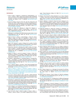
Figure 7. The anti-tumor efficacy of the
SET135 and SET171 in Huh7 bearing animal
model
Huh7 cells were transplanted subcutaneously to the
BALB/nude mice and subjected to SET171 (10 and
20 mg/kg), SET135 (5 and 10 mg/kg), JNK-IN-5A
(10 mg/kg), sorafenib (10 mg/kg), and vehicle
control.
(A) Body weight changes for 21 days measured
every other day.
(B) Tumor volume changes for 21 days measured
every other day.
(C and D) Isolated tumor weights and image after
21 days treatment.
(E and F) Statistic and image of IHC staining (ki67
and cleaved PARP) and fluorescence staining
TUNEL positive cells. Scale bars, 100 μm.
(G) Represented H&E staining images of the major
organs of the mice after 21 days treatment. Scale
bars, 100 μm. Data are represented as mean ± SD.
Significance was tested using t test (*p < 0.05,
**p < 0.005, ***p < 0.0005). Scale bars, 100 μm.
12 iScience 29, 116005, June 19, 2026
iScience
Article
ll
OPEN ACCESS
multi-target kinase inhibitors such as sorafenib and regorafenib;
however, the need for therapeutic options is underscored by
their modest efficacy,63
and the development of chemoresist­
ance in some patient64,65
highlights the need for therapeutic
options.
In our earlier research, we identified the PKLR gene as a po­
tential drug target for HCC treatment and proposed JNK-IN-5A
as a potent inhibitor/modulator of PKLR protein expression, sug­
gesting it as a potential therapeutic agent for HCC.14,16
Building
on this, we have now introduced six novel drug candidates
derived from JNK-IN-5A. Beyond their role in decreasing PKL
expression, the JNK-IN-5A and six derivatives have demon­
strated anticancer effects via p-JNKs activation and interfering
TNF-α signaling pathway. JNK-IN-5A induces the expression
of p53-dependent tumor suppressor proteins, decreases c-
Myc levels, and leads to a significant increase in p21 expression
across all treatment groups. The compounds’ ability to selec­
tively enhance nuclear p21 expression provides strong evidence
of their capacity to induce G2/M phase cell-cycle arrest. Specif­
ically, SET135 and SET171 have shown remarkable anticancer
effects, inducing both apoptotic and necrotic cell death in
HepG2 cells. Moreover, these drugs significantly augment nu­
clear p53 expression and reduce c-Myc expression, surpassing
the effects of JNK-IN-5A, sorafenib, and regorafenib. Therefore,
the mechanisms by which SET135 and SET171 elicit apoptotic
cell death, necroptosis, and ferroptosis merit further exploration.
SET135 has been observed to induce the overexpression of
p62 and its phosphorylated form at serine 349 (p-S349 p62),
which is recognized as a potent oncogenic protein.66
The phos­
phorylation of p62 at serine 349 enhances its binding affinity to
the Keap1 protein, leading to the activation of the Nrf2 pathway.
This pathway is well-known for its cytoprotective functions in
normal cells. However, it also plays a role in tumorigenesis in
pre-malignant cells, contributing to drug resistance and facili­
tating tumor growth. p62, which is a crucial component of protein
aggregates forming Mallory-Denk bodies (MDBs) or intracellular
hyaline bodies (IHBs), is considered both a prognostic and diag­
nostic marker for NASH and HCC.67–69
A recent study has differ­
entiated two isoforms of p62: p62-H1, the full-length isoform,
and p62-H2, which is partially devoid of the PB1 domain.70
These
isoforms differ in their aggregation potential. Notably, p62-H2 is
more predominantly expressed in human liver tissue than p62-
H1. Intriguingly, in our study, the SET135 treatment group dis­
played a significant increase in p62-H1 expression exclusively,
as shown in Figure 2B. This finding positions SET135 as a critical
agent for further research into the role of p62/SQSTM1 as a
target protein in disease mechanisms. The distinct expression
pattern elicited by SET135 treatment provides valuable insights
that could contribute to understanding the molecular underpin­
nings of diseases associated with p62 dysregulation.
In animal studies, SET135 and SET171 showed promising
anti-HCC effects with conventional HCC drug sorafenib. For
21 days, SET135 and SET171 inhibited the transplanted Huh7
cells’ tumor growth, and the TUNEL assay revealed that they
strongly induced apoptotic cell death. 10 mg/kg SET135 showed
stronger tumor growth inhibition and a higher rate of apoptotic
cell death. More interestingly, SET135 and SET171 did not
show toxic side effects on the major five organs, heart, liver,
spleen, lung, and kidney, and also hematologic and serum
biochemical tests.
We repositioned the drug candidates based on their ability to
modulate PKL expression levels. However, we found that siRNA-
mediated inhibition of PKL did not manifest the desired anti­
cancer effect, as shown in Figure S4, since WT HepG2 cells
also express PKM2. We transfected three distinct siRNAs into
HepG2 cells and, after 3 days, observed no statistically signifi­
cant change in cell viability (Figure S4A). Furthermore, western
blot analysis revealed that the suppression of PKL expression
via siRNAs did not result in increased cleaved-PARP or
decreased Bcl-2 expression levels, unlike the effects seen with
JNK-IN-5A and six derivatives (Figure S4B). This suggests the
presence of alternative pathways, potentially involving p53 and
c-Myc, that lead to apoptosis.
Open MoA is a previously developed robust tool for eluci­
dating the MoA of drugs by leveraging network topology and hi­
erarchy.21
Utilizing drug-specific DEGs data, Open MoA con­
structs drug-specific subnetworks and identifies the most
probable pathways from the drugs to p53 or c-Myc. All identified
interactions have a high confidence score, surpassing 0.99
(Figure S5). For the JNK-IN-5A, MAPK8 or MAPK9, the known
target of JNK-IN-5A, was selected as the starting point.18
JUN
was identified as the sole intermediate target in the projected
MoA, with its strong association with p53 and c-Myc supported
by prior research.26
Regarding regorafenib, of its 18 direct down­
stream targets, ABL1 was pinpointed as having the highest likeli­
hood of interacting with p53 and c-Myc. For sorafenib, among its
eight downstream targets, PDGFRB and FGFR1 were deemed
the most likely to influence the mechanisms leading to p53 and
c-Myc, with STAT1 and SOS1 acting as intermediate proteins,
respectively.
Although the predicted MoAs for sorafenib, regorafenib, and
JNK-IN-5A showed significant variation, none included PKL,
although all compounds inhibited PKL protein expression
(Figure 1D). The intermediate proteins identified within these
MoAs may serve as pivotal targets through which the drugs
impact apoptosis by modulating p53 and c-Myc activation.
The differing MoAs might contribute to the observed variations
in cell apoptosis. To further increase our understanding and
improve the efficacy of the JNK-IN-5A and six derivatives, addi­
tional in vivo studies are needed. These investigations should
aim to fully characterize the drugs’ MoAs and their effects on
cellular processes, particularly those related to p53 and c-Myc
signaling pathways.
In summary, we characterized the anti-HCC effect of JNK-IN-
5A and six potent derivatives, SET135, SET156, SET158,
SET159, SET171, and SET172. Mechanism study revealed that
six derivatives induce apoptotic cell death and cell-cycle arrest
through activation of the p53 signaling pathway by activating
p-JNK pathway and autophagy via mitochondrial stress by
Bcl2 inhibition. SET135 and SET171 uniquely triggered necrop­
totic and ferroptotic pathways, indicating their multifaceted po­
tential to overcome apoptosis resistance in HCC. Furthermore,
transcriptional profiling demonstrated that these compounds
modulate cell-cycle and metabolism-related pathways distinct
from those targeted by sorafenib and regorafenib. SET135 and
SET171 also exhibited superior in vivo anti-tumor efficacy
iScience 29, 116005, June 19, 2026 13
iScience
Article
ll
OPEN ACCESS
compared to sorafenib, while showing safety profiles in animal
model. Collectively, our findings establish SET135 and SET171
as promising multi-mechanistic therapeutic candidates for
HCC treatment, providing new insights into the p-JNK pathway
as a therapeutic target for HCC.
Limitations of the study
Our study extensively showed that JNK-IN-5A and its deriva­
tives, specifically SET135 and SET171, induces stronger
apoptotic cell death compare to conventional drugs, sorafenib
and regorafenib, additional cell-cycle arrest and also necrotic
cell death. These results are supported with computational
biology and also in vivo study. Despite this vast range of anti-
HCC effect, there are still works to prove to develop new drug.
First, much more strong MoA between target protein and deriv­
atives. We showed interaction between compounds and p-JNKs
with CETSA and cellular protein expression, and also activation
of c-JUN and nuclear translocation but these results may have
considered as an indirect evidence. CETSA showed significant
thermal shift at 55◦
C. Though, complete CETSA melting curves
across a temperature gradient or microscale thermophoresis
(MST) with purified p-JNK will convince binding affinity between
compounds and p-JNK. We showed strong necrotic cell death
by SET135 and SET171 and described as necroptosis and fer­
roptosis. However, still needed more detailed experiment to
prove how SET135 and SET171 induce apoptosis necrosis at
the same time. Immune microenvironment is considered as a
critical role in regulating tumor progression and resistance to
anti-HCC compounds.71,72
We have needed to perform further
study that how our new six derivative can show anti-HCC effect
on tumor microenvironment (TME) in vitro and in vivo animal
model. Major organ histology of 21 days mouse treatment with
10 to 20 mg/kg dose and GLP-like toxicity test of 7 days rat treat­
ment with 30–300 mg/kg dose showed JNK-IN-5A derivatives
are not strong toxic. However, to develop new drug pharmaco­
kinetics analysis in vivo is demanded.
RESOURCE AVAILABILITY
Lead contact
Further information and requests for resources and reagents should be
directed to and will be fulfilled by the lead contact, Adil Mardinoglu (adilm@
scilifelab.se).
Materials availability
This study did not generate new unique reagents.
Data and code availability
• RNA-sequencing data generated in this study have been deposited in
the Gene Expression Omnibus and are publicly available under acces­
sion number GSE255617. This study did not generate original codes.
• Microscopy data reported in this paper will be shared by the lead con­
tact upon request. Any additional information required to reanalyze the
data reported in this paper is available from the lead contact upon
request.
SIGNIFICANCE STATEMENT
This study identifies JNK/c-Jun pathway as a therapeutic target
to hepatocellular carcinoma and demonstrates that JNK-IN-5A
derivatives induce distinct apoptotic and necrotic cell death
mechanisms, offering a promising therapeutic strategy beyond
current kinase inhibitors.
ACKNOWLEDGMENTS
The authors would like to acknowledge financial support from ScandiEdge
Therapeutics and the Knut and Alice Wallenberg Foundation.
AUTHOR CONTRIBUTIONS
Conceptualization, W.K. and A.M.; methodology, W.K., H.J., M.O., P.M., X.L.,
M.L., S.I., J.S., S.A, and B.B.; writing – original draft, W.K., H.J., M.O., P.M., X.
L., and M.L.; writing – review and editing, all authors involved editing and
approved the final version of the article; supervision, A.M., M.U., C.Z., X.S.,
J.B., and H.T.
DECLARATION OF INTERESTS
A.M., J.B., and M.U. are the shareholders of ScandiEdge Therapeutics AB.
STAR★METHODS
Detailed methods are provided in the online version of this paper and include
the following:
• KEY RESOURCES TABLE
• EXPERIMENTAL MODEL AND STUDY PARTICIPANT DETAILS
○ Cell culture, cell viability and cytotoxicity assay
• METHOD DETAILS
○ Western blot and CETSA analysis
○ FACs analysis
○ Mitochondria membrane potential, autophagy, ROS, and lipid per­
oxidation assay
○ Caspase-3 activity assay
○ Invasion assay and wound healing assay
○ RNA extraction and sample preparation for RNA sequencing
○ In vivo anti-tumor activity evaluation
○ Immunohistochemistry (IHC) experiment
○ TUNEL staining
○ Pathological examinations
○ Haematological tests
• QUANTIFICATION AND STATISTICAL ANALYSIS
○ RNA-sequencing
○ RNA-seq data pre-processing
○ Differential expression analysis
○ GLP-like toxicity study in rats
○ Principal component analysis
○ Gene set analysis
○ Comparison of DEGs
○ Mechanism of action (MoA) prediction
○ Statistical analysis for RNA-seq and experimental data
SUPPLEMENTAL INFORMATION
Supplemental information can be found online at https://doi.org/10.1016/j.isci.
2026.116005.
Received: August 6, 2025
Revised: November 14, 2025
Accepted: April 30, 2026
14 iScience 29, 116005, June 19, 2026
iScience
Article
ll
OPEN ACCESS
REFERENCES
1. Sung, H., Ferlay, J., Siegel, R.L., Laversanne, M., Soerjomataram, I., Je­
mal, A., and Bray, F. (2021). Global Cancer Statistics 2020: GLOBOCAN
Estimates of Incidence and Mortality Worldwide for 36 Cancers in 185
Countries. CA. Cancer J. Clin. 71, 209–249. https://doi.org/10.3322/
caac.21660.
2. Tan, D.J.H., Wong, C., Ng, C.H., Poh, C.W., Jain, S.R., Huang, D.Q., and
Muthiah, M.D. (2021). A Meta-Analysis on the Rate of Hepatocellular Car­
cinoma Recurrence after Liver Transplant and Associations to Etiology,
Alpha-Fetoprotein, Income and Ethnicity. J. Clin. Med. 10, 238. https://
doi.org/10.3390/jcm10020238.
3. Llovet, J.M., Kelley, R.K., Villanueva, A., Singal, A.G., Pikarsky, E.,
Roayaie, S., Lencioni, R., Koike, K., Zucman-Rossi, J., and Finn, R.S.
(2021). Hepatocellular carcinoma. Nat. Rev. Dis. Primer 7, 6. https://doi.
org/10.1038/s41572-020-00240-3.
4. Mardinoglu, A., and Palsson, B.Ø. (2025). Genome-scale models in human
metabologenomics. Nat. Rev. Genet. 26, 123–140. https://doi.org/10.
1038/s41576-024-00768-0.
5. Reig, M., Forner, A., Rimola, J., Ferrer-Fàbrega, J., Burrel, M., Garcia-
Criado, Á., Kelley, R.K., Galle, P.R., Mazzaferro, V., Salem, R., et al.
(2022). BCLC strategy for prognosis prediction and treatment recommen­
dation: The 2022 update. J. Hepatol. 76, 681–693. https://doi.org/10.
1016/j.jhep.2021.11.018.
6. Marin, J.J.G., Herraez, E., Lozano, E., Macias, R.I.R., and Briz, O. (2019).
Models for Understanding Resistance to Chemotherapy in Liver Cancer.
Cancers 11, 1677. https://doi.org/10.3390/cancers11111677.
7. Swierz, M.J., Storman, D., Riemsma, R.P., Wolff, R., Mitus, J.W., Pedzi­
wiatr, M., Kleijnen, J., and Bala, M.M. (2020). Transarterial (chemo)embo­
lisation versus no intervention or placebo for liver metastases. Cochrane
Database Syst. Rev. 3, CD009498. https://doi.org/10.1002/14651858.
CD009498.pub4.
8. Wilhelm, S., Carter, C., Lynch, M., Lowinger, T., Dumas, J., Smith, R.A.,
Schwartz, B., Simantov, R., and Kelley, S. (2006). Discovery and develop­
ment of sorafenib: a multikinase inhibitor for treating cancer. Nat. Rev.
Drug Discov. 5, 835–844. https://doi.org/10.1038/nrd2130.
9. Fondevila, F., Méndez-Blanco, C., Fernández-Palanca, P., González-Gal­
lego, J., and Mauriz, J.L. (2019). Anti-tumoral activity of single and
combined regorafenib treatments in preclinical models of liver and gastro­
intestinal cancers. Exp. Mol. Med. 51, 1–15. https://doi.org/10.1038/
s12276-019-0308-1.
10. Zhu, Y., Zheng, B., Wang, H., and Chen, L. (2017). New knowledge of the
mechanisms of sorafenib resistance in liver cancer. Acta Pharmacol. Sin.
38, 614–622. https://doi.org/10.1038/aps.2017.5.
11. Hotta, K., Ueyama, J., Tatsumi, Y., Tsukiyama, I., Sugiura, Y., Saito, H.,
Matsuura, K., and Hasegawa, T. (2015). Lack of Contribution of Multidrug
Resistance-associated Protein and Organic Anion-transporting Polypep­
tide to Pharmacokinetics of Regorafenib, a Novel Multi-Kinase Inhibitor,
in Rats. Anticancer Res. 35, 4681–4689.
12. Liu, S., Du, Y., Ma, H., Liang, Q., Zhu, X., and Tian, J. (2019). Preclinical
comparison of regorafenib and sorafenib efficacy for hepatocellular carci­
noma using multimodality molecular imaging. Cancer Lett 453, 74–83.
https://doi.org/10.1016/j.canlet.2019.03.037.
13. Al-Humaidi, J.Y., Riyadh, S.M., Farag, B., Zaki, M.E.A., Abolibda, T.Z., and
Gomha, S.M. (2025). Synthesis of Bis-thiazoles Tethered 1,4-Dihydropyr­
idine and Pyridine Linkers via Simple Oxidation and their Molecular Dock­
ing as VEGFR -TK Inhibitors. Curr. Med. Chem. 32, 1789–1802. https://doi.
org/10.2174/0109298673266044231002071238.
14. Lee, S., Zhang, C., Liu, Z., Klevstig, M., Mukhopadhyay, B., Bergentall, M.,
Cinar, R., Ståhlman, M., Sikanic, N., Park, J.K., et al. (2017). Network an­
alyses identify liver-specific targets for treating liver diseases. Mol. Syst.
Biol. 13, 938. https://doi.org/10.15252/msb.20177703.
15. Yuan, M., Shi, M., Yang, H., Ashraf, S., Iqbal, S., Turkez, H., Boren, J.,
Zhang, C., Uhlén, M., Altay, O., et al. (2025). Targeting PKLR in liver dis­
eases. Trends Endocrinol. Metab. 36, 1099–1110. https://doi.org/10.
1016/j.tem.2025.03.009.
16. Zhang, C., Shi, M., Kim, W., Arif, M., Klevstig, M., Li, X., Yang, H., Bayram,
C., Bolat, I., Tozlu, Ö.Ö., et al. (2022). Discovery of therapeutic agents tar­
geting PKLR for NAFLD using drug repositioning. eBioMedicine 83,
104214. https://doi.org/10.1016/j.ebiom.2022.104214.
17. Iqbal, S., Sebhaoui, J., Ashraf, S., Ozcan, M., Kim, W., Belmen, B., Ye­
s
‚ilyurt, G., Hanashalshahaby, E., Zhang, C., Uhlen, M., et al. (2024). Design
and synthesis of novel JNK inhibitors targeting liver pyruvate kinase for the
treatment of non-alcoholic fatty liver disease and hepatocellular carci­
noma. Bioorganic Chem 147, 107425. https://doi.org/10.1016/j.bioorg.
2024.107425.
18. Angell, R.M., Atkinson, F.L., Brown, M.J., Chuang, T.T., Christopher, J.A.,
Cichy-Knight, M., Dunn, A.K., Hightower, K.E., Malkakorpi, S., Musgrave,
J.R., et al. (2007). N-(3-Cyano-4,5,6,7-tetrahydro-1-benzothien-2-yl)am­
ides as potent, selective, inhibitors of JNK2 and JNK3. Bioorg. Med.
Chem. Lett. 17, 1296–1301. https://doi.org/10.1016/j.bmcl.2006.12.003.
19. Kim, W., Li, M., Jin, H., Yang, H., Türkez, H., Uhlén, M., Zhang, C., and
Mardinoglu, A. (2023). Characterization of an in vitro steatosis model simu­
lating activated de novo lipogenesis in MAFLD patients. iScience 26,
107727. https://doi.org/10.1016/j.isci.2023.107727.
20. Chiyo, T., Fujita, K., Iwama, H., Fujihara, S., Tadokoro, T., Ohura, K., Mat­
sui, T., Goda, Y., Kobayashi, N., Nishiyama, N., et al. (2019). Galectin-9 In­
duces Mitochondria-Mediated Apoptosis of Esophageal Cancer In Vitro
and In Vivo in a Xenograft Mouse Model. Int. J. Mol. Sci. 20, 2634.
https://doi.org/10.3390/ijms20112634.
21. Liao, X., Ozcan, M., Shi, M., Kim, W., Jin, H., Li, X., Turkez, H., Achour, A.,
Uhlén, M., Mardinoglu, A., et al. (2023). Open MoA: Revealing the Mecha­
nism of Action (MoA) based on Network Topology and Hierarchy. Bioinfor­
matics 39, btad666. https://doi.org/10.1093/bioinformatics/btad666.
22. Seki, E., Brenner, D.A., and Karin, M. (2012). A Liver Full of JNK: Signaling
in Regulation of Cell Function and Disease Pathogenesis, and Clinical Ap­
proaches. Gastroenterology 143, 307–320. https://doi.org/10.1053/j.gas­
tro.2012.06.004.
23. Benedetti, G., Fredriksson, L., Herpers, B., Meerman, J., Van De Water, B.,
and De Graauw, M. (2013). TNF-α-mediated NF-κB survival signaling
impairment by cisplatin enhances JNK activation allowing synergistic
apoptosis of renal proximal tubular cells. Biochem. Pharmacol. 85,
274–286. https://doi.org/10.1016/j.bcp.2012.10.012.
24. Jang, W.Y., Lee, J.Y., Lee, S.T., Jun, D.Y., and Kim, Y.H. (2014). Inhibition
of JNK2 and JNK3 by JNK inhibitor IX induces prometaphase arrest-
dependent apoptotic cell death in human Jurkat T cells. Biochem. Bio­
phys. Res. Commun. 452, 845–851. https://doi.org/10.1016/j.bbrc.2014.
09.015.
25. Shi, J., Yang, X., Kang, Q., Lu, J., Denzinger, M., Kornmann, M., and Traub,
B. (2022). JNK inhibitor IX restrains pancreatic cancer through p53 and
p21. Front. Oncol. 12, 1006131. https://doi.org/10.3389/fonc.2022.
1006131.
26. Eferl, R., Ricci, R., Kenner, L., Zenz, R., David, J.-P., Rath, M., and Wagner,
E.F. (2003). Liver Tumor Development. Cell 112, 181–192. https://doi.org/
10.1016/S0092-8674(03)00042-4.
27. Ho, J.S.L., Ma, W., Mao, D.Y.L., and Benchimol, S. (2005). p53-Dependent
Transcriptional Repression of c-myc Is Required for G 1 Cell Cycle Arrest.
Mol. Cell. Biol. 25, 7423–7431. https://doi.org/10.1128/MCB.25.17.7423-
7431.2005.
28. Jindal, A., A Palejwala, V., Ciomei, M., Degrassi, A., Texido, G., and Shai­
lubhai, K. (2021). Milciclib and sorafenib synergistically downregulate c-
Myc to suppress tumor growth in an orthotopic murine model of human
hepatocellular carcinoma. J. Transl. Sci. 7. https://doi.org/10.15761/
JTS.1000416.
29. Cai, M.-H., Xu, X.-G., Yan, S.-L., Sun, Z., Ying, Y., Wang, B.-K., and Tu, Y.-
X. (2018). Regorafenib suppresses colon tumorigenesis and the genera­
tion of drug resistant cancer stem-like cells via modulation of miR-34a
iScience 29, 116005, June 19, 2026 15
iScience
Article
ll
OPEN ACCESS
associated signaling. J. Exp. Clin. Cancer Res. 37, 151. https://doi.org/10.
1186/s13046-018-0836-x.
30. el-Deiry, W.S., Tokino, T., Velculescu, V.E., Levy, D.B., Parsons, R., Trent,
J.M., Lin, D., Mercer, W.E., Kinzler, K.W., and Vogelstein, B. (1993). WAF1,
a potential mediator of p53 tumor suppression. Cell 75, 817–825. https://
doi.org/10.1016/0092-8674(93)90500-p.
31. Heslop, K.A., Rovini, A., Hunt, E.G., Fang, D., Morris, M.E., Christie, C.F.,
Gooz, M.B., DeHart, D.N., Dang, Y., Lemasters, J.J., et al. (2020). JNK
activation and translocation to mitochondria mediates mitochondrial
dysfunction and cell death induced by VDAC opening and sorafenib in
hepatocarcinoma cells. Biochem. Pharmacol. 171, 113728. https://doi.
org/10.1016/j.bcp.2019.113728.
32. Sha, B., Sun, Y., Zhao, S., Li, M., Huang, W., Li, Z., Shi, J., Han, X., Li, P.,
Hu, T., et al. (2023). USP8 inhibitor–induced DNA damage activates cell
cycle arrest, apoptosis, and autophagy in esophageal squamous cell car­
cinoma. Cell Biol. Toxicol. 39, 2011–2032. https://doi.org/10.1007/
s10565-021-09686-x.
33. Ozaki, T., and Nakagawara, A. (2011). Role of p53 in Cell Death and Human
Cancers. Cancers 3, 994–1013. https://doi.org/10.3390/cancers3010994.
34. Shen, Y., and White, E. (2001). p53-Dependent apoptosis pathways. In
Advances in Cancer Research, G.F. Vande Woude and G. Klein, eds.
(Elsevier), pp. 55–84. https://doi.org/10.1016/S0065-230X(01)82002-9.
35. Mrakovcic, M., and Fröhlich, L. (2018). p53-Mediated Molecular Control of
Autophagy in Tumor Cells. Biomolecules 8, 14. https://doi.org/10.3390/
biom8020014.
36. Lei, K., and Davis, R.J. (2003). JNK phosphorylation of Bim-related mem­
bers of the Bcl2 family induces Bax-dependent apoptosis. Proc. Natl.
Acad. Sci. 100, 2432–2437. https://doi.org/10.1073/pnas.0438011100.
37. Zhang, S., Wang, Y., Cao, Y., Wu, J., Zhang, Z., Ren, H., Xu, X., Kazna­
cheyeva, E., Li, Q., and Wang, G. (2022). Inhibition of the PINK1-Parkin
Pathway Enhances the Lethality of Sorafenib and Regorafenib in Hepato­
cellular Carcinoma. Front. Pharmacol. 13, 851832. https://doi.org/10.
3389/fphar.2022.851832.
38. Wei, Y., Sinha, S.C., and Levine, B. (2008). Dual Role of JNK1-mediated
phosphorylation of Bcl-2 in autophagy and apoptosis regulation. Auto­
phagy 4, 949–951. https://doi.org/10.4161/auto.6788.
39. Wei, Y., Pattingre, S., Sinha, S., Bassik, M., and Levine, B. (2008). JNK1-
Mediated Phosphorylation of Bcl-2 Regulates Starvation-Induced Auto­
phagy. Mol. Cell 30, 678–688. https://doi.org/10.1016/j.molcel.2008.
06.001.
40. Lin, C.-Y., Chang, T.-W., Hsieh, W.-H., Hung, M.-C., Lin, I.-H., Lai, S.-C.,
and Tzeng, Y.-J. (2016). Simultaneous induction of apoptosis and necrop­
tosis by Tanshinone IIA in human hepatocellular carcinoma HepG2 cells.
Cell Death Discov 2, 16065. https://doi.org/10.1038/cddiscovery.2016.65.
41. Dhuriya, Y.K., and Sharma, D. (2018). Necroptosis: a regulated inflamma­
tory mode of cell death. J. Neuroinflammation 15, 199. https://doi.org/10.
1186/s12974-018-1235-0.
42. Galluzzi, L., Kepp, O., Chan, F.K.-M., and Kroemer, G. (2017). Necroptosis:
Mechanisms and Relevance to Disease. Annu. Rev. Pathol. Mech. Dis. 12,
103–130. https://doi.org/10.1146/annurev-pathol-052016-100247.
43. Goodall, M.L., Fitzwalter, B.E., Zahedi, S., Wu, M., Rodriguez, D., Mul­
cahy-Levy, J.M., Green, D.R., Morgan, M., Cramer, S.D., and Thorburn,
A. (2016). The Autophagy Machinery Controls Cell Death Switching be­
tween Apoptosis and Necroptosis. Dev. Cell 37, 337–349. https://doi.
org/10.1016/j.devcel.2016.04.018.
44. Cao, L., and Mu, W. (2021). Necrostatin-1 and necroptosis inhibition: Path­
ophysiology and therapeutic implications. Pharmacol. Res. 163, 105297.
https://doi.org/10.1016/j.phrs.2020.105297.
45. Rodrı́guez-Vargas, J.M., Ruiz-Magaña, M.J., Ruiz-Ruiz, C., Majuelos-Mel­
guizo, J., Peralta-Leal, A., Rodrı́guez, M.I., Muñoz-Gámez, J.A., De Almo­
dóvar, M.R., Siles, E., Rivas, A.L., et al. (2012). ROS-induced DNA damage
and PARP-1 are required for optimal induction of starvation-induced auto­
phagy. Cell Res 22, 1181–1198. https://doi.org/10.1038/cr.2012.70.
46. Filomeni, G., De Zio, D., and Cecconi, F. (2015). Oxidative stress and auto­
phagy: the clash between damage and metabolic needs. Cell Death Differ
22, 377–388. https://doi.org/10.1038/cdd.2014.150.
47. Anuradha, C.D., Kanno, S., and Hirano, S. (2001). Oxidative damage to
mitochondria is a preliminary step to caspase-3 activation in fluoride-
induced apoptosis in HL-60 cells. Free Radic. Biol. Med. 31, 367–373.
https://doi.org/10.1016/S0891-5849(01)00591-3.
48. Yan, B., Ai, Y., Sun, Q., Ma, Y., Cao, Y., Wang, J., Zhang, Z., and Wang, X.
(2021). Membrane Damage during Ferroptosis Is Caused by Oxidation of
Phospholipids Catalyzed by the Oxidoreductases POR and CYB5R1. Mol.
Cell 81, 355–369.e10. https://doi.org/10.1016/j.molcel.2020.11.024.
49. Hirata, Y., Cai, R., Volchuk, A., Steinberg, B.E., Saito, Y., Matsuzawa, A.,
Grinstein, S., and Freeman, S.A. (2023). Lipid peroxidation increases
membrane tension, Piezo1 gating, and cation permeability to execute fer­
roptosis. Curr. Biol. 33, 1282–1294.e5. https://doi.org/10.1016/j.cub.
2023.02.060.
50. Yang, W.S., SriRamaratnam, R., Welsch, M.E., Shimada, K., Skouta, R.,
Viswanathan, V.S., Cheah, J.H., Clemons, P.A., Shamji, A.F., Clish, C.B.,
et al. (2014). Regulation of Ferroptotic Cancer Cell Death by GPX4. Cell
156, 317–331. https://doi.org/10.1016/j.cell.2013.12.010.
51. SharathBabu, B.N., Shivangi, S., Valiyaveettil, A., Annaraj, B., and Mu­
gesh, G. (2025). Small molecule selenium-based glutathione peroxidase
4 mimetic inhibits lipid peroxidation and protects cultured neurons from
ferroptosis. Free Radic. Biol. Med. 238, 275–292. https://doi.org/10.
1016/j.freeradbiomed.2025.06.047.
52. Gong, Y., Fan, Z., Luo, G., Yang, C., Huang, Q., Fan, K., Cheng, H., Jin, K.,
Ni, Q., Yu, X., et al. (2019). The role of necroptosis in cancer biology and
therapy. Mol. Cancer 18, 100. https://doi.org/10.1186/s12943-019-
1029-8.
53. Jordan, A., Hadfield, J.A., Lawrence, N.J., and McGown, A.T. (1998).
Tubulin as a target for anticancer drugs: Agents which interact with the
mitotic spindle. Med. Res. Rev. 18, 259–296. https://doi.org/10.1002/
(SICI)1098-1128(199807)18:4<259::AID-MED3>3.0.CO;2-U.
54. Khwaja, S., Kumar, K., Das, R., and Negi, A.S. (2021). Microtubule associ­
ated proteins as targets for anticancer drug development. Bioorganic
Chem. 116, 105320. https://doi.org/10.1016/j.bioorg.2021.105320.
55. Lee, C.-C., Cheng, Y.-C., Chang, C.-Y., Lin, C.-M., and Chang, J.-Y.
(2018). Alpha-tubulin acetyltransferase/MEC-17 regulates cancer cell
migration and invasion through epithelial–mesenchymal transition sup­
pression and cell polarity disruption. Sci. Rep. 8, 17477. https://doi.org/
10.1038/s41598-018-35392-6.
56. Izdebska, M., Zieli�
nska, W., Grzanka, D., and Gagat, M. (2018). The Role of
Actin Dynamics and Actin-Binding Proteins Expression in Epithelial-to-
Mesenchymal Transition and Its Association with Cancer Progression
and Evaluation of Possible Therapeutic Targets. BioMed Res. Int. 2018,
4578373. https://doi.org/10.1155/2018/4578373.
57. Simiczyjew, A., Mazur, A.J., Dratkiewicz, E., and Nowak, D. (2017).
Involvement of β- and γ-actin isoforms in actin cytoskeleton organization
and migration abilities of bleb-forming human colon cancer cells. PLOS
ONE 12, e0173709. https://doi.org/10.1371/journal.pone.0173709.
58. Yu, G., Li, F., Qin, Y., Bo, X., Wu, Y., and Wang, S. (2010). GOSemSim: an R
package for measuring semantic similarity among GO terms and gene
products. Bioinformatics 26, 976–978. https://doi.org/10.1093/bioinfor­
matics/btq064.
59. Subramanian, A., Tamayo, P., Mootha, V.K., Mukherjee, S., Ebert, B.L.,
Gillette, M.A., Paulovich, A., Pomeroy, S.L., Golub, T.R., Lander, E.S.,
et al. (2005). Gene set enrichment analysis: A knowledge-based approach
for interpreting genome-wide expression profiles. Proc. Natl. Acad. Sci.
102, 15545–15550. https://doi.org/10.1073/pnas.0506580102.
60. Kanehisa, M., Furumichi, M., Sato, Y., Kawashima, M., and Ishiguro-Wa­
tanabe, M. (2023). KEGG for taxonomy-based analysis of pathways and
genomes. Nucleic Acids Res 51, D587–D592. https://doi.org/10.1093/
nar/gkac963.
16 iScience 29, 116005, June 19, 2026
iScience
Article
ll
OPEN ACCESS
61. Schubert, M., Klinger, B., Klünemann, M., Sieber, A., Uhlitz, F., Sauer, S.,
Garnett, M.J., Blüthgen, N., and Saez-Rodriguez, J. (2018). Perturbation-
response genes reveal signaling footprints in cancer gene expression. Nat.
Commun. 9, 20. https://doi.org/10.1038/s41467-017-02391-6.
62. Marin, J.J.G., Macias, R.I.R., Monte, M.J., Romero, M.R., Asensio, M.,
Sanchez-Martin, A., Cives-Losada, C., Temprano, A.G., Espinosa-Escu­
dero, R., Reviejo, M., et al. (2020). Molecular Bases of Drug Resistance
in Hepatocellular Carcinoma. Cancers 12, 1663. https://doi.org/10.3390/
cancers12061663.
63. Pelosof, L., Lemery, S., Casak, S., Jiang, X., Rodriguez, L., Pierre, V., Bi,
Y., Liu, J., Zirkelbach, J.F., Patel, A., et al. (2018). Benefit-Risk Summary
of Regorafenib for the Treatment of Patients with Advanced Hepatocellular
Carcinoma That Has Progressed on Sorafenib. The Oncologist 23,
496–500. https://doi.org/10.1634/theoncologist.2017-0422.
64. Wang, Z., Zhu, Q., Li, X., Ren, X., Li, J., Zhang, Y., Zeng, S., Xu, L., Dong,
X., and Zhai, B. (2022). TOP2A inhibition reverses drug resistance of hepa­
tocellular carcinoma to regorafenib. Am. J. Cancer Res 12, 4343–4360.
65. Tang, W., Chen, Z., Zhang, W., Cheng, Y., Zhang, B., Wu, F., Wang, Q.,
Wang, S., Rong, D., Reiter, F.P., et al. (2020). The mechanisms of sorafenib
resistance in hepatocellular carcinoma: theoretical basis and therapeutic
aspects. Signal Transduct. Target. Ther. 5, 87. https://doi.org/10.1038/
s41392-020-0187-x.
66. Ichimura, Y., and Komatsu, M. (2018). Activation of p62/SQSTM1–Keap1–
Nuclear Factor Erythroid 2-Related Factor 2 Pathway in Cancer. Front. On­
col. 8, 210. https://doi.org/10.3389/fonc.2018.00210.
67. Stumptner, C., Heid, H., Fuchsbichler, A., Hauser, H., Mischinger, H.-J.,
Zatloukal, K., and Denk, H. (1999). Analysis of Intracytoplasmic Hyaline
Bodies in a Hepatocellular Carcinoma. Am. J. Pathol. 154, 1701–1710.
https://doi.org/10.1016/S0002-9440(10)65426-0.
68. Lahiri, P., Schmidt, V., Smole, C., Kufferath, I., Denk, H., Strnad, P., Rü­
licke, T., Fröhlich, L.F., and Zatloukal, K. (2016). p62/Sequestosome-1 Is
Indispensable for Maturation and Stabilization of Mallory-Denk Bodies.
PLOS ONE 11, e0161083. https://doi.org/10.1371/journal.pone.0161083.
69. Tan, C.T., Soh, N.J.H., Chang, H., and Yu, V.C. (2023). p62/SQSTM1 in
liver diseases: the usual suspect with multifarious identities. FEBS J
290, 892–912. https://doi.org/10.1111/febs.16317.
70. Somlapura, M., Gottschalk, B., Lahiri, P., Kufferath, I., Pabst, D., Rülicke,
T., Graier, W.F., Denk, H., and Zatloukal, K. (2021). Different Roles of p62
(SQSTM1) Isoforms in Keratin-Related Protein Aggregation. Int. J. Mol.
Sci. 22, 6227. https://doi.org/10.3390/ijms22126227.
71. Meng, L., Wu, H., Wu, J., Ding, P., He, J., Sang, M., and Liu, L. (2024).
Mechanisms of immune checkpoint inhibitors: insights into the regulation
of circular RNAS involved in cancer hallmarks. Cell Death Dis 15, 3. https://
doi.org/10.1038/s41419-023-06389-5.
72. Akbulut, Z., Aru, B., Aydın, F., and Yanıkkaya Demirel, G. (2024). Immune
checkpoint inhibitors in the treatment of hepatocellular carcinoma. Front.
Immunol. 15, 1379622. https://doi.org/10.3389/fimmu.2024.1379622.
73. Bray, N.L., Pimentel, H., Melsted, P., and Pachter, L. (2016). Near-optimal
probabilistic RNA-seq quantification. Nat. Biotechnol. 34, 525–527.
https://doi.org/10.1038/nbt.3519.
74. Soneson, C., Love, M.I., and Robinson, M.D. (2016). Differential analyses
for RNA-seq: transcript-level estimates improve gene-level inferences.
F1000Research 4, 1521. https://doi.org/10.12688/f1000research.7563.2.
75. Love, M.I., Huber, W., and Anders, S. (2014). Moderated estimation of fold
change and dispersion for RNA-seq data with DESeq2. Genome Biol 15,
550. https://doi.org/10.1186/s13059-014-0550-8.
76. Stacklies, W., Redestig, H., Scholz, M., Walther, D., and Selbig, J. (2007).
pcaMethods—a bioconductor package providing PCA methods for
incomplete data. Bioinformatics 23, 1164–1167. https://doi.org/10.1093/
bioinformatics/btm069.
77. Liberzon, A., Birger, C., Thorvaldsdóttir, H., Ghandi, M., Mesirov, J.P., and
Tamayo, P. (2015). The Molecular Signatures Database Hallmark Gene Set
Collection. Cell Syst 1, 417–425. https://doi.org/10.1016/j.cels.2015.
12.004.
78. Wu, T., Hu, E., Xu, S., Chen, M., Guo, P., Dai, Z., Feng, T., Zhou, L., Tang,
W., Zhan, L., et al. (2021). clusterProfiler 4.0: A universal enrichment tool
for interpreting omics data. The Innovation 2, 100141. https://doi.org/10.
1016/j.xinn.2021.100141.
79. Yu, G., Wang, L.-G., Han, Y., and He, Q.-Y. (2012). clusterProfiler: an R
Package for Comparing Biological Themes Among Gene Clusters. OMICS
J. Integr. Biol. 16, 284–287. https://doi.org/10.1089/omi.2011.0118.
80. Badia-i-Mompel, P., Vélez Santiago, J., Braunger, J., Geiss, C., Dimitrov,
D., Müller-Dott, S., Taus, P., Dugourd, A., Holland, C.H., Ramirez Flores,
R.O., et al. (2022). decoupleR: ensemble of computational methods to
infer biological activities from omics data. Bioinforma. Adv 2, vbac016.
https://doi.org/10.1093/bioadv/vbac016.
iScience 29, 116005, June 19, 2026 17
iScience
Article
ll
OPEN ACCESS
STAR★METHODS
KEY RESOURCES TABLE
REAGENT or RESOURCE SOURCE IDENTIFIER
Antibodies
PARP Cell signaling Cat# 9542S; RRID: AB_2160739
Caspase 3 Abcam Cat# ab32042; RRID: AB_725947
Caspase 9 Abcam Cat# ab2324; RRID: AB_302981
Bcl-2 Abcam Cat# ab182858; RRID: AB_2715467
STAT1 Sigma-Aldrich Cat# HPA000982; RRID: AB_1080099
PKL Sigma-Aldrich Cat# HPA006653; RRID: AB_2667632
PKM Cell signaling Cat# 4053S; RRID: AB_1904096
JNKs Abcam Cat# ab179461; RRID: AB_2744672
JNK1 Abcam Cat# ab199380; RRID: AB_3095624
JNK2 Abcam Cat# ab76125; RRID: AB_1310369
phospho-JNKs Abcam Cat# ab124956; RRID: AB_10973183
c-Jun Sigma-Aldrich Cat# HPA059474; RRID: AB_2684030
phospho-c-Jun Invitrogen Cat# MA5-15115; RRID: AB_10979594
GAPDH Abcam Cat# ab8245; RRID: AB_2107448
p53 Abcam Cat# ab32389; RRID: AB_776981
p21 Abcam Cat# ab109199; RRID: AB_10861551
p-S349 p62 Abcam Cat# ab211324; RRID: AB_2885114
p62 Abcam Cat# ab109012; RRID: AB_2810880
c-Myc Abcam Cat# ab32072; RRID: AB_731658
GPX4 Abcam Cat# ab125066; RRID: AB_10973901
MMP9 Invitrogen Cat# MA5-32705; RRID: AB_2809982
β-actin Abcam Cat# ab8227; RRID: AB_2305186
α-tubulin Abcam Cat# ab7291; RRID: AB_2241126
Goat Anti-Rabbit HRP Abcam Cat# ab205718; RRID: AB_2819160
Goat Anti-mouse IgG-HRP Santa Cruz Biotechology, Inc. Cat# sc2005; RRID: AB_631736
Lamin B1 Abcam Cat# ab16048; RRID: AB_443298
LC3A/LC3B Invitrogen Cat# PA1-16931; RRID: AB_2137583
ki-67 Proteintech Cat# 27309-1-AP; RRID: AB_2756525
Cleaved-PARP Proteintech Cat# 60555-1-Ig; RRID: AB_3743221
Chemicals, peptides, and recombinant proteins
DMEM Sigma-Aldrich Catalog #: D0819
RPMI Sigma-Aldrich Catalog #: R2405
Fetal bovine serum Sigma-Aldrich Catalog #: F7524
Penicillin-Streptomycin Sigma-Aldrich Catalog #: P4333
JNK-IN-5A MedChemExpress Catalog #: HY-15881
Necrostatin-1 Santa Cruz Biotechnology Catalog #: sc-200142
Ebselen MedChemExpress Catalog #: HY-13750
Sorafenib MedChemExpress Catalog #:HY-10201
Regorafenib MedChemExpress Catalog #:HY-10331
Rat tail collagen Sigma-Aldrich Catalog #: C3867
Propodium iodide Sigma-Aldirich Catalog #: P3566
(Continued on next page)
e1 iScience 29, 116005, June 19, 2026
iScience
Article
ll
OPEN ACCESS
EXPERIMENTAL MODEL AND STUDY PARTICIPANT DETAILS
Cell culture, cell viability and cytotoxicity assay
HepG2 human liver cancer cell lines were maintained and used with RPMI 1640 (R2405, Sigma-Aldrich) supplemented with 10% fetal
bovine serum (F7524, Sigma-Aldrich), 1% P/S (P4333, Sigma-Aldrich). Huh7 liver cancer cell line and THLE-2 non-tumorigenic hu­
man liver epithelial cell line were cultured using DMEM high glucose (D0819, Sigma-Aldrich) with 10% FBS and 1% P/S. 20,000 cells
Continued
REAGENT or RESOURCE SOURCE IDENTIFIER
Critical commercial assays
MTT assay ThermoFisher Catalog #: M6494
LDH assay kit Abcam Catalog #: ab65393
CelLytic M Sigma-Aldrich Catalog #: C2978
2x Laemmli Sample Buffer Biorad Catalog #: 1610737
TMRE-Mitochondrial Membrane
Potential Assay Kit
Abcam Catalog #: ab113852
Autophagy Assay Kit Abcam Catalog #: ab139484
DCFDA/H2DCFDA - Cellular ROS Assay Kit Abcam Catalog #: ab113851
Lipid Peroxidation Assay Kit (Cell-based) Abcam Catalog #: ab243377
RNeasy Plus Mini Kit QIAGEN Catalog #: 74134
RNA 6000 Nano kit Agilent Catalog #: 5067-1511
TUNEL BrightGreen Apoptosis Detection Kit Vazyme Catalog #: A112-02
Caspase-3 assay kit Abcam Catalog #: ab39401
Universal two-step detection kit ZSGB-BIO Catalog #: PV-9000
enhanced Enzyme-Labeled Sheep
Anti-Mouse/Rabbit IgG Polymer
ZSGB-BIO Catalog #: PV-6000
Deposited data
RNA sequencing data of the
treated HepG2 cell line
This study GEO: GSE255617
Experimental models: Cell lines
HepG2 ATCC ATCC HB-8065ä
Huh7 iCell Bioscience Inc iCell-h080
THLE-2 ATCC ATCC-CRL-2706
Experimental models: Organisms/strains
BALB/c nude mice (male) Hangzhou Ziyuan Laboratory
Animal Technology Co., Ltd.
Balb/c NU
Wistar rat (male) Envigo N/A
Oligonucleotides
siPKLR origen Origene Catalog #:SR321329
Software and algorithms
FastQC (v0.11.9) N/A https://www.bioinformatics.babraham.ac.uk/
projects/fastqc/
kallisto (v0.48.0) Bray et al.73
https://pachterlab.github.io/kallisto/
tximport (v1.28.0) Soneson et al.74
https://github.com/thelovelab/tximport
DESeq2 (v1.40.2) Love et al.75
https://github.com/thelovelab/DESeq2
pcaMethods (v1.92.0) Stacklies et al.76
https://github.com/hredestig/pcaMethods
msigdbr (v7.5.1) Liberzon et al.77
https://github.com/igordot/msigdbr
clusterProfiler (v4.8.2) Wu et al.78,79
https://github.com/YuLab-SMU/clusterProfiler
progeny (v1.24.0) Schubert et al.61
https://github.com/saezlab/progeny
decoupleR (v2.8.0) Badia-i-Mompel et al.80
https://github.com/saezlab/decoupleR
GOSemSim (v2.26.1) Yu et al.58
https://github.com/YuLab-SMU/GOSemSim
R (v4.3.1) N/A https://www.r-project.org/
iScience 29, 116005, June 19, 2026 e2
iScience
Article
ll
OPEN ACCESS
per well of HepG2 cells were seeded into a 96-well plate as triplicated for MTT assay and LDH assay. Cell viability was measured by
MTT assay (M6494, ThermoFisher) and cytotoxicity was measured by LDH assay kit (ab65393, Abcam) by following the manufac­
turer’s instructions. Optical density (O.D) had been enumerated with a microplate reader (Hidex Sense Meta Plus).
METHOD DETAILS
Western blot and CETSA analysis
For CETSA, one million HepG2 cells were treated with 20 μM compounds for 1hrs. Cells were washed and prepared into 100 μL PBS
in 1.5 mL Eppendorf tube. Heat shock was given by MultiTherm Shaker (H5000-HC, Benchmark scientific) at 50◦
C for 3min 300rpm.
Proteins were extracted with freeze thaw method using liquid nitrogen. After three times freeze and thaw, samples were centrifuged
at 4◦
C for 20min max speed. HepG2 cells were seeded into a 6-well plate at 400,000. After two days of 10 μM small molecule treat­
ment, lysates were harvested with CelLytic M (C2978, Sigma-Aldrich). 20 μg lysate was prepared with 2x Laemmli Sample Buffer
(1610737, Biorad). SDS PAGE were performed using Mini-PROTEAN TGX Precast Gels (Bio-Rad) and transferred using Trans-
Blot Turbo Transfer System (Bio-Rad). Primary antibody, PARP (95425, Cell signaling), Caspase 3 (ab32042, Abcam), Bcl-2
(ab182858), STAT1 (HPA000982, Merk), PKL (06653, Sigma), PKM (4053S, Cell signaling), JNKs (ab179461, Abcam), JNK1
(ab199380, Abcam), JNK2 (ab76125, Abcam), phospho-JNKs (ab124956, Abcam), c-Jun (HPA059474), phospho-c-Jun (MA6-
15115, Invitrogen), GAPDH (ab8245, Abcam), p53 (ab32389, Abcam), p21 (ab109199, Abcam), p-S349 p62 (ab211324, Abcam),
p62 (ab109012, Abcam), LC3A/LC3B (PA1-16931, Invitrogen), c-Myc (ab32072, Abcam), GPX4 (ab125066, Abcam), MMP9 (MA5-
32705, Invitrogen), β-actin (ab8227, Abcam), Lamin B1 (ab16048, Abcam), and α-tubulin (ab7291, Abcam) were blotted for overnight
and Goat Anti-Rabbit HRP (ab205718) and goat anti-mouse IgG-HRP (sc2005, Santa Cruz Biotechnology, Inc.) were blotted for sec­
ondary antibody 1 h. ImageQuantTMLAS 500 (29-0050-63, GE) has been used to detect protein bands.
FACs analysis
Compounds were treated to 400,000 HepG2 cells at 10 μM for 2 days in 6 well plates. Cells were fixed with 70% ethanol and stained
with 50 μg/ml PI. Prepared cells were analyzed using BD FACS Canto II Flow Cytometry system at PE-A channel 10,000 cells per
group. Data were histogram plotted with BD FACSDiva 9.0.
Mitochondria membrane potential, autophagy, ROS, and lipid peroxidation assay
HepG2 cells were seeded into 6-well plate at 400,000 cells per well. After the desired concentration and time of compound treatment,
TMRE (ab113852, Abcam), Autophagy (ab139484, Abcam), ROS (ab113851, Abcam), and Lipid peroxidation (ab243377, Abcam)
assay were performed by manufacturer’s instruction. The fluorescence image was taken using ZOE Fluorescent Cell Imager (Biorad)
and the ROI of fluorescence intensity was measured and calculated by ImageJ. For the microplate reader fluorescence assay, 40,000
HepG2 cells were seeded into a 96-well plate as triplicated. GFP (Ex480nm, Em530nm) and RFP (Ex549nm, Em574nm) were
measured with a microplate reader (Hidex Sense Meta Plus).
Caspase-3 activity assay
400,000 HepG2 cells were seeded into 6-well plate and compounds were treated for 2days with 10 μM. Proteins were extracted and
caspase-3 activity were measure based on substrated labeled DEVD-pNA catalysis (ab39401, Abcam). We measured activity with
microplate reader (Hidex Sense Meta Plus) at O.D 400 nm.
Invasion assay and wound healing assay
The effects of the drugs on the invasion ability of HepG2 cells were determined by an 8 μm pores transwell chamber (Corning 3464,
USA). The transwell membrane was coated with 100 μL collagen (Sigma C3867, USA) that was diluted with PBS at 1 mg/ml concen­
tration. After overnight incubation at 4◦
C, discard the collagen solution and washed with PBS. 100,000 cells in 200 μL serum-free
media with drugs were added into the upper well and 600 μL of 10% FBS-supplemented growth media were added to the lower
well as an invasive attractant in 24 well plates. Cells were incubated in a 37◦
C CO2 incubator for one day. Cells were fixed with
3.7% formaldehyde for 30 min at room temperature and washed with PBS twice. Cells were stained with 1% crystal violet in PBS
for 20 min and residual cells in the inner membrane were swabbed gently with a cotton swab. Invaded cells located outer membrane
were imaged by ZOE Fluorescent Cell Imager (Bio-Rad, USA) and counted by image. For the wound healing assay, 100,000 cells
were seeded into 24-well plates for each well and grown until 95–100% confluent. The linear wound was created by using a
200 μL pipette tip. Image was recorded at the time point of day 0 until control group wound closure with 1 μM drugs by ZOE Fluo­
rescent Cell Imager (Bio-Rad, USA). Wound areas were measured using ImageJ software.
RNA extraction and sample preparation for RNA sequencing
RNA samples for RNA sequencing were extracted with RNeasy Plus Mini Kit (74134, QIAGEN). RIN score of total RNA samples was
measured using RNA 6000 Nano kit (5067-1511, Agilent). 2100 Bioanalyzer Instrument (G2939BA, Agilent) was used for RIN score
test using electrophoresis. Samples with RIN score more than 8 were submitted to SZA Omics, Istanbul for RNA sequencing analysis.
e3 iScience 29, 116005, June 19, 2026
iScience
Article
ll
OPEN ACCESS
In vivo anti-tumor activity evaluation
All animal experiments were conducted in accordance with the guidelines and approved by the Welfare Ethics Committee of Zhengz­
hou University, Laboratory Animal Center (Approval number: ZZU-LAC20240322[01]). Male BALB/c nude mice (aged 5 weeks) were
purchased from Hangzhou Ziyuan Laboratory Animal Technology Co., Ltd. (Zhejiang, China). To establish hepatocellular carcinoma
xenograft tumor models, 0.2 mL serum-free cell culture medium containing 1×10⁷
Huh7 cells were injected into the subcutaneous
area near the right scapular region of mice. Once the tumor volume reached 100 mm3, the mice were randomly divided into 7 groups
and each group of mice was intraperitoneal administered daily with corresponding compound solutions including SET171(10 mg/kg,
20 mg/kg), SET135 (5 mg/kg,10 mg/kg), JNK-5A (10 mg/kg), Sorafenib (10 mg/kg) and equal volume of solvent control (45% saline,
40%PEG300, 5%Tween-80, and 10%DMSO). The tumor weight and body weight were measured every two days. After 21 days, the
mice were euthanized, and the tumors were isolated and weighed. The blood and major organs were collected for pathological
examinations.
Immunohistochemistry (IHC) experiment
The expression of Ki-67 and Cleaved-PARP in tumor tissues was detected by IHC assay according to the manufacturer’ instructions
(PV-9000, ZSGB-BIO). Briefly, 4 μm paraffin sections were dewaxed in xylene, dehydrated in gradient ethanol and boiled in sodium
citrate antigen retrieval solution for 20 min. After cooling to room temperature, the slide was incubated with endogenous peroxidase
blockers for 20 min at room temperature and treated with primary antibodies Ki-67 (27309-1-AP, Proteintech) and Cleaved-PARP
(60555-1-Ig, 1:400, Proteintech, China) at 4 ◦
C overnight. On the following day, the sections were incubated with reaction enhance­
ment solution for 60 min and enhanced Enzyme-Labeled Sheep Anti-Mouse/Rabbit IgG Polymer (PV-6000, ZSGB-BIO) for 60 min at
37◦
C. Finally, these sections were stained with 3,3-diaminobenzidine (ZLI-9017, ZSGB-BIO) for 8 min and the nuclear was counter­
stained with hematoxylin.
TUNEL staining
The apoptotic cells in tumor tissues were detected using the TUNEL BrightGreen Apoptosis Detection Kit (Vazyme A112-02, Jiangsu,
China) following the manufacturer’s instructions. Briefly, after dewaxing and rehydration, each slide was incubated with 100 μL of the
diluted Proteinase K solution at room temperature for 20 min. Then the slices were washed with PBS for 3 times, and immersed with
Equilibration Buffer and TdT incubation buffer. Finally, the nuclear were stained using fresh DAPI solution at room temperature for
5 min, and with Mounting Medium, antifading (Solarbio S2100, China). All slides were observed using the 3DHISTECH/Pannoramic
MIDI II microscope. ImageJ software was used to count the number of positive cells.
Pathological examinations
The histomorphology of the organ tissues was examined by Hematoxylin and Eosin (H&E) staining. Briefly, major organs including
heart, liver, spleen, lung, kidney and tumor tissues were fixed in 4% paraformaldehyde for 48 h, then dehydrated with gradient ethanol
and xylene, and embedded in paraffin. 4 μm thick paraffin sections were obtained with a microtome, deparaffinized and then sub­
jected to H&E staining on a CW-100 tissue stainer (Ningbo Chiwell Biotechnology Co., Ltd, Zhejiang, China) as recommended by the
manufacturer of the dye (Zhuhai Baso Biotech Co., Guangzhou, China).
Haematological tests
Haematological tests include routine blood tests and blood biochemistry tests. About 50 μL of fresh mouse blood conducted routine
blood tests was firstly treated with EDTA-2K anticoagulation, and the complete blood cell counts were obtained using a veterinary
haematology analyser (Hemavet950FS, Drew Scientific, Waterbury, CT, USA). For blood biochemistry tests, approximately 300 μL of
fresh blood was collected in 1.5 mL centrifuge tube (without anticoagulant or preservative), allowed to stand for 30min at room tem­
perature, and centrifuged at a relative centrifugal force (RCF) of 14,000 g for 10min. The upper layer of serum was then transferred to a
clean centrifuge and tested on an automatic biochemical analyser (Indiko, Thermo, Massachusetts, USA) using commercial kits
(Zhongtuo Biological Co., Shandong, China) for liver and kidney function testing.
QUANTIFICATION AND STATISTICAL ANALYSIS
RNA-sequencing
The quality, quantity, and RNA integrity number (RIN) of the samples were assessed with NanoDrop (Thermo Fisher, USA), and Bio­
analyzer (2100, Agilent Technology, USA). For the RNA samples, the ratio of absorbance at 260 and 280 nm (A260/280) of ∼2, A260/
230 of 2.0–2.2, and RIN >8.0 are acceptable. All samples passed the quality control.
The Illumina Stranded Total RNA Prep, Ligation with Ribo-Zero Plus kit was used for the construction of NGS libraries from the
samples with acceptable quality parameters. In short, RNA samples were sequenced with 2x100 paired-end reads by the
NovaSeq 6000 system. Raw sequencing data (.bcl) were demultiplexed and converted to FASTQ files with DRAGEN Software
(v3.9.5). The data was delivered in FASTQ format using Illumina 1.8 quality scores.
iScience 29, 116005, June 19, 2026 e4
iScience
Article
ll
OPEN ACCESS
RNA-seq data pre-processing
The quality of FASTQ files was assessed by FastQC (v0.11.9) software to ensure the quality of every sequenced sample. Kallisto
(v0.48.0)73
was used to quantify the count and transcripts per million (TPM) values of transcripts based on the Homo sapiens refer­
ence cDNA (version GRCh38, Ensembl release 110). The transcript read counts and TPM values were assembled to gene level using
the R package tximport (v1.28.0),74
with only protein-coding transcripts and genes included. The lowly expressed genes with an
average count below 5 were filtered out, resulting in a total of 14,592 genes for the downstream analysis.
Differential expression analysis
Differential expression analysis was conducted using the R package DESeq2 (v1.40.2)75
followed by the Benjamini-Hochberg pro­
cedure to correct p-values. Adjusted p-value <0.05 was chosen as the threshold for the significance of differentially expressed genes
(DEGs), with log2 fold change >1 for up-regulation and log2 fold change < − 1 for down-regulation.
GLP-like toxicity study in rats
A 7-day oral toxicity study was conducted in male Wistar rats (supplier: Envigo, Venray, Netherlands) to preliminarily assess the toler­
ability of SET135 and SET171 at dose levels of 30, 100, and 300 mg/kg body weight. The study included a vehicle control group (4
males and 4 females) and three dose groups for each compound, with each dose group consisting of 3 male and 3 female rats. The
vehicle comprised 1.5% (w/w) hydroxypropyl methylcellulose (HPMC) and 1.5% (w/w) polysorbate 80 (PS80) in 10 mM phosphate-
buffered saline (PBS, pH 7), administered at a dosing volume of 5 mL/kg. Dosing was performed once daily in the morning for seven
consecutive days. For hematology, 0.5 mL of blood was collected into K2EDTA tubes per animal; for plasma chemistry, 0.6 mL was
collected into lithium heparin tubes. All samples were analyzed within 60 min of collection. The Exigo hematology analyzer was cali­
brated against a reference sample provided by the manufacturer, and a full control sample analysis cycle was performed prior to
analysis. All the animal procedures and ethical review were performed according to the 2010/63/EU Directive on the protection of
animals used for biomedical research.
Principal component analysis
Principal component analysis (PCA) was applied to visualize the distribution of the samples, which was performed by the R package
pcaMethods (v1.92.0)76
based on the gene expression profiles after variance stabilizing transformation by DESeq2.
Gene set analysis
Gene set overrepresentation analysis (GSOA) was applied to determine whether a list of DEGs of interest was significantly associated
with specific Gene Ontology (GO) biological process terms in the RNA-seq dataset, with all 14,592 analyzed genes as the back­
ground. In addition, we performed gene set enrichment analysis (GSEA)59
to assess the activation or inhibition of biological pathways
in response to the treatments. Based on this method, genes were sorted based on the log2 fold change in descending order, and the
50 hallmark gene sets from the Molecular Signatures Database (MSigDB)77
were tested for their significance. Here, a positive normal­
ized enrichment score (NES) indicates that a hallmark gene set is activated in the corresponding gene list ranked by log2 fold change,
and vice versa. The hallmark gene sets were retrieved from the R package msigdbr (v7.5.1).77
GSEA was also performed based on the
KEGG pathways60
in the same manner. The R package clusterProfiler (v4.8.2)78,79
was used for both GSOA and GSEA, with p-values
adjusted by the Benjamini-Hochberg procedure. An adjusted p-value <0.05 was considered significant.
In addition, the PROGENy analysis61
was performed to infer the activity scores of the 14 cancer-related pathways. For this, the log2
fold change values of each treatment vs. control were used as input, and the relative scores across all nine treatments were inferred
using the R package progeny (v1.24.0) and decoupleR (v2.8.0).80
Comparison of DEGs
We used three approaches to estimate the similarity between DEGs under different comparisons. First, we calculated the Jaccard
index (in other words, Jaccard similarity) defined as the ratio of the size of the intersection relative to the size of the union of the two
sets of DEGs. Second, we calculated the p-value for the overlap between DEGs by hypergeometric testing. The p-values were then
corrected by the Benjamini-Hochberg procedure. Lastly, we used the R package GOSemSim (v2.26.1)58
to evaluate the semantic
similarity between the significant GO biological process terms that are associated with different sets of DEGs.
Mechanism of action (MoA) prediction
We used our previously developed computational pipeline Open MoA21
for predicting the potential MoAs of the sorafenib, regora­
fenib, JNK-IN-5A and six derivatives. The original integrated network (IN) provided by Open MoA study was used as the reference
network. Among all treatment groups and control groups, only genes with TPM value maxima greater than 1.00 were kept and
matched to the IN for constructing a study-specific IN. Next, for each drug, significant DEGs with false discovery rate (FDR) smaller
than 0.05 are filtered. Since JNK-IN-5A and six derivatives are novel drugs which not included in the DrugBank database and the
original IN, their direct downstream target MAPK8 and MAPK9 were chosen as the starting protein. The drug-specific subnetworks
were generated by setting MAPK8 or MAPK9, Sorafenib, and Regorafenib as the starting points and their significant DEGs as the
endpoints respectively, followed by the confidence score calculation for edge weights in each subnetwork via Open MoA. The
e5 iScience 29, 116005, June 19, 2026
iScience
Article
ll
OPEN ACCESS
most potential MoA for each drug was predicted through the shortest paths analysis from drugs, MAPK8, or MAPK9 (starting points)
to genes TP53 or MYC (endpoints) by Open MoA pipeline. All procedures including drug-specific subnetwork construction, confi­
dence scores calculation, and shortest path analysis are performed according to the Open MoA pipeline by R (v4.3.1).
Statistical analysis for RNA-seq and experimental data
Error bars shown in the graphs are presented as the mean ± standard deviation (SD). For RNA-seq data analysis, p-values were
adjusted using the Benjamini-Hochberg procedure where applicable. Statistical significance was defined as p-value <0.05 (or
adjusted p-value <0.05), indicated by an asterisk (*).
iScience 29, 116005, June 19, 2026 e6
iScience
Article
ll
OPEN ACCESS