INTRODUCTION
• Bleeding disordersconstitute an important group of disorders in haematology.
• Bleeding disorders result when the blood's ability to form a clot at the site of
blood vessel injury is impaired.
• They are classified as disorders of primary hemostasis (platelet or vascular
disorders) or disorders of secondary hemostasis (coagulation protein disorders).
2
3.
3
Bleeding disorders:
• Excessivebleeding can result from :
1)Increased fragility of vessels
2)Platelet deficiency or dysfunction
3)Derangement of coagulation
• The evaluation of a patient suspected of having a bleeding disorder
can be divided into two parts:
- Clinical evaluation–history, physical examination, family history
- Laboratory evaluation–screening tests, specific tests.
4.
4
CLASSIFICATION OF BLEEDINGDISORDERS
Bleeding disorders are classified as:
- Disorders of primary hemostasis
- Disorders of secondary hemostasis
5.
5
A) DISORDERS OFPRIMARY HEMOSTASIS:
1) Vessels wall abnormalities
- Congenital e.g. Ehlers-Danlos syndrome
- Acquired e.g. Henoch-Schönlein purpura
2) Platelet abnormalities:
- Quantitative: Thrombocytopenia (e.g. ITP, drug-induced, congenital)
- Qualitative: Platelet function disorders
i) Inherited, e.g. Glanzmann thrombasthenia, Wiskott-Aldrich syndrome,
Bernard Soulier syndrome
ii) Acquired, e.g. Uremia drugs
6.
6
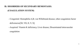
B) DISORDERS OFSECONDARY HEMOSTASIS:
(COAGULATION SYSTEM)
- Congenital: Hemophilia A,B; von Willebrand disease; other coagulation factor
deficiencies[XI, VII, II, V, X]
- Acquired: Vitamin K deficiency, Liver disease, Disseminated intravascular
coagulation
7.
7
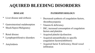
AQUIRED BLEEDING DISORDERS
DISEASE
•Liver disease and cirrhosis
• Gastrointestinal malabsorption
• Shock/Sepsis/Malignancy
• Renal disease
• Lymphoproliferative disorders
• Amyloidosis
PATHOPHYSIOLOGY
- Decreased synthesis of coagulation factors,
thrombocytopenia
- Vitamin K deficiency
- DIC, increased consumption of coagulation
factors and platelets
- Acquired platelet dysfunction
- Acquired autoantibodies to specific
coagulation factors(inhibitors)
- Acquired factor X deficiency, blood vessel
infiltration
8.
8
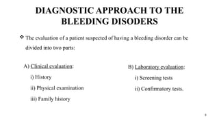
DIAGNOSTIC APPROACH TOTHE
BLEEDING DISODERS
A) Clinical evaluation:
i) History
ii) Physical examination
iii) Family history
B) Laboratory evaluation:
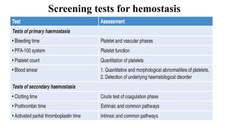
i) Screening tests
ii) Confirmatory tests.
The evaluation of a patient suspected of having a bleeding disorder can be
divided into two parts:
9.
9
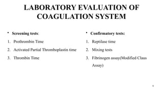
LABORATORY EVALUATION OF
COAGULATIONSYSTEM
• Screening tests:
1. Prothrombin Time
2. Activated Partial Thromboplastin time
3. Thrombin Time
• Confirmatory tests:
1. Reptilase time
2. Mixing tests
3. Fibrinogen assay(Modified Claus
Assay)
13
SPECIMEN COLLECTION
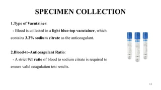
1.Type ofVacutainer:
- Blood is collected in a light blue-top vacutainer, which
contains 3.2% sodium citrate as the anticoagulant.
2.Blood-to-Anticoagulant Ratio:
- A strict 9:1 ratio of blood to sodium citrate is required to
ensure valid coagulation test results.
14.
14
3.Venipuncture Technique:
- Bloodshould be drawn using a clean, atraumatic venipuncture to avoid hemolysis
and activation of coagulation pathways.
- For accurate coagulation studies, it's also important to avoid traumatic
venipuncture, which may release tissue thromboplastin and interfere with test results.
15.
15
Special Note– Torniquet use:
- In sample collection for bleeding disorders, tourniquet application is generally
avoided or minimized, and if used, it should be applied briefly and gently.
- Prolonged or tight tourniquet application can cause hemoconcentration, activate
platelets, and alter coagulation test results (especially PT, aPTT, and D-dimer).
- If necessary, a tourniquet may be applied loosely for no more than 1 minute and
released immediately after venipuncture begins.
- For platelet function tests or bleeding time, tourniquets are strictly avoided, as they
can activate platelets artificially.
16.
16
4. Sample Handling:
-Tubes must be gently inverted several times to mix and transported promptly to
the lab; clotting or delay can lead to inaccurate results.
5. Plasma Preparation:
- Samples are centrifuged at 1500–2000g for 10–15 minutes to obtain platelet-
poor plasma, essential for tests like PT, aPTT, and factor assays.
17.
17
6. Storage Guidelines:
-If not tested within 2 hours, plasma should be frozen at -20°C or lower to preserve
coagulation factor stability.
19
1. PROTHROMBIN TIME
•Principle:
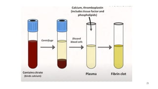
- Tissue thromboplastin and calcium
are added to plasma & clotting time is
determined.
- Assesses the extrinsic and common
coagulation pathways, mainly factors I,
II, V, VII, X.
• Collection:
- Light blue-top vacutainer (3.2%
sodium citrate).
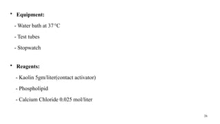
• Equipment:
- Water bath at 37°C
- Test tubes
- Stopwatch
20.
20
• Reagents:
- Thromboplastinreagent: contains tissue factor and phospholipids
- Calcium Chloride 0.025 moL/liter
• Method:
- Deliver 0.1ml of plasma in a glass test tube kept in water bath at 37°C.
- Add 0.1ml of thromboplastin reagent and mix.
- After 1 minute, add 0.1ml of calcium chloride solution. Immediately start the
stopwatch record the time required for clot formation.
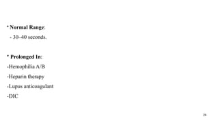
• Normal Range:
- 11–16 seconds; INR: 0.8–1.2.
22
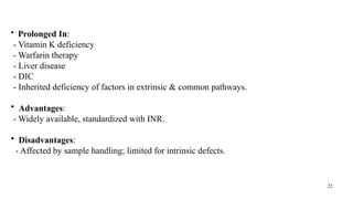
• Prolonged In:
-Vitamin K deficiency
- Warfarin therapy
- Liver disease
- DIC
- Inherited deficiency of factors in extrinsic & common pathways.
• Advantages:
- Widely available, standardized with INR.
• Disadvantages:
- Affected by sample handling; limited for intrinsic defects.
23.
23
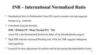
INR – InternationalNormalized Ratio
• Standardized form of Prothrombin Time (PT) used to monitor oral anticoagulant
therapy (e.g., warfarin).
• Calculated using the formula:
INR = (Patient PT / Mean Normal PT) ^ ISI,
where ISI is the International Sensitivity Index of the thromboplastin reagent.
• High INR indicates increased bleeding risk, while low INR suggests inadequate
anticoagulation.
• Essential for dose adjustment of warfarin and for preventing thromboembolic events.
24.
24
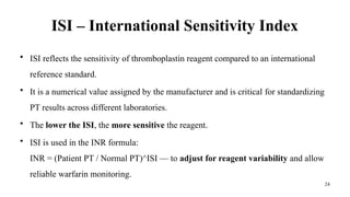
ISI – InternationalSensitivity Index
• ISI reflects the sensitivity of thromboplastin reagent compared to an international
reference standard.
• It is a numerical value assigned by the manufacturer and is critical for standardizing
PT results across different laboratories.
• The lower the ISI, the more sensitive the reagent.
• ISI is used in the INR formula:
INR = (Patient PT / Normal PT)^ISI — to adjust for reagent variability and allow
reliable warfarin monitoring.
25.
25
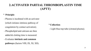
2.ACTIVATED PARTIAL THROMBOPLASTINTIME
(APTT)
• Principle:
- Plasma is incubated with an activator
(which initiates intrinsic pathway of
coagulation by contact activation).
- Phospholipid and calcium are then
added & clotting time is measured.
- Evaluates intrinsic and common
pathways (factors VIII, IX, XI, XII).
• Collection:
- Light blue-top tube (citrated plasma).
27
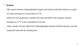
• Method:
- Mixequal volumes of phospholipid reagent and calcium chloride solution in a glass
test tube and keep in a water bath at 37°C.
- Deliver 0.1ml of plasma in another test tube and add 0.1ml of kaolin solution.
- Incubate at 37°C in the waterbath for 10 mins.
- After exactly 10 mins, add 0.2ml of phospholipid-calcium chloride mixture, start the
stopwatch and note the clotting time.
29
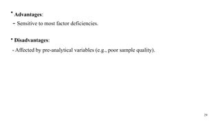
• Advantages:
- Sensitiveto most factor deficiencies.
• Disadvantages:
- Affected by pre-analytical variables (e.g., poor sample quality).
30.
30
3)Thrombin Time (TT)
•Principle:
- Assesses the final step of coagulation i.e.
measures conversion of fibrinogen to
fibrin by thrombin.
- Thrombin is added to patient’s plasma &
time required for clot formation is noted.
• Collection:
- Citrated plasma.
• Equipment:
- Water bath at 37°C
- Test tubes
- Stopwatch
31.
31
• Reagent:
- Thrombinsolution
• Method:
- Take 0.1ml of buffered saline in a test tube and add 0.1ml of plasma.
- Note clotting time after addition of 0.1ml of thrombin solution.
• Normal range:
- +/- 3 seconds of control
32.
32
• Prolonged In:
•Hypofibrinogenemia, dysfibrinogenemia, afibrinogenemia
• Heparin therapy
• DIC
• Advantages: Simple, rapid.
• Disadvantages: Non-specific; abnormal in presence of heparin or FDPs.
33.
33
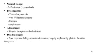
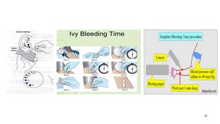
4)Bleeding Time (BT)
•Principle:
-Measures primary hemostasis—the time taken for bleeding to stop after a standardized
skin incision; reflects platelet function and vascular integrity.
•Collection:
- Requires capillary blood, usually using a template method (Ivy's method preferred).
•Procedure:
- Standardized incision made on the forearm; blotting paper used every 30 seconds until
bleeding stops.
34.
34
• Normal Range:
-2–7 minutes (Ivy method).
• Prolonged In:
- Thrombocytopenia
- von Willebrand disease
- Uremia
- Aspirin use
• Advantages:
- Simple, inexpensive bedside test.
• Disadvantages:
- Poor reproducibility, operator dependent, largely replaced by platelet function
analyzers.
36
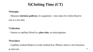
5)Clotting Time (CT)
•Principle:
-Measures intrinsic pathway of coagulation—time taken for whole blood to
clot in a test tube.
•Collection:
- Venous or capillary blood in a glass tube, no anticoagulant.
•Procedure:
- Capillary method (Duke's) or tube method (Lee-White); observe clot formation
at intervals.
37.
37
•Normal Range:
- 6–10minutes (Lee-White method).
•Prolonged In:
- Hemophilia A/B
- Severe liver disease
- DIC
•Advantages:
- Inexpensive.
•Disadvantages:
- Non-specific, poor sensitivity and standardization; obsolete in modern practice.
38.
38
6) PLATELET COUNT
Various methods are used to perform platelet count:
1. Manual Platelet Count (Hemocytometer Method)
• Uses a Neubauer chamber and diluted blood (usually 1% ammonium oxalate).
• Platelets are counted under a phase contrast microscope.
• Time-consuming and less accurate; used in resource-limited settings or when automated
counts are doubtful.
39.
39
2. Peripheral BloodSmear Estimation:
• Semi-quantitative method: Estimate platelet count by counting platelets per oil-
immersion field.
• 10–20 platelets/HPF = ~150,000–400,000/µL.
• Useful for confirming automated count accuracy, detecting giant platelets, or platelet
clumps.
40.
40
3. Impedance (Coulter)Method
• Most common method in automated hematology analyzers.
• Based on electrical resistance: as cells pass through an aperture, each platelet causes
a pulse.
• Differentiates platelets by size (2–20 fL).
41.
41
4. Optical/Light ScatterMethod:
• Uses laser light scatter and flow cytometry principles.
• Differentiates platelets from other cells based on size, granularity, and light scatter
patterns.
• More accurate in distinguishing platelets from RBC fragments or microcytes.
42.
42
5. Fluorescence FlowCytometry
• Uses fluorescent dyes that bind specifically to platelets.
• Highly sensitive and accurate; useful in cases with platelet clumping, giant platelets,
or thrombocytopenia.
6. Immunological Methods (e.g., CD41/CD61 tagging)
• Flow cytometry-based method using monoclonal antibodies against platelet-specific
antigens (e.g., GPIIb/IIIa).
• Very accurate, especially in diagnosing immune thrombocytopenia.
43.
43
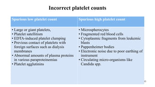
Incorrect platelet counts
Spuriouslow platelet count Spurious high platelet count
• Large or giant platelets,
• Platelet satellitism
• EDTA-induced platelet clumping
• Previous contact of platelets with
foreign surfaces such as dialysis
membranes
• Abnormal amounts of plasma proteins
in various paraproteinemias
• Platelet agglutinins
• Microspherocytes
• Fragmented red blood cells
• Cytoplasmic fragments from leukemic
blasts
• Pappenheimer bodies
• Electronic noise due to poor earthing of
instrument
• Circulating micro-organisms like
Candida spp.
46
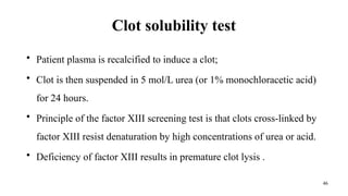
Clot solubility test
•Patient plasma is recalcified to induce a clot;
• Clot is then suspended in 5 mol/L urea (or 1% monochloracetic acid)
for 24 hours.
• Principle of the factor XIII screening test is that clots cross-linked by
factor XIII resist denaturation by high concentrations of urea or acid.
• Deficiency of factor XIII results in premature clot lysis .
47.
47
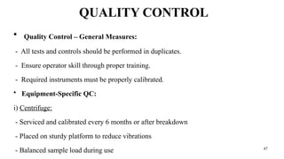
QUALITY CONTROL
• QualityControl – General Measures:
- All tests and controls should be performed in duplicates.
- Ensure operator skill through proper training.
- Required instruments must be properly calibrated.
• Equipment-Specific QC:
i) Centrifuge:
- Serviced and calibrated every 6 months or after breakdown
- Placed on sturdy platform to reduce vibrations
- Balanced sample load during use
48.
48
ii) Water Baths:
-Maintain temperature at 37°C (check in all 4 corners)
- Use transparent walls for better visualization
- Fill with distilled water to indicated level
iii) Pipettes:
- Must be calibrated
- Use disposable tips to avoid contamination
Editor's Notes
#10 This can be divided into two parts—screening tests and specific tests. Screening tests
are initial tests that are simple to perform, rapid and assess the integrity of primary or
secondary haemostasis. These tests are non-specific and do not pinpoint the nature of
the defect, but assess the function of one phase of the haemostatic system. Directed by the results of the screening tests,
appropriate specific tests are performed to define the precise nature of the defect

 Most read
 Most read
 Most read