Cerebrum Workbook | 1
ANATOMY OF THE HUMAN CEREBRUM
An Interactive Workbook for Identification, Labelling & Clinical Application
Course / Year: ___________________________________
Student Name: ___________________________________
Roll No.: ___________________________________
Conceptualized and Developed by
Dr. Rupal Shah
Associate Professor
Department of Rachana Sharira
Tilak Ayurved Mahavidyalaya, Pune
Version 1.0 | August 2026
Cerebrum Workbook | 2
About This Workbook
This workbook turns the study of the cerebrum into a sequence of observe-identify-mark-relate activities.
Rather than learning cortical anatomy only as a list of names, you will repeatedly return to the same
hemisphere from different viewpoints and build a three-dimensional understanding of its surfaces,
landmarks, functional areas and blood supply.
Use the illustrations as working diagrams. Trace boundaries, add labels, develop your own colour key and
write brief anatomical notes in the spaces provided. The final section asks you to use these observations to
localise lesions in short clinical situations, so that structural anatomy is connected with function and
vascular territory.
The workbook is intended for supervised internal academic and laboratory use. Standard anatomical terms
have been retained deliberately; the activities and explanatory text are organised for practical learning.
Copyright © 2026 Dr. Rupal Shah. All rights reserved. No part of this workbook may be reproduced,
redistributed or uploaded without prior written permission of the author.
Cerebrum Workbook | 3
SHEET 1 | Getting Oriented: Surfaces, Borders, Lobes & Poles
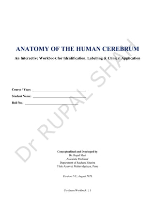
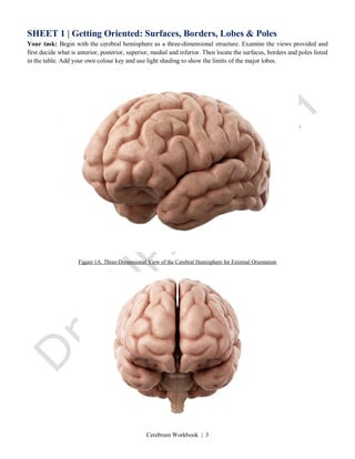
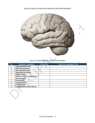
Your task: Begin with the cerebral hemisphere as a three-dimensional structure. Examine the views provided and
first decide what is anterior, posterior, superior, medial and inferior. Then locate the surfaces, borders and poles listed
in the table. Add your own colour key and use light shading to show the limits of the major lobes.
Figure 1A. Three-Dimensional View of the Cerebral Hemisphere for External Orientation
Cerebrum Workbook | 4
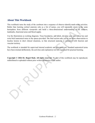
Figure 1B. Anterior View showing the Separation of the Cerebral Hemispheres
Figure 1C. External Landmarks of the Cerebral Hemisphere
No. Structure / landmark Colour Key One-Line Anatomical Note
1 Superomedial border
2 Inferolateral border
3 Inferomedial border
4 Superolateral surface
5 Medial surface
6
Inferior surface - orbital and
tentorial parts
7 Frontal pole
8 Temporal pole
9 Occipital pole
10 Longitudinal cerebral fissure
Cerebrum Workbook | 5
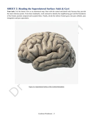
SHEET 2 | Reading the Superolateral Surface: Sulci & Gyri
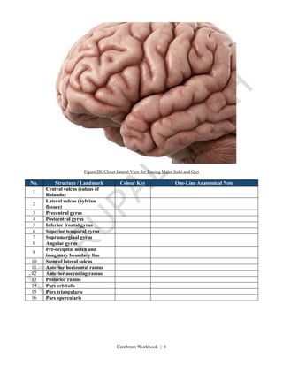
Your task: Use the lateral view as an anatomical map. Start with the central and lateral sulci because they provide
the main reference points. From these landmarks, work outward to identify the neighbouring gyri and the boundaries
of the frontal, parietal, temporal and occipital lobes. Finally, divide the inferior frontal gyrus into pars orbitalis, pars
triangularis and pars opercularis.
Figure 2A. Superolateral Surface of the Cerebral Hemisphere
Cerebrum Workbook | 6
Figure 2B. Closer Lateral View for Tracing Major Sulci and Gyri
No. Structure / Landmark Colour Key One-Line Anatomical Note
1
Central sulcus (sulcus of
Rolando)
2
Lateral sulcus (Sylvian
fissure)
3 Precentral gyrus
4 Postcentral gyrus
5 Inferior frontal gyrus
6 Superior temporal gyrus
7 Supramarginal gyrus
8 Angular gyrus
9
Pre-occipital notch and
imaginary boundary line
10 Stem of lateral sulcus
11 Anterior horizontal ramus
12 Anterior ascending ramus
13 Posterior ramus
14 Pars orbitalis
15 Pars triangularis
16 Pars opercularis
Cerebrum Workbook | 7
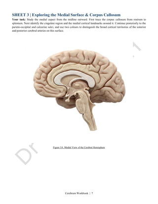
SHEET 3 | Exploring the Medial Surface & Corpus Callosum
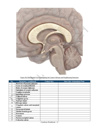
Your task: Study the medial aspect from the midline outward. First trace the corpus callosum from rostrum to
splenium. Next identify the cingulate region and the medial cortical landmarks around it. Continue posteriorly to the
parieto-occipital and calcarine sulci, and use two colours to distinguish the broad cortical territories of the anterior
and posterior cerebral arteries on this surface.
Figure 3A. Medial View of the Cerebral Hemisphere
Cerebrum Workbook | 8
Figure 3B. Mid-Sagittal View Highlighting the Corpus Callosum and Neighbouring Structures
No. Structure / Landmark Colour Key One-Line Anatomical Note
1 Rostrum of corpus callosum
2 Genu of corpus callosum
3 Body of corpus callosum
4 Splenium of corpus callosum
5 Lamina terminalis
6 Indusium griseum
7 Callosal sulcus
8 Cingulate gyrus
9 Forceps major
10 Forceps minor
11
Cingulate sulcus and marginal
ramus
12 Paracentral lobule
13 Medial frontal gyrus
14 Precuneus
15 Cuneus
16 Parieto-occipital sulcus
17 Calcarine sulcus
Cerebrum Workbook | 9
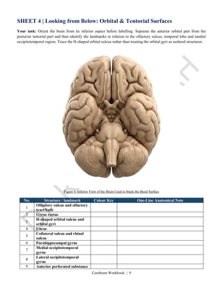
SHEET 4 | Looking from Below: Orbital & Tentorial Surfaces
Your task: Orient the brain from its inferior aspect before labelling. Separate the anterior orbital part from the
posterior tentorial part and then identify the landmarks in relation to the olfactory sulcus, temporal lobe and medial
occipitotemporal region. Trace the H-shaped orbital sulcus rather than treating the orbital gyri as isolated structures.
Figure 4. Inferior View of the Brain Used to Study the Basal Surface
No. Structure / landmark Colour Key One-Line Anatomical Note
1
Olfactory sulcus and olfactory
tract/bulb
2 Gyrus rectus
3
H-shaped orbital sulcus and
orbital gyri
4 Uncus
5
Collateral sulcus and rhinal
sulcus
6 Parahippocampal gyrus
7
Medial occipitotemporal
gyrus
8
Lateral occipitotemporal
gyrus
9 Anterior perforated substance
Cerebrum Workbook | 10
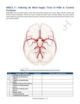
SHEET 5 | Following the Blood Supply: Circle of Willis & Cerebral
Territories
Your task: Trace the arterial pathway rather than memorising isolated vessel names. Begin with the vertebral and
internal carotid contributions, follow the vessels forming the arterial circle, and then identify the major cerebral
arteries. Return to your earlier surface sheets and create a consistent colour key for the broad cortical territories of the
ACA, MCA and PCA.
Figure 5. Inferior View of the Cerebral Arterial Circulation and Circle of Willis
No. Structure / landmark Colour Key One-Line Anatomical Note
1 Internal carotid artery (ICA)
2
Anterior cerebral artery
(ACA)
3
Anterior communicating
artery
4 Middle cerebral artery (MCA)
5
Posterior communicating
artery
6
Posterior cerebral artery
(PCA)
7 Basilar artery
8 Vertebral arteries
9 Lenticulostriate branches
Cerebrum Workbook | 11
SHEET 6 | From Surface Anatomy to Clinical Localisation
Your task: For each patient, work in the same order: identify the neurological deficit, decide which cortical function is
disturbed, localise the likely cortical region, and only then relate it to the most probable arterial territory. Support each
answer with a short anatomical reason.
CASE 1 | Speech Becomes Effortful
A 64-year-old man with hypertension suddenly finds it difficult to express himself. He remains alert and clearly
understands what is said to him. When asked to close his eyes or point towards the ceiling, he performs the task
correctly. His own speech, however, is slow and laboured, with short phrases such as “want... water...”. He becomes
visibly frustrated while trying to communicate. Examination also shows mild weakness of the right lower face and
right hand.
Work through the localisation:
1. Localise the lesion as precisely as possible. Name the lobe, gyrus and relevant Brodmann areas.
2. Which cerebral artery and branch would most likely supply this region? Would you expect the lesion in the
dominant or non-dominant hemisphere?
3. How does the functional organisation of the language cortex explain preserved comprehension with impaired
fluent speech?
Anatomical reasoning: ___________________________________________________________________________
______________________________________________________________________________________________
______________________________________________________________________________________________
CASE 2 | Fluent Words Without Meaning
A 71-year-old woman is brought by her family after a sudden change in her speech. She talks rapidly, with normal
rhythm and clear pronunciation, yet her sentences do not convey meaningful information. She does not appear
concerned about the problem. During examination she cannot carry out a simple spoken instruction such as “show
me your tongue”, and reading and writing are also disturbed.
Work through the localisation:
1. What language disorder does this pattern suggest? Identify the cortical region, gyri and relevant Brodmann area(s).
2. Which arterial territory supplies this region, and how is it related to the classical Wernicke language area?
3. Compare this patient's speech fluency, comprehension and awareness with the findings in Case 1.
Anatomical reasoning: ___________________________________________________________________________
______________________________________________________________________________________________
______________________________________________________________________________________________
Cerebrum Workbook | 12
CASE 3 | A Deficit Confined to the Lower Limb
A 58-year-old marathon runner collapses and is brought to the emergency department. Examination shows complete
loss of voluntary movement and sensation in the right leg and foot. Motor power and sensation in the right face, arm
and hand are preserved. Her speech is normal and she is fully oriented.
Work through the localisation:
1. Where on the cerebral cortex would you localise the lesion? Specify the gyrus/lobule and whether the affected
representation is mainly medial or lateral.
2. Use the motor and sensory homunculi to explain why the face and upper limb are spared.
3. Which major cerebral artery is most likely involved, and which cortical branches supply this region?
Anatomical reasoning: ___________________________________________________________________________
______________________________________________________________________________________________
______________________________________________________________________________________________
CASE 4 | Half the Field Is Lost, the Centre Is Spared
A 67-year-old retired literature professor suddenly develops difficulty seeing objects on her right side. Visual-field
testing demonstrates a right homonymous hemianopia. More detailed testing shows preservation of central macular
vision. MRI confirms a localised ischaemic lesion in the left occipital lobe.
Work through the localisation:
1. Which sulcus and primary cortical visual area are most likely involved? State the relevant Brodmann area.
2. Why can a lesion in this location produce contralateral homonymous hemianopia while central vision remains
preserved?
3. Which cerebral artery is most likely occluded, and how can overlapping arterial supply at the occipital pole
contribute to macular sparing?
Anatomical reasoning: ___________________________________________________________________________
______________________________________________________________________________________________
______________________________________________________________________________________________
Cerebrum Workbook | 13
End-of-Workbook Reflection
Before closing the workbook, return to your labelled sheets and check whether you can move between views of the
same hemisphere without relying on the answer table. Choose any one clinical case and trace the reasoning
backwards: symptom → function → cortical location → surface landmark → arterial territory.
One structure I can now identify confidently: __________________________________________________
One relationship I still need to revise: ______________________________________________________
One clinical correlation that helped me understand the anatomy: ______________________________________
Cerebrum Workbook | 14
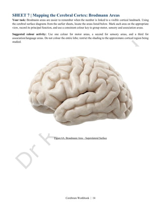
SHEET 7 | Mapping the Cerebral Cortex: Brodmann Areas
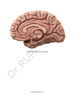
Your task: Brodmann areas are easier to remember when the number is linked to a visible cortical landmark. Using
the cerebral surface diagrams from the earlier sheets, locate the areas listed below. Mark each area on the appropriate
view, record its principal function, and use a consistent colour key to group motor, sensory and association areas.
Suggested colour activity: Use one colour for motor areas, a second for sensory areas, and a third for
association/language areas. Do not colour the entire lobe; restrict the shading to the approximate cortical region being
studied.
Figure 6A. Broadmann Area – Superolateral Surface
Cerebrum Workbook | 15
Figure 6B. Broadmann Area – Medial Surface
Cerebrum Workbook | 16
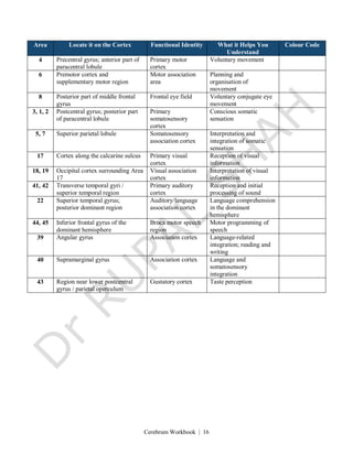
Area Locate it on the Cortex Functional Identity What it Helps You
Understand
Colour Code
4 Precentral gyrus; anterior part of
paracentral lobule
Primary motor
cortex
Voluntary movement
6 Premotor cortex and
supplementary motor region
Motor association
area
Planning and
organisation of
movement
8 Posterior part of middle frontal
gyrus
Frontal eye field Voluntary conjugate eye
movement
3, 1, 2 Postcentral gyrus; posterior part
of paracentral lobule
Primary
somatosensory
cortex
Conscious somatic
sensation
5, 7 Superior parietal lobule Somatosensory
association cortex
Interpretation and
integration of somatic
sensation
17 Cortex along the calcarine sulcus Primary visual
cortex
Reception of visual
information
18, 19 Occipital cortex surrounding Area
17
Visual association
cortex
Interpretation of visual
information
41, 42 Transverse temporal gyri /
superior temporal region
Primary auditory
cortex
Reception and initial
processing of sound
22 Superior temporal gyrus;
posterior dominant region
Auditory/language
association cortex
Language comprehension
in the dominant
hemisphere
44, 45 Inferior frontal gyrus of the
dominant hemisphere
Broca motor speech
region
Motor programming of
speech
39 Angular gyrus Association cortex Language-related
integration; reading and
writing
40 Supramarginal gyrus Association cortex Language and
somatosensory
integration
43 Region near lower postcentral
gyrus / parietal operculum
Gustatory cortex Taste perception
Cerebrum Workbook | 17
Apply What You Have Mapped
1. Contralateral hand weakness follows a small cortical lesion. Which Brodmann area and gyrus would you examine
first?
Reasoning: ______________________________________________________________________
2. A patient understands speech but speaks slowly and with effort. Which Brodmann areas are likely to be involved?
Reasoning: ______________________________________________________________________
3. A patient produces fluent speech but cannot understand spoken language. Which posterior cortical area should be
considered?
Reasoning: ______________________________________________________________________
4. Which Brodmann area lies along the calcarine sulcus, and what is its principal function?
Reasoning: ______________________________________________________________________
5. A patient can feel an object in the hand but cannot recognise it by touch with the eyes closed. Which parietal
association areas may be involved?
Reasoning: ______________________________________________________________________