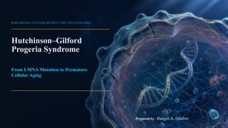
Hutchinson–Gilford Progeria Syndrome : From LMNA Mutation to Premature
One mutation. One abnormal protein. A lifetime of premature cellular aging.
Discover how a seemingly silent change in the LMNA gene can reshape the nucleus, disrupt cellular function, and reveal unexpected insights into how human aging works.
Hutchinson–Gilford Progeria Syndrome : From LMNA Mutation to Premature
1.
RARE DISEASE •NUCLEAR ARCHITECTURE • CELLULAR AGING
Hutchinson–Gilford
Progeria Syndrome
From LMNA Mutation to Premature
Cellular Aging
Prepared by : Rangin A. Ghafwr
2.
HGPS • SCIENTIFICOVERVIEW
What is Progeria?
A segmental premature-aging disorder caused by a toxic form of
lamin A protien-progerin.
Early onset. Children are typically healthy at birth; growth
failure and distinctive physical findings emerge during the
first year of life.
Selective vulnerability. Skin, bone, connective tissue,
and especially arteries are affected, while motor and
intellectual development are generally preserved.
Molecular clue. The abnormal lamin A protein, progerin,
destabilizes the nuclear lamina and accelerates cellular
decline.
A single splicing error can disrupt the cell’s mechanical and genomic control
center.
Progerin-driven disruption of the nuclear envelope is the molecular
hallmark of HGPS.
Sources: [1] GeneReviews® (2025) • [2] StatPearls (2025) 1
3.
HGPS • SCIENTIFICOVERVIEW
Epidemiology & Rarity
Ultra-rare in frequency; globally important for what it reveals about nuclear biology and vascular aging.
ESTIMATED INCIDENCE
1 in 4 million
Reported estimate at live birth in a
pivotal clinical-trial publication.
ESTIMATED PREVALENCE
1 in 20 million
Commonly cited estimate for living
individuals with HGPS.
ORIGIN OF THE VARIANT
≈98% de novo
Usually autosomal dominant;
parental gonadal mosaicism
explains a small minority.
Estimates vary by study design, case ascertainment, and whether incidence or living prevalence is measured.
Rare does not mean biologically unimportant.
Sources: [1] GeneReviews® (2025) • [3] Gordon et al., JAMA (2018) 2
4.
HGPS • CLINICALPHENOTYPE
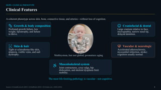
Clinical Features
A coherent phenotype across skin, bone, connective tissue, and arteries—without loss of cognition.
Growth & body composition
Profound growth failure, low
weight, lipoatrophy, and failure
to thrive.
Skin & hair
Tight or scleroderma-like skin,
alopecia, visible veins, and nail
dystrophy.
Craniofacial & dental
Large cranium relative to face,
micrognathia, narrow nasal tip,
delayed dentition.
Vascular & neurologic
Accelerated atherosclerosis,
myocardial infarction, stroke;
cognition usually normal.
Multisystem, but not global, premature aging
Musculoskeletal system
Joint contractures, coxa valga, hip
dislocation, and skeletal dysplasia limit
mobility.
The most life-limiting pathology is vascular—not cognitive.
Sources: [1] GeneReviews® (2025) • [2] StatPearls (2025) • [3] Gordon et al., JAMA (2018) 3
5.
HGPS • PHYSIOLOGICALIMPACT
Physiological Impact of HGPS
HGPS is multisystem disease, but the most life-limiting physiology is vascular.
Cardiovascular
Arterial stiffness, accelerated atherosclerosis, and
vascular smooth-muscle-cell dysfunction drive
myocardial infarction and stroke risk.
Musculoskeletal
Reduced bone mineralization, skeletal dysplasia,
joint contractures, and progressive mobility
limitation.
Metabolic / adipose
Severe loss of subcutaneous fat, abnormal body
composition, low body weight, and profound
growth failure.
Nervous system
Cognitive development is generally preserved;
neurological injury is mainly secondary to vascular
disease, especially stroke.
A systemic phenotype with selective tissue vulnerability.
Sources: [1] GeneReviews® (2025) • [2] StatPearls (2025) • [3] Gordon et al., PNAS (2012) • [5] Gonzalo et al. (2016) 4
6.
HGPS • CARDIOVASCULARPHYSIOLOGY
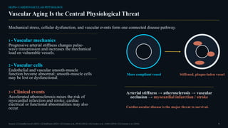
Vascular Aging Is the Central Physiological Threat
Mechanical stress, cellular dysfunction, and vascular events form one connected disease pathway.
1 • Vascular mechanics
Progressive arterial stiffness changes pulse-
wave transmission and increases the mechanical
load on vulnerable vessels.
2 • Vascular cells
Endothelial and vascular smooth-muscle
function become abnormal; smooth-muscle cells
may be lost or dysfunctional.
3 • Clinical events
Accelerated atherosclerosis raises the risk of
myocardial infarction and stroke; cardiac
electrical or functional abnormalities may also
occur.
More compliant vessel Stiffened, plaque-laden vessel
Arterial stiffness → atherosclerosis → vascular
occlusion → myocardial infarction / stroke
Cardiovascular disease is the major threat to survival.
Sources: [1] GeneReviews® (2025) • [2] StatPearls (2025) • [3] Gordon et al., PNAS (2012) • [4] Gordon et al., JAMA (2018) • [5] Gonzalo et al. (2016) 5
7.
HGPS • MUSCULOSKELETAL& METABOLIC PHYSIOLOGY
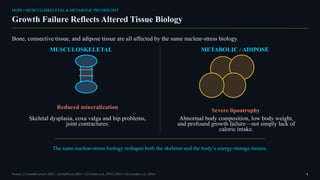
Growth Failure Reflects Altered Tissue Biology
Bone, connective tissue, and adipose tissue are all affected by the same nuclear-stress biology.
MUSCULOSKELETAL
Reduced mineralization
Skeletal dysplasia, coxa valga and hip problems,
joint contractures.
METABOLIC / ADIPOSE
Severe lipoatrophy
Abnormal body composition, low body weight,
and profound growth failure—not simply lack of
caloric intake.
The same nuclear-stress biology reshapes both the skeleton and the body’s energy-storage tissues.
Sources: [1] GeneReviews® (2025) • [2] StatPearls (2025) • [3] Gordon et al., PNAS (2012) • [5] Gonzalo et al. (2016) 6
8.
HGPS • NERVOUSSYSTEM
The Brain Is Not the Primary Target
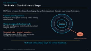
HGPS does not cause global neurological aging; the cerebral circulation is the major route to neurologic injury.
Cognition generally preserved.
Intellectual development is usually not the primary
site of disease.
Motor development often intact early.
Mobility later becomes limited mainly by skeletal
and joint disease.
Neurologic injury is mainly secondary.
Stroke can produce deficits when accelerated
vascular disease reaches the brain.
Cognition
preserved
Stroke
cerebrovascular injury
The brain is not the primary target—the cerebral circulation is.
Sources: [1] GeneReviews® (2025) • [2] StatPearls (2025) • [4] Gordon et al., JAMA (2018) 7
9.
HGPS • GENETICORIGIN
LMNA Gene
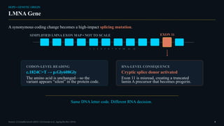
A synonymous coding change becomes a high-impact splicing mutation.
SIMPLIFIED LMNA EXON MAP • NOT TO SCALE
1 2 3 4 5 6 7 8 9 10 11 12
EXON 11
CODON-LEVEL READING
c.1824C>T → p.Gly608Gly
The amino acid is unchanged—so the
variant appears “silent” in the protein code.
RNA-LEVEL CONSEQUENCE
Cryptic splice donor activated
Exon 11 is misread, creating a truncated
lamin A precursor that becomes progerin.
Same DNA letter code. Different RNA decision.
Sources: [1] GeneReviews® (2025) • [5] Gonzalo et al., Ageing Res Rev (2016) 8
10.
HGPS • NUCLEARARCHITECTURE
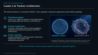
Lamin A & Nuclear Architecture
The nuclear lamina is a structural scaffold—and a regulator of genome organization and cellular signaling.
Mechanical support
Lamin A/C helps the nucleus resist deformation
and couples physical forces to cell behavior.
Genome organization
It anchors chromatin at the nuclear periphery,
shaping transcriptional programs and DNA
transactions.
Nuclear signaling
Lamina interactions influence gene expression,
replication, DNA repair, and
mechanotransduction.
ORDERLY LAMINA HGPS-LIKE STRESS
Conceptual illustration: smooth nuclear geometry contrasts with
blebbing, lamina disruption, and chromatin disorganization.
When the lamina fails, structure, genome regulation, and force sensing fail together.
Sources: [5] Gonzalo et al., Ageing Res Rev (2016) • [6] Cao et al., J Clin Invest (2011) 9
11.
HGPS • CAUSALBIOLOGY
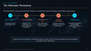
The Molecular Mechanism
One altered RNA-processing decision initiates a cascade from nuclear damage to premature tissue aging.
1
LMNA c.1824C>T
Activates a cryptic
splice site in exon
11.
2
Aberrant splicing
Removes 150
nucleotides / 50
amino acids from
prelamin A.
3
ZMPSTE24 site
lost
The farnesyl tail
cannot be removed
from progerin.
4
Progerin
accumulates
Persistent membrane
anchoring distorts the
nuclear lamina.
5
Nuclear stress
Chromatin disruption,
DNA damage,
senescence, and tissue
injury.
Large arteries are particularly vulnerable because vascular cells face chronic mechanical stress and depend
on durable nuclear architecture.
Sources: [3] Gordon et al., JAMA (2018) • [5] Gonzalo et al. (2016) • [6] Cao et al. (2011) 10
12.
HGPS • PROTEINPROCESSING
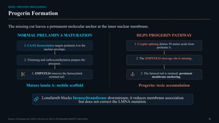
Progerin Formation
The missing cut leaves a permanent molecular anchor at the inner nuclear membrane.
NORMAL PRELAMIN A MATURATION
1. CAAX farnesylation targets prelamin A to the
nuclear envelope.
2. Trimming and carboxymethylation prepare the
precursor.
3. ZMPSTE24 removes the farnesylated
terminal tail.
Mature lamin A: mobile scaffold
HGPS PROGERIN PATHWAY
1. Cryptic splicing deletes 50 amino acids from
prelamin A.
2. The ZMPSTE24 cleavage site is missing.
3. The farnesyl tail is retained: persistent
membrane anchoring.
Progerin: toxic accumulation
Lonafarnib blocks farnesyltransferase downstream; it reduces membrane association
but does not correct the LMNA mutation.
Sources: [5] Gonzalo et al. (2016) • [6] Cao et al. (2011) • [9] DailyMed ZOKINVY label (2024) 11
13.
HGPS • CELLULARAGING
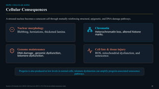
Cellular Consequences
A stressed nucleus becomes a senescent cell through mutually reinforcing structural, epigenetic, and DNA-damage pathways.
Nuclear morphology
Blebbing, herniations, thickened lamina.
Chromatin
Heterochromatin loss, altered histone
marks.
Genome maintenance
DNA damage , genomic dysfunction,
telomere dysfunction.
Cell fate & tissue injury
ROS, mitochondrial dysfunction, and
senescence.
Progerin is also produced at low levels in normal cells; telomere dysfunction can amplify progerin-associated senescence
pathways.
Sources: [5] Gonzalo et al. (2016) • [6] Cao et al. (2011) • [7] J Clin Invest cellular-senescence study 12
14.
HGPS • CAUSALPATHWAY
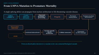
From LMNA Mutation to Premature Mortality
A single splicing defect can propagate from nuclear architecture to life-threatening vascular disease.
LMNA
c.1824C>T
Cryptic
splice-site
activation
Abnormal
prelamin A
processing Progerin
Persistent
farnesylation
Nuclear-envelope
deformation
Chromatin
& DNA damage
Cellular senescence
Vascular smooth-
muscle
dysfunction
Accelerated atherosclerosis
Myocardial
infarction
From an altered splice decision to a vascular event: one connected biological cascade.
Sources: [1] GeneReviews® (2025) • [5] Gonzalo et al. (2016) • [6] Cao et al. (2011) 13
stroke
15.
HGPS • PROGNOSIS
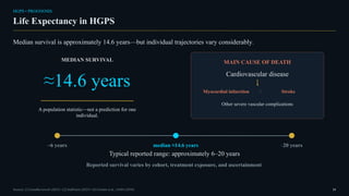
LifeExpectancy in HGPS
Median survival is approximately 14.6 years—but individual trajectories vary considerably.
MEDIAN SURVIVAL
≈14.6 years
A population statistic—not a prediction for one
individual.
MAIN CAUSE OF DEATH
Cardiovascular disease
Myocardial infarction / Stroke
Other severe vascular complications
~6 years median ≈14.6 years ~20 years
Typical reported range: approximately 6–20 years
Reported survival varies by cohort, treatment exposure, and ascertainment.
Sources: [1] GeneReviews® (2025) • [2] StatPearls (2025) • [4] Gordon et al., JAMA (2018) 14
16.
HGPS • CLINICALTRAJECTORY
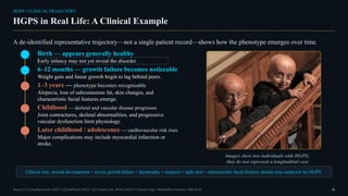
HGPS in Real Life: A Clinical Example
A de-identified representative trajectory—not a single patient record—shows how the phenotype emerges over time.
Birth — appears generally healthy
Early infancy may not yet reveal the disorder.
6–12 months — growth failure becomes noticeable
Weight gain and linear growth begin to lag behind peers.
1–3 years — phenotype becomes recognizable
Alopecia, loss of subcutaneous fat, skin changes, and
characteristic facial features emerge.
Childhood — skeletal and vascular disease progresses
Joint contractures, skeletal abnormalities, and progressive
vascular dysfunction limit physiology.
Later childhood / adolescence — cardiovascular risk rises
Major complications may include myocardial infarction or
stroke.
Images show two individuals with HGPS;
they do not represent a longitudinal case.
Clinical clue: normal development + severe growth failure + lipoatrophy + alopecia + tight skin + characteristic facial features should raise suspicion for HGPS.
Sources: [1] GeneReviews® (2025) • [2] StatPearls (2025) • [3] Gordon et al., PNAS (2012) • Clinical image: MedlinePlus Genetics, NIH/NLM 15
17.
HGPS • CLINICALRECOGNITION
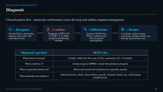
Diagnosis
Clinical pattern first—molecular confirmation closes the loop and enables targeted management.
1. Recognize
Growth failure, lipoatrophy,
alopecia, skin, joint, and
craniofacial clues.
2. Confirm
Sequence LMNA for
c.1824C>T or other
progerin-producing
variants.
3. Differentiate
Separate HGPS from
Werner syndrome and
other progeroid
laminopathies.
4. Monitor
Coordinate cardiovascular,
neurologic, skeletal, dental, eye,
hearing, and nutrition care.
Diagnostic question HGPS clue
When does it emerge? Usually within the first year of life, commonly at 6–12 months.
What confirms it? A heterozygous LMNA variant that produces progerin.
What is generally preserved? Motor and mental development are typically normal.
What demands surveillance?
Atherosclerosis, stroke, heart failure, growth, skeletal, dental, eye, and hearing
complications.
Sources: [1] GeneReviews® (2025) • [2] StatPearls (2025) 16
18.
HGPS • CURRENTMANAGEMENT
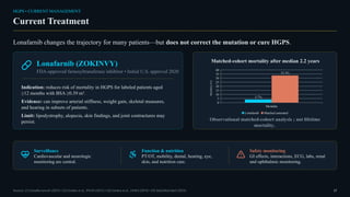
Current Treatment
Lonafarnib changes the trajectory for many patients—but does not correct the mutation or cure HGPS.
Lonafarnib (ZOKINVY)
FDA-approved farnesyltransferase inhibitor • Initial U.S. approval 2020
Indication: reduces risk of mortality in HGPS for labeled patients aged
≥12 months with BSA ≥0.39 m².
Evidence: can improve arterial stiffness, weight gain, skeletal measures,
and hearing in subsets of patients.
Limit: lipodystrophy, alopecia, skin findings, and joint contractures may
persist.
Matched-cohort mortality after median 2.2 years
Mortality
0
5
10
15
20
25
30
35
40
3.7%
33.3%
Lonafarnib Matched untreated
Mortality
(%)
Observational matched-cohort analysis ; not lifetime
mortality..
Surveillance
Cardiovascular and neurologic
monitoring are central.
Function & nutrition
PT/OT, mobility, dental, hearing, eye,
skin, and nutrition care.
Safety monitoring
GI effects, interactions, ECG, labs, renal
and ophthalmic monitoring.
Sources: [1] GeneReviews® (2025) • [3] Gordon et al., PNAS (2012) • [4] Gordon et al., JAMA (2018) • [9] DailyMed label (2024) 17
19.
HGPS • TRANSLATIONALFRONTIER
Research & Future Therapies
The field is moving from moderating progerin toxicity toward suppressing, editing, or correcting its source.
Reduce
Antisense oligonucleotides.
Correct
Base editing.
Block
Progerin membrane interaction.
Molecular correction is an aspiration—not yet a proven human therapy.
HGPS reveals a general principle: nuclear architecture is not passive—it is a determinant of cellular aging.
SELECTED REFERENCES
[1] GeneReviews® (2025). HGPS. NCBI Bookshelf.
[3] Gordon et al. PNAS. 2012;109:16666–16671. doi:10.1073/pnas.1202529109
[4] Gordon et al. JAMA. 2018;319:1687–1695. doi:10.1001/jama.2018.3264
[5] Gonzalo et al. Ageing Res Rev. 2016;33:18–29. doi:10.1016/j.arr.2016.06.007
[6] Cao et al. J Clin Invest. 2011;121:2833–2844. doi:10.1172/JCI43578
[8] Puttaraju et al. Nat Med. 2021;27:526–535. doi:10.1038/s41591-021-01262-4
[9] DailyMed. ZOKINVY (lonafarnib) label, 2024.
[10] Koblan et al. Nature. 2021;589:608–614. doi:10.1038/s41586-020-03086-7 • [11] NCT06775041,
ClinicalTrials.gov (updated 2026-02-09)
Sources: [5] [8] [10] [11] • Full references are provided above. 18
20.
THANK YOU
Thank youfor reading!
For more presentations on
Genetics, Cancer Biology & Biomedical Science
follow my profile.
Genetics • Cancer Biology • Biomedical Science