HEMODYNAMICS &
ELECTROPHYSIOLOGY OF
HEART
BY:
N J V S PAVAN
M.PHARMACY
HINDU COLLEGE OF PHARMACY
GUNTUR, A.P, INDIA., 522002
HEMODYNAMICS
• Hemodynamics is how your blood flows through your arteries and veins and the
forces that affect your blood flow.
• Normally, your blood flows in a laminar (streamlined) pattern. It flows fastest in the
middle of a blood vessel, where there’s no friction with blood vessel walls.
• However, the flow is turbulent in your ventricles (lower heart chambers) and in
places where a vessel branches off or changes in diameter. You’ll need to use
more energy to move blood that has a turbulent flow because it’s not moving
efficiently.
FLOW
OF
BLOOD
IN
HUMAN
BODY
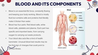
BLOOD AND ITS COMPONENTS
• Blood is an essential life force, constantly flowing
and keeping your body working. Blood is mostly
fluid but contains cells and proteins that literally
make it thicker than water.
• Blood has four parts: Red blood cells, white
blood cells, platelets and plasma. Each part has
specific and important tasks, from carrying
oxygen to carrying out waste products.
• Your blood also acts like a kind of health
barometer. Unusual blood test results may be
the first sign of changes that could point to
serious illness.
Functions of BLOOD
Blood flowing through your blood vessels is responsible for many things:
•It carries oxygen and nutrients throughout your body.
•It forms blood clots to manage bleeding.
•It protects your body from infections.
•It carries waste products.
•It regulates your body temperature.
Cardiac Output
Cardiac output is how many liters of blood your heart pumps in one minute.
Your healthcare provider can figure this out with this cardiac output equation:
multiply stroke volume by heart rate.
HEMODYNAMIC FACTORS
Hemodynamic factors are things that affect how well your blood flows. They
can make it easier or harder for your blood to get to your organs and tissues.
Your body makes constant adjustments to give your cells what they need.
Hemodynamic factors include:
• The size (diameter) of a blood vessel. It’s easier for your blood to move through a larger or more
open vessel. It’s harder for blood to flow through an artery that’s become narrow because of a
plaque deposit containing cholesterol.
• The amount of friction your blood must overcome to keep moving. The friction of blood flowing
against a blood vessel wall slows it down. Blood closer to your artery wall moves more slowly
because of this friction. Blood in the middle of your artery moves faster.
• Blood vessel expansion and contraction in response to blood pressure changes. Your blood
vessels have the ability to become wider or narrower to control blood flow and keep it
consistent.
• Pressure differences between how forcefully your heart pumps and how much resistance
your blood vessels provide. The blood your heart pumps has to push against your blood
vessels’ force. This is why having high blood pressure makes your heart work harder.
• Cardiac output. Your heart can pump out more or less blood with each heartbeat depending
on what you’re doing at the time. When you exercise, your heart sends more blood to your
cells because they need more oxygen than when they’re resting.
• Heart rate. Your heart can beat more times per minute when you’re exercising than when
you’re resting. This helps your body get the increased amount of oxygen it needs for
exercise.
• How well your heart’s ventricles are working. If you have a heart condition that makes your
ventricles unable to pump blood well, this affects how much blood they can send to your
body.
HEMODYNAMIC INSTABILITY
Hemodynamic instability means your body can’t get enough blood flow. This is known as shock.
There are several types, depending on the cause.
Types of shock
• Cardiogenic.
• Hypovolemic.
• Obstructive.
• Distributive.
• Neurogenic.
ELECTROPHYSIOLOGY
OF HEART
Cardiac electrophysiology is a branch of cardiology that
studies, diagnoses, and treats heart conditions that affect
the heart's electrical activity. This includes heart rhythm
abnormalities, or cardiac arrhythmias, such as skipped
beats, atrial fibrillation, and SVTs.
Your heart’s conduction system is the network of nodes (groups of cells that can be either nerve
or muscle tissue), specialized cells and electrical signals that keep your heart beating.
Two types of cells control your heartbeat:
•Conducting cells carry the electric signals.
•Muscle cells control your heart’s contractions.
Your heart (cardiac) conduction system sends the signal to start a heartbeat. It also sends
signals that tell different parts of your heart to relax and contract (squeeze). This process of
contracting and relaxing controls blood flow through your heart and to the rest of your body.
Your heart is a pump that sends blood through your body. For each heartbeat, electrical signals
travel through the conduction pathway of your heart. It starts when your sinoatrial (SA) node
creates an excitation signal. This electrical signal is like electricity traveling through wires to an
appliance in your home.
The excitation signal travels to:
1.Your atria (top heart chambers), telling them to contract.
2.The atrioventricular (AV) node, delaying the signal until your atria are empty of blood.
3.The bundle of His (center bundle of nerve fibers), carrying the signal to the Purkinje fibers.
4.The Purkinje fibers to your ventricles (bottom heart chambers), causing them to contract.
These steps make up one full contraction of your heart muscle. Your heart conduction system
sends out thousands of signals per day to keep your heart beating.
ELECTRICAL CONDUCTION IN HEART
Repolarization is the process by which electrical
charges in a cell return to their resting state. It's a stage
of an action potential that occurs after depolarization,
when the cell reaches its highest voltage. During
repolarization, the cell's voltage decreases due to
potassium (K+) ions moving out of the cell along its
electrochemical gradient. This restores the difference in
charge between the inside and outside of the cell
membrane.
Depolarization is caused by a rapid rise in membrane
potential, which opens sodium channels in the cellular
membrane and allows a large influx of sodium ions. This
process can occur in excitable cells, such as neurons
and muscle cells, which respond to electrical signals.
Hyperpolarization is a phase in electrophysiology when
the membrane potential of a neuron becomes more
negative at a specific point. This happens when
potassium ions leave the cell and potassium channels
close, which can be caused by an increase in potassium
permeability. Hyperpolarization can also occur during
the final part of an action potential or in response to
inhibitory neural messages.
RELATIONSHIP BETWEEN ECG AND CARDIAC CYCLE
hemodynamics and electrophysiology of heart.pptx