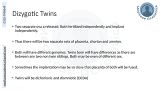
medicoseacademics@gmail.com
0310-7990649
Extraembryonic Membranes &
Multiple Gestation
Dr. Muhammad Ali Rabbani
Asst. Professor Anatomy
MBBS (NMC, Multan)
FCPS (Anatomy)
CHPE (Certificate in Health Professional Education)
DHPE (Diploma in Health Professional Education)
MBA (Masters of Business Administration)
MPH (Masters of Public Health)
medicoseacademics@gmail.com
0310-7990649
Learning Objectives
By the end of this lecture students should be able to
• Describe the development and components of the placenta.
• Explain placental circulation, exchange and major functions.
• Describe the development and functions of amnion & amniotic fluid.
• Explain the development & significance of the yolk sac and allantois.
• Describe the formation and main features of the umbilical cord.
• Relate common abnormalities of the placenta, membranes, amniotic
fluid and umbilical cord to their developmental basis.
2
medicoseacademics@gmail.com
0310-7990649
3
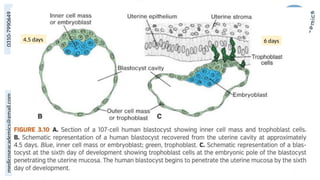
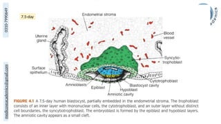
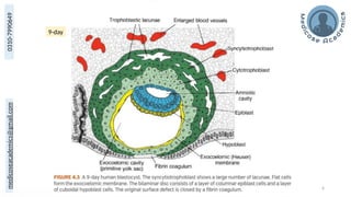
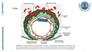
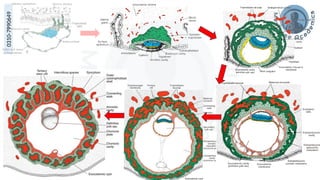
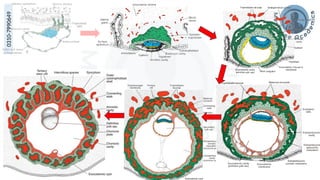
Early Development
medicoseacademics@gmail.com
0310-7990649
4
4.5 days 6 days
medicoseacademics@gmail.com
0310-7990649
5
7.5-day
medicoseacademics@gmail.com
0310-7990649
6
9-day
medicoseacademics@gmail.com
0310-7990649
7
12-day
medicoseacademics@gmail.com
0310-7990649
8
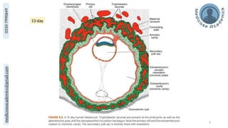
13-day
medicoseacademics@gmail.com
0310-7990649
9
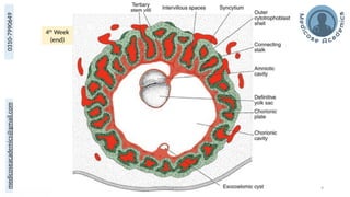
4th
Week
(end)
medicoseacademics@gmail.com
0310-7990649
10
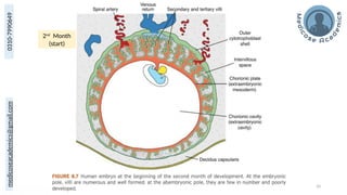
2nd
Month
(start)
medicoseacademics@gmail.com
0310-7990649
medicoseacademics@gmail.com
0310-7990649
12
Placenta
medicoseacademics@gmail.com
0310-7990649
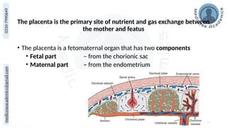
The placenta is the primary site of nutrient and gas exchange between
the mother and featus
• The placenta is a fetomaternal organ that has two components
• Fetal part – from the chorionic sac
• Maternal part – from the endometrium
13
medicoseacademics@gmail.com
0310-7990649
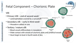
Fetal Component – Chorionic Plate
Villi
• Primary Villi – end of second week1
• cytotrophoblast covered by a syncytium
• Secondary villi – early in third week1
• Mesoderm added at core
• Tertiary / mature villi
• Blood vessels differentiate in mesoderm
• Make contact with vessels of chorionic plate and umbilical vessels
• Heart begin to beat in fourth week of dev.
14
medicoseacademics@gmail.com
0310-7990649
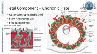
Fetal Component – Chorionic Plate
• Outer Cytotrophoblastic Shell
• Stem / Anchoring Villi
• Free Terminal Villi
15
end of the third week
end of the fourth week
medicoseacademics@gmail.com
0310-7990649
medicoseacademics@gmail.com
0310-7990649
17
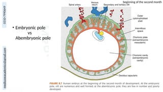
• Embryonic pole
vs
Abembryonic pole
beginning of the second month
medicoseacademics@gmail.com
0310-7990649
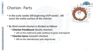
Chorion- Parts
• In the early weeks (till beginning of 8th
week)1
, villi
cover the entire surface of the chorion
• By third month chorion is divided as follows
• Chorion frondosum (bushy chorion)
• villi on the embryonic pole continue to grow and expand
• Chorion laeve (smooth chorion)
• Villi on the abembryonic pole degenerate
18
medicoseacademics@gmail.com
0310-7990649
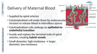
Delivery of Maternal Blood
• Supplied by spiral arteries
• Cytotrophoblast cell erode these by endovascular
invasion to release blood in intervillous spaces
• Cytotrophoblast cells undergo an epithelial-to-
endothelial transition
• Invade and replace the terminal ends of spiral
arteries, creating hybrid vessels
• Small-diameter, high-resistance → larger
diameter, low-resistance
19
medicoseacademics@gmail.com
0310-7990649
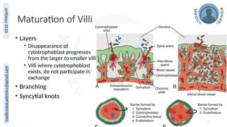
Maturation of Villi
• Layers
• Disappearance of
cytotrophoblast progresses
from the larger to smaller villi
• Villi where cytotrophoblast
exists, do not participate in
exchange
• Branching
• Syncytial knots
20
medicoseacademics@gmail.com
0310-7990649
22
medicoseacademics@gmail.com
0310-7990649
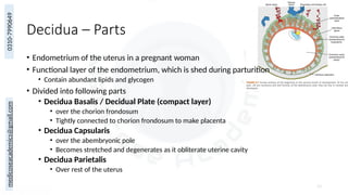
Decidua – Parts
• Endometrium of the uterus in a pregnant woman
• Functional layer of the endometrium, which is shed during parturition
• Contain abundant lipids and glycogen
• Divided into following parts
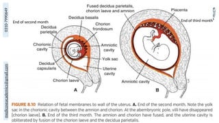
• Decidua Basalis / Decidual Plate (compact layer)
• over the chorion frondosum
• Tightly connected to chorion frondosum to make placenta
• Decidua Capsularis
• over the abembryonic pole
• Becomes stretched and degenerates as it obliterate uterine cavity
• Decidua Parietalis
• Over rest of the uterus
23
medicoseacademics@gmail.com
0310-7990649
24
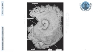
End of second month End of third month
medicoseacademics@gmail.com
0310-7990649
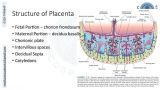
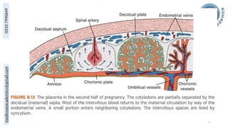
Structure of Placenta
25
• Fetal Portion – chorion frondosum
• Maternal Portion – decidua basalis
• Chorionic plate
• Intervillous spaces
• Decidual Septa
• Cotyledons
medicoseacademics@gmail.com
0310-7990649
28
medicoseacademics@gmail.com
0310-7990649
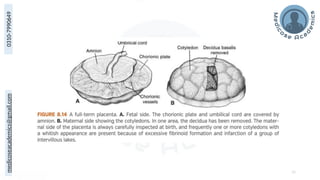
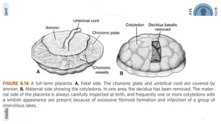
Full Term Placenta
• Covers approximately 15% to 30% of internal surface of the uterus
• Diameter of 15 to 25 cm
• Thickness 3cm
• Weighs 500-600g (1/6th
of fetal weight)
• Expelled 30 min after birth as afterbirth
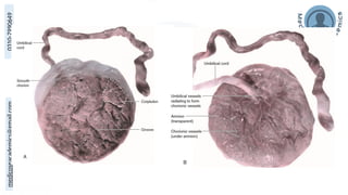
• Maternal side
• Cobblestone appearance, 15-20 cotyledons, covered by thin layers of decidua basalis
• Fetal Surface
• Chorionic plate, chorionic vessels, covered by amnion (shining)

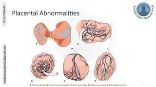
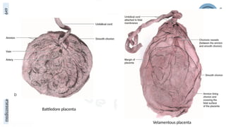
• Attachment of the umbilical cord is usually eccentric and occasionally even marginal
• velamentous insertion – insertion into chorionic membranes
30
medicoseacademics@gmail.com
0310-7990649
31
medicoseacademics@gmail.com
0310-7990649
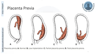
Placenta Previa
32
medicoseacademics@gmail.com
0310-7990649
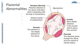
Placental
Abnormalities
33
medicoseacademics@gmail.com
0310-7990649
Placental Abnormalities
34
medicoseacademics@gmail.com
0310-7990649
medicoseacademics@gmail.com
0310-7990649
Battledore placenta
Velamentous placenta
medicoseacademics@gmail.com
0310-7990649
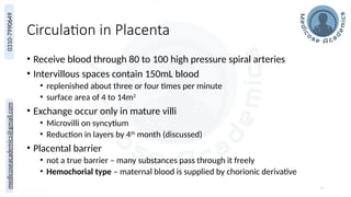
Circulation in Placenta
• Receive blood through 80 to 100 high pressure spiral arteries
• Intervillous spaces contain 150mL blood
• replenished about three or four times per minute
• surface area of 4 to 14m2
• Exchange occur only in mature villi
• Microvilli on syncytium
• Reduction in layers by 4th
month (discussed)
• Placental barrier
• not a true barrier – many substances pass through it freely
• Hemochorial type – maternal blood is supplied by chorionic derivative
37
medicoseacademics@gmail.com
0310-7990649
38
medicoseacademics@gmail.com
0310-7990649
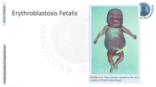
Erythroblastosis Fetalis
39
medicoseacademics@gmail.com
0310-7990649
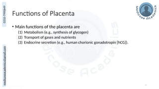
Functions of Placenta
• Main functions of the placenta are
(1) Metabolism (e.g., synthesis of glycogen)
(2) Transport of gases and nutrients
(3) Endocrine secretion (e.g., human chorionic gonadotropin [hCG]).
40
medicoseacademics@gmail.com
0310-7990649
Metabolism
• The placenta, particularly during early pregnancy, synthesizes
• Glycogen
• Cholesterol
• fatty acids
• which serve as sources of nutrients and energy for the embryo/fetus
41
medicoseacademics@gmail.com
0310-7990649
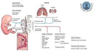
Transfer / Exchange
• Gases
• oxygen, carbon dioxide, and carbon monoxide
• At term, the fetus extracts 20 to 30 mL of oxygen per minute
• O2 delivery is flow limited, not diffusion limited
• Nutrients and Electrolytes
• Occurs bi-directionally via various transport mechanism
• Hormones
• Thyroxine, triiodothyronine exception for water soluble
• Unconjugated steroids pass freely
• Maternal Antibodies and proteins
• IgG begin transported at about 14 weeks
• Complement system at the end of first trimester
• IgG production reaches adult levels at 3 years of age
• Waste
• Drugs
• Infectious Agents 42
medicoseacademics@gmail.com
0310-7990649
medicoseacademics@gmail.com
0310-7990649
Hormone Production
All hormones are synthesized in the syncytial trophoblast
• Progesterone – sufficient to support pregnancy by 4th
month
• Estriol – until just before the end of pregnancy
• hCG – during first two months
• Human chorionic Somatomammotropin (human placental lactogen)
• gives the fetus priority on maternal blood glucose
• makes the mother somewhat diabetogenic
• It also promotes breast development for milk production
• Human chorionic thyrotropin
• Human chorionic corticotropin
44
medicoseacademics@gmail.com
0310-7990649
45
Amnion
medicoseacademics@gmail.com
0310-7990649
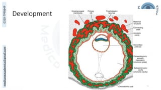
Development
46
medicoseacademics@gmail.com
0310-7990649
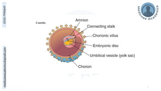
47
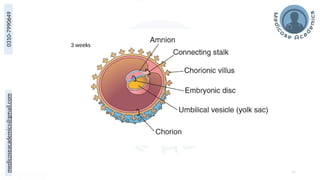
3 weeks
medicoseacademics@gmail.com
0310-7990649
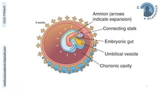
48
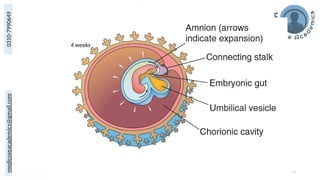
4 weeks
medicoseacademics@gmail.com
0310-7990649
49
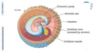
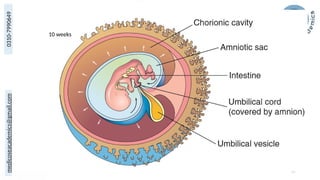
10 weeks
medicoseacademics@gmail.com
0310-7990649
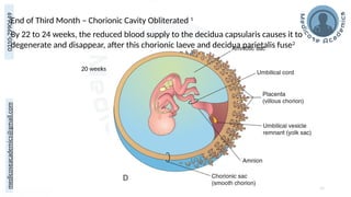
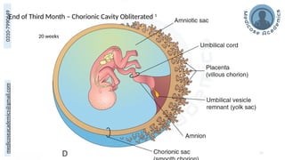
End of Third Month – Chorionic Cavity Obliterated 1
50
20 weeks
By 22 to 24 weeks, the reduced blood supply to the decidua capsularis causes it to
degenerate and disappear, after this chorionic laeve and decidua parietalis fuse2
medicoseacademics@gmail.com
0310-7990649
Amniotic Bands
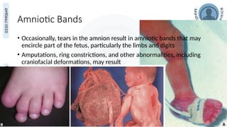
• Occasionally, tears in the amnion result in amniotic bands that may
encircle part of the fetus, particularly the limbs and digits
• Amputations, ring constrictions, and other abnormalities, including
craniofacial deformations, may result
51
medicoseacademics@gmail.com
0310-7990649
Premature Rupture of Membranes (PROM)
• Rupture of the membranes before uterine contractions (10%)
• Preterm PROM occurs before 37 completed weeks of pregnancy (3%)
• Risk factors
• previous pregnancy affected by prematurity or PROM, black race, smoking,
infections, and severe polyhydramnios
52
medicoseacademics@gmail.com
0310-7990649
Amniotic Fluid
• A clear, watery fluid that fills the amniotic cavity
• 7.2 and specific gravity 1.0069 to 1.008
• Increases with gestational age1
• 30 mL at 10 weeks of gestation
• 450 mL at 20 weeks
• 800 to 1,000 mL at 37 weeks
• Produced in part by amniotic cells but is derived primarily from maternal blood
• Replaced every 3 hours1,2
• Mostly through amniochorionic membrane, others umbilical cord, fetal side placenta
• From the beginning of the 5 month, the fetus swallow 400mL a day, absorbed by GIT
• Almost same amount excreted in urine
53
medicoseacademics@gmail.com
0310-7990649
Amniotic Fluid – Composition
• Aqua
• Undissolved material
• desquamated fetal epithelial cells
• Organic compounds
• carbohydrates, fats, enzymes, hormones, and pigment
• Inorganic salts
• As pregnancy advances, the composition of the amniotic fluid changes as
fetal excreta is added
• This is also basis of tests based on amniocentesis
54
medicoseacademics@gmail.com
0310-7990649
Amniotic Fluid- Functions
1) Permits symmetric external growth of the embryo and fetus
2) Acts as a barrier to infection
3) Permits normal fetal lung development
4) absorbs jolts
5) prevents adherence of the embryo to the amnion
6) Maintain relatively uniform and constant temp around fetus
7) allows for fetal movements, aiding in muscular development
8) Maintain hemostasis of fluid electrolyte
9) amniochorionic membrane forms a hydrostatic wedge that helps to dilate the
cervical canal
55
medicoseacademics@gmail.com
0310-7990649
Hydroamnios / Polyhydroamnios
• Excess of amniotic fluid (1,500 to 2,000 mL)
• Causes
• idiopathic causes (35%)
• maternal diabetes (25%]
• congenital malformations, that prevent the infant from swallowing the fluid
• central nervous system disorders (e.g., anencephaly)
• gastrointestinal defects (atresias, e.g., esophageal)
• Complications / Associations
• severe defects of the central nervous system, such as meroencephaly (anencephaly)
56
medicoseacademics@gmail.com
0310-7990649
Oligohydroamnios
• Decreased amount of amniotic fluid (< 400 mL]
• Cause
• Placental Insufficiency
• PrePROM
• Renal agenesis
• Obstructive Uropathy
• Complication
• constrict the fetus
• there may be too little fluid for the fetus to “ breathe” into its lungs resulting in lung
hypoplasia
• facial defects, and limb defects
57
medicoseacademics@gmail.com
0310-7990649
58
Yolk Sac (Umbilical Vesicle)
Umbilical vesicle preferred in humans as it does not contain yolk
medicoseacademics@gmail.com
0310-7990649
59
3 weeks
medicoseacademics@gmail.com
0310-7990649
60
4 weeks
medicoseacademics@gmail.com
0310-7990649
61
10 weeks
medicoseacademics@gmail.com
0310-7990649
End of Third Month – Chorionic Cavity Obliterated 1
62
20 weeks
medicoseacademics@gmail.com
0310-7990649
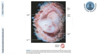
• Primary and Secondary Yolk sac (discussed)
• Can be observed sonographically early in the fifth week
• By 10 weeks, the umbilical vesicle has shrunk to a pear-shaped
remnant approximately 5 mm in diameter
• By 20 weeks, the umbilical vesicle is very small
• Thereafter, it is usually not visible (very rarely persists, not sig.)
• Omphaloenteric duct usually detaches from the midgut loop by the
end of the sixth week
• Failure – Meckel diverticulum
63
medicoseacademics@gmail.com
0310-7990649
medicoseacademics@gmail.com
0310-7990649
Significance
• Transfer of nutrients to the embryo during the second and third
weeks
• Blood development first occurs in the well-vascularized
extraembryonic mesoderm covering the wall of the umbilical vesicle
beginning in the third week
• During the fourth week, the endoderm of the umbilical vesicle is
incorporated into the embryo as the primordial gut
• Primordial germ cells appear in the endodermal lining of the wall of
the umbilical vesicle in the third week and subsequently migrate
65
medicoseacademics@gmail.com
0310-7990649
66
Allantois
medicoseacademics@gmail.com
0310-7990649
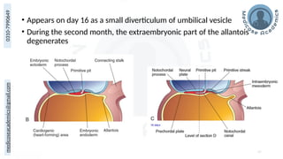
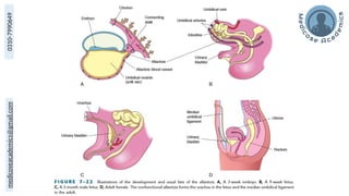
• Appears on day 16 as a small diverticulum of umbilical vesicle
• During the second month, the extraembryonic part of the allantois
degenerates
67
medicoseacademics@gmail.com
0310-7990649
medicoseacademics@gmail.com
0310-7990649
Function
• Other animals (reptiles, birds, and most mammals)
• respiratory function and/or
• acts as a reservoir for urine during embryonic life
• In Humans
• Surrounding mesenchyme forms umbilical blood vessels
• Blood formation occurs in its wall during the third to fifth weeks.
• Proximal part persist throughout dev. as urachus
Urachus in adults is represented as median umbilical ligament
69
medicoseacademics@gmail.com
0310-7990649
Allantoic Cysts
• Allantoic cysts, remnants of the extraembryonic portion of the
allantois, are usually found between the fetal umbilical vessels
• can be detected by ultrasonography
• They are most commonly detected in the proximal part of the
umbilical cord, near its attachment to the anterior abdominal wall
• The cysts are generally asymptomatic until childhood or adolescence,
when they can become infected and inflamed.
70
medicoseacademics@gmail.com
0310-7990649
71
Umbilical Cord
medicoseacademics@gmail.com
0310-7990649
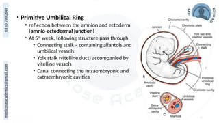
• Primitive Umbilical Ring
• reflection between the amnion and ectoderm
(amnio-ectodermal junction)
• At 5th
week, following structure pass through
• Connecting stalk – containing allantois and
umbilical vessels
• Yolk stalk (vitelline duct) accompanied by
vitelline vessels
• Canal connecting the intraembryonic and
extraembryonic cavities
72
medicoseacademics@gmail.com
0310-7990649
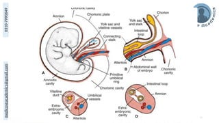
• By the end of third month amniotic cavity enlarges rapidly
• Amnion fuses with chorion obliterating chorionic cavity
• Amnion begins to envelop the connecting and yolk sac stalks,
crowding them together and giving rise to the primitive umbilical
cord
• Distally – yolk sac and umbilical vesicles
• Proximally – intestinal loops and remnants of allantois
• Cavity in the cord is obliterated after retraction of physiological
umbilical hernia
• Later allantois and the vitelline duct and its vessels are also
obliterated, all that remains in the cord are the umbilical vessels
surrounded by Wharton jelly
73
medicoseacademics@gmail.com
0310-7990649
74
medicoseacademics@gmail.com
0310-7990649
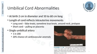
Umbilical Cord Abnormalities
• At birth 2 cm in diameter and 50 to 60 cm long
• Length of cord reflects intrauterine movements
• Long cord – false knots, sometime true knots, around neck, prolapse
• Short cord – pulling on placenta
• Single umbilical artery
• 1 in 200
• 20% chance of cardiovascular anomaly
75
medicoseacademics@gmail.com
0310-7990649
Stages of Labour
76
medicoseacademics@gmail.com
0310-7990649
77
Multiple Pregnancies
medicoseacademics@gmail.com
0310-7990649
Learning Objectives
• Differentiate between monozygotic and dizygotic twins.
• Explain different types of monozygotic twins.
• Define conjoint twins
• State the different anomalies associated with twin pregnancies?
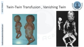
(vanishing twin, fetus papyraceus, twin-twin transfusion syndrome)
medicoseacademics@gmail.com
0310-7990649
Dr Muhammad Ali Rabbani
medicoseacademics@gmail.com
0310-7990649
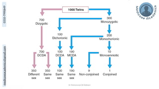
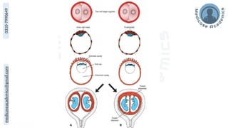
Dizygotic Twins
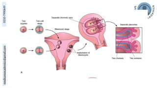
• Two separate ova a released. Both fertilized independently and implant
independently.
• Thus there will be two separate sets of placenta, chorion and amnion.
• Both will have different genomes. Twins born will have differences as there are
between any two non twin siblings. Both may be even of different sex.
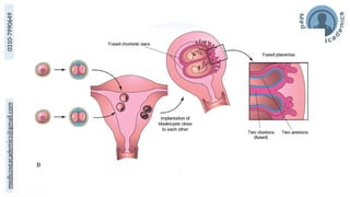
• Sometimes the implantation may be so close that placenta of both will be fused.
• Twins will be dichorionic and diamniotic (DCDA)
medicoseacademics@gmail.com
0310-7990649
medicoseacademics@gmail.com
0310-7990649
medicoseacademics@gmail.com
0310-7990649
medicoseacademics@gmail.com
0310-7990649
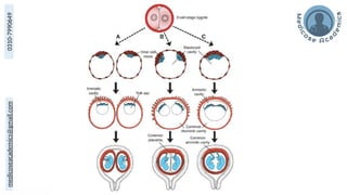
Monozygotic Twins
• One ovum is released and is fertilized.
• Fertilized ovum (zygote) may then split and the two fragments grow
independently to form twins.
• The time when the split occurs, determines which type of twins will
form.
medicoseacademics@gmail.com
0310-7990649
medicoseacademics@gmail.com
0310-7990649
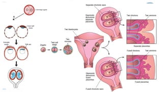
Dichorionic Diamniotic (DCDA)
• When separation occurs within 3 days of conception. Earliest
separation occur at two cell stage and later in the morula stage.
• Thus two blastocysts form. Grow independently.
• Each Zygote will have its own placenta and its own amniotic cavity.
medicoseacademics@gmail.com
0310-7990649
medicoseacademics@gmail.com
0310-7990649
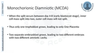
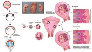
Monochorionic Diamniotic (MCDA)
• When the split occurs between day 4-8 (early blastocyst stage), inner
cell mass split into two, outer cell mass will not split.
• Thus only one trophoblast grows, leading to only One Placenta
• Two separate embryoblast grows, leading to two different embryos
with two different amniotic cavity.
medicoseacademics@gmail.com
0310-7990649
medicoseacademics@gmail.com
0310-7990649
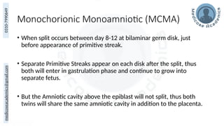
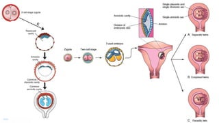
Monochorionic Monoamniotic (MCMA)
• When split occurs between day 8-12 at bilaminar germ disk, just
before appearance of primitive streak.
• Separate Primitive Streaks appear on each disk after the split, thus
both will enter in gastrulation phase and continue to grow into
separate fetus.
• But the Amniotic cavity above the epiblast will not split, thus both
twins will share the same amniotic cavity in addition to the placenta.
medicoseacademics@gmail.com
0310-7990649
medicoseacademics@gmail.com
0310-7990649
Conjoined or Siamese Twins
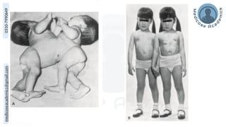
• If splitting is delayed beyond day 12, the embryonic disk has formed
and conjoined or Siamese Twins are formed.
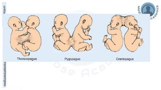
• Different types of Siamese Twins are named according to the body
parts from which they are fused.
medicoseacademics@gmail.com
0310-7990649
medicoseacademics@gmail.com
0310-7990649
medicoseacademics@gmail.com
0310-7990649
medicoseacademics@gmail.com
0310-7990649
Twin-Twin Transfusion , Vanishing Twin
96
medicoseacademics@gmail.com
0310-7990649
Thank You
Questions?
Suggestions?
https://www.medicoseacademics.com/
https://www.facebook.com/medicoseacademics
https://www.youtube.com/@MedicoseAcademics
Lecture Recording & Slides