Bioorganic Chemistry 151 (2024) 107597
Available online 6 July 2024
0045-2068/Crown Copyright © 2024 Published by Elsevier Inc. All rights are reserved, including those for text and data mining, AI training, and similar technologies.
Design and evaluation of novel inhibitors for the treatment of clear cell
renal cell carcinoma
Younos Bouzian a,1
, Mohamed El Hafi b,1
, Negar Parvizi a,1
, Woonghee Kim c,1
,
Mine Subaşioğlu a
, Mehmet Ozcan d
, Hasan Turkez e
, Adil Mardinoglu c,f,*
a
Trustlife Labs Drug Research & Development Center, 34774 Istanbul, Turkiye
b
Faculty of Medicine and Pharmacy, Mohammed First University, Oujda, Morocco
c
Science for Life Laboratory, KTH – Royal Institute of Technology, Stockholm SE-17165, Sweden
d
Department of Medical Biochemistry, Faculty of Medicine, Zonguldak Bulent Ecevit University, Zonguldak, Turkey
e
Department of Medical Biology, Faculty of Medicine, Atatürk University, Erzurum, Turkey
f
Centre for Host-Microbiome Interactions, Faculty of Dentistry, Oral & Craniofacial Sciences, King’s College London, London SE1 9RT, UK
A R T I C L E I N F O
Keywords:
Drug design
ccRCC
BUB1B
Caki-1
TG-101209
A B S T R A C T
The efficacy of conventional chemotherapies in treating clear cell renal cell carcinoma (ccRCC) is often limited
due to its high molecular diversity, generally low response rates to standard treatments, and prevalent drug
resistance. Recent advancements in the molecular understanding of ccRCC, alongside the discovery of novel
therapeutic agents targeting specific proteins, have significantly altered the treatment landscape for ccRCC. Here,
we synthesized 27 new compounds that are derivatives of TG-101209 to modulate BUB1B (BUB1 mitotic
checkpoint serine/threonine kinase B). BUB1B has been recently identified as a drug target for the development
of effective ccRCC treatment based on global transcriptomics profiling of ccRCC tumours and gene co-expression
network analysis. We characterized the molecular structures of these 27 compounds by 1
H and 13
C NMR and
Mass spectrometry. We evaluated the effect of these 27 compounds by analysing the modulation of the BUB1B
expression. Our primary objective was to design and assess the efficacy of these new compounds in reducing the
viability of Caki-1 cells, a ccRCC cell line. We performed the computational docking studies by the Schrödinger
Maestro software and demonstrated that three of these compounds (13a, 5i, and 5j) effectively downregulated
BUB1B expression and eventually triggered necrosis and apoptosis in the Caki-1 cell line based on the struc­
ture–activity relationship (SAR) analysis. The IC50 values for compounds 13a, 5i, and 5j were calculated as
2.047 µM, 10.046 µM, and 6.985 µM, respectively, indicating their potent inhibitory effects on cell viability. Our
study suggests that these compounds targeting BUB1B could offer a more effective and promising approach for
ccRCC treatment compared to the conventionally used tyrosine kinase inhibitors. Our study underscores the
potential of leveraging targeted therapies against specific molecular pathways in ccRCC may open new avenues
for the development of effective treatment strategies against ccRCC.
1. Introduction
Clear cell renal cell carcinoma (ccRCC) is the most common histo­
logical subtype of renal cell carcinomas (RCC), accounting for approx­
imately 70 % of all RCC cases [1]. In recent years, the treatment
landscape for ccRCC has undergone significant changes, offering new
hope for patients. The introduction of tyrosine kinase inhibitors (TKIs)
such as axitinib, sorafenib, pazopanib, and sunitinib [1–3] has played a
pivotal role in this transformation, alongside immune checkpoint
inhibitors such as ipilimumab, nivolumab, pembrolizumab, and atezo­
lizumab, [4,5] and mammalian target of rapamycin (mTOR) inhibitors
such as everolimus and temsirolimus [6,7]. These advancements have
markedly improved outcomes for ccRCC patients. However, the benefits
of these treatments are not universal, with only a subset of patients
responding to treatment [8,9]. This variability in patient response may
be explained by the high molecular heterogeneity of ccRCC, coupled
with generally low response rates to conventional treatments and the
development of drug resistance, which poses significant challenges in
* Corresponding author.
E-mail address: adilm@scilifelab.se (A. Mardinoglu).
1
These authors contributed equally.
Contents lists available at ScienceDirect
Bioorganic Chemistry
journal homepage: www.elsevier.com/locate/bioorg
https://doi.org/10.1016/j.bioorg.2024.107597
Received 2 March 2024; Received in revised form 23 June 2024; Accepted 25 June 2024
Bioorganic Chemistry 151 (2024) 107597
2
the effective management of ccRCC. Hence, there is a vital need to
develop innovative and effective treatment strategies. Computational
drug repositioning along with novel drug discovery efforts are crucial for
developing well-tolerated and more effective drugs [10,11]. Such ap­
proaches have the potential to diversify and enhance the therapeutic
options available to ccRCC patients, to address the urgent need for
personalized and effective treatment strategies [12,13].
Systems biology methods have become instrumental in computa­
tional drug repositioning, serving as a powerful means of detecting po­
tential interactions between drugs and their targets and the nexus
between drugs and diseases [14–16]. The strategy of repurposing well-
characterized compounds can lead to cost savings and accelerate the
development of effective drugs [17]. We have previously identified four
genes, including BUB1B (BUB1 mitotic checkpoint serine/threonine
kinase B), RRM2, ASF1B, and CCNB2 that may contribute to the
unfavourable survival rates observed in ccRCC patients based on co-
expression network analysis of global transcriptomic profiling of
ccRCC patients. We have also performed drug repositioning and iden­
tified TG-101209 as a drug candidate to modulate the expression of
BUB1B. We have also tested the efficacy of TG-101209 on the Caki-1 cell
line and showed that the protein level of BUB1B and cell viability
significantly decreased. These findings positioned TG-101209 as a po­
tential drug candidate for effective ccRCC treatment [18].
Drug discovery is an intricate and multifaceted endeavour focused on
the development and synthesis of increasingly efficacious drug candi­
dates. In a prior investigation, TG-101209 was identified as an effective
drug candidate for the treatment of ccRCC. Nonetheless, there is a need
to refine the molecular structure of TG-101209 to enhance its impact on
the target protein, BUB1B, and to improve its toxic effect on cell
viability. The objective of the current study was to design and synthesize
novel derivatives of TG-101209 that are more specific and potent. The
effectiveness of these derivatives on cell viability and their ability to
target protein were evaluated by performing in vitro experiments. The
study has yielded several promising compounds that could potentially
lead to more effective drug candidates for the treatment of ccRCC.
2. Results and discussion
2.1. Chemistry
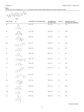
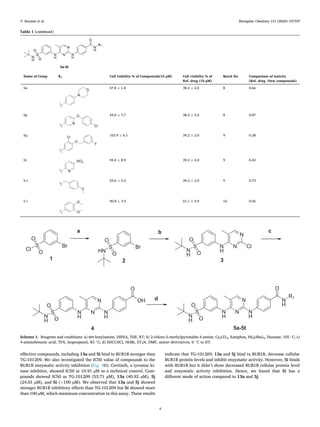
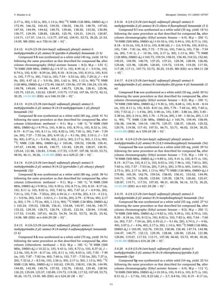
The compounds 5a-5 t presented in Table 1 were prepared as shown
in Scheme 1. 3-bromobenzenesulfonyl chloride 1 was reacted with tert-
butylamine in THF to yield N-(tert-butyl)-3-bromobenzenesulfonamide
2. Furthermore, compound 2 underwent to the Buchwald-Hartwig re­
action with 2-chloro-5-methylpyrimidin-4-amine to produce compound
3. Subsequently, compound 3 was treated with 4-aminobenzoic acid to
obtain product 4. In the final step, compound 4 was reacted with
different kinds of amine derivatives to generate 20 target compounds
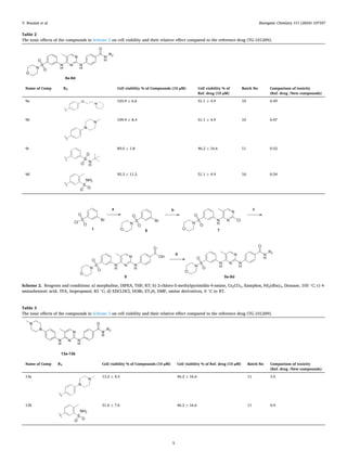
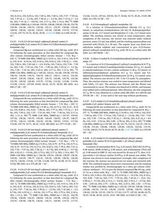
5a-5 t. The compounds 9a-9d, shown in Table 2, were prepared as
detailed in Scheme 2. 3-bromobenzenesulfonyl chloride 1 was reacted
with morpholine in THF to form 4-((3-bromophenyl)sulfonyl)morpho­
line 6. In addition, compound 6 reacted with 2-chloro-5-methylpyrimi­
din-4-amine via Buchwald-Hartwig reaction to yield compound 7.
Subsequently, compound 7 was treated with 4-aminobenzoic acid to
produce product 8. In the final step, compound 8 was reacted with
different kinds of amine derivatives to generate the target compounds
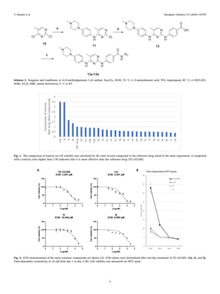
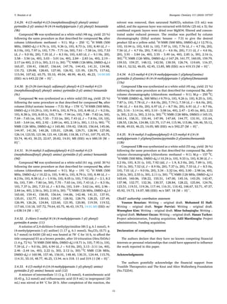
9a-9d. The compounds 13a and 13b, presented in Table 3, were pre­
pared according to Scheme 3. 2,4-dichloro-5-methylpyrimidine 10 was
reacted with 4-(4-methylpiperazin-1-yl) aniline in EtOH in the presence
of Na2CO3 to produce compound 11. Compound 11 reacted with 4-ami­
nobenzoic acid to obtain product 12. Finally, compound 12 was reacted
with two different amines to yield the target compounds 13a and 13b.
2.2. Biology
MTT assay was performed to evaluate the effect of newly synthesized
27 compounds on cell viability. The toxicity values of the reference drug
(TG-101209) and the new compounds at 10 μM concentration on the
Caki-1 cell line are shown in Tables 1-3.
Since MTT tests of newly synthesized compounds were performed at
different times and in different batches, the toxic effect on cell viability
was calculated by comparing the cell viability of each drug to the
reference drug tested in the same experiments, thus eliminating external
factors between the experiments. The comparable toxic effect of each
compound on cell viability was demonstrated in the last column in Ta­
bles 1-3. A compound with a toxicity ratio higher than 1.00 indicates
greater effectiveness than the reference drug in terms of cell viability.
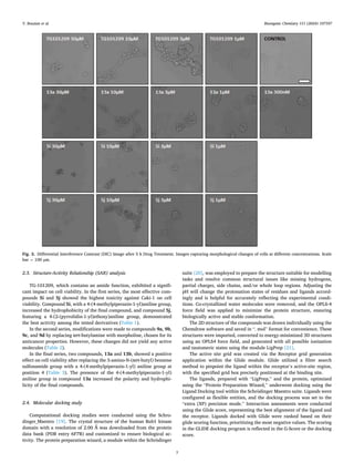
Consequently, it was determined that the toxic effect of compounds 13a,
5j, 5i and 5 k on cell viability was 3.50, 3.47, 1.68 and 1.37 times that of
the ref drug, respectively (Fig. 1).
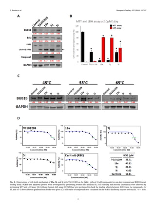
We have selected the 3 compounds with the most toxic effect on
Caki-1 cell viability as follows, 13a (13.2 %), 5i (28.2 %), and 5j (13.6
%). For further investigation, we examined the IC50 values of the three
most effective compounds compared to the reference drug (TG-101209).
The compounds were treated in triplicate at concentrations of 30 µM, 10
µM, 3 µM, 1 µM, 300 nM, 1 nM, and 30 nM for one day. The MTT assay
revealed IC50 values of TG-101209 (2.881 µM), 13a (2.047 µM), 5i
(10.046 µM), and 5j (6.985 µM), respectively (Fig. 2 A). We also
observed the time-dependent toxicity of these 3 compounds. We treated
the cells with 10 µM of the 3 most toxic compounds and TG-101209 and
collected the data from day 1 to day 4 independently (Fig. 2 B). 13a, 5i,
and 5j showed decreased cell viability at day 1 (<10 %), and a more
toxic effect on days 2,3 and 4 (<5%) on cell viability. Reference drug
(TG-101209) also showed decreased cell viability at day 1 (32.9 %), day
2 (11.9 %), day 3 (5.1 %) and day 3 (<5%).
Subsequently, we studied the short-term cytotoxicity by analysing
the cell morphological changes after 5 h of treatment using Differential
Interference Contrast (DIC) imaging. The different concentrations
influencing morphological changes were captured in DIC images
(Fig. 3). TG-101209 exhibited circular and aggregated cell morphology
up to 1 µM during the 5-hour treatment. Similarly, 13a (up to 300 nM),
5i (up to 1 µM), and 5j (up to 1 µM) demonstrated circular, shrunken,
and aggregated cell morphology during the 5-hour treatment.
We also investigated the cell death mechanism of 13a, 5i, and 5j
compared to TG-101209 (Fig. 4). Caki-1 cells were treated with the
compounds at 10 µM for one day. Western blot analysis showed a
significantly decreased protein level of BUB1B with TG-101209 (30 %),
13a (20 %), and 5j (37 %) treatments. Interestingly, 5i treated cells only
decreased the protein level of BUB1B by 14 % (Fig. 4A). We also
observed that the protein level of Bcl2, which is involved in apoptosis via
mitochondrial membrane potential was significantly decreased by TG-
101209 (33 %), 13a (54 %), 5j (59 %), and 5i (66 %). 13a and 5j
significantly increased the protein level of Cleaved PARP and Caspase3,
which are DNA fragmentation apoptosis pathways, compared to TG-
101209. Compound 5i showed a moderate effect. For further study,
we performed MTT and LDH assay to check necrotic cell death (Fig. 4B).
Our most effective compounds 13a, 5j, and 5i dramatically increased
the cell necrosis. Interestingly, TG-101209 did not induce cell necrosis.
These data indicate that 13a, 5j, and 5i have necrotic cell death ability
and 13a and 5j induce stronger apoptosis than TG-101209.
Cellular thermal shift assay (CETSA) showed the binding affinity of
compounds to BUB1B (Fig. 4C). We give heat shock to 20 µM compounds
cells after 1 h treatment at 45 ◦
C, 55 ◦
C, and 65 ◦
C for 3 min. 13a, and 5i
showed 1.32– and 1.20-times heat resistance remained soluble protein
than DMSO treated Control group at 45 ◦
C on BUB1B. All compounds
including TG-101209 showed higher heat resistance at 55 ◦
C. 13a (3.50
times) and 5i (2.06 times) showed much stronger heat resistance of
BUB1B than TG-101209. At the 65 ◦
C heat shock group, most of the
soluble BUB1B disappeared in DMSO treated control group. However,
TG-101209 (2.23 times), 13a (3.23 times), 5i (3.07 times), and 5j (2.68
times) treated groups are still showing protein bands. Our results indi­
cated that all 3 compounds and TG-101209 bind to BUB1B and our most
Y. Bouzian et al.
Bioorganic Chemistry 151 (2024) 107597
3
Table 1
The toxic effects of the compounds in Scheme 1 on cell viability and their relative effect compared to the reference drug (TG-101209).
Name of Comp R1 Cell viability % of Compounds(10 µM) Cell viability % of
Ref. drug (10 µM)
Batch No Comparison of toxicity
(Ref. drug /New compounds)
5a 68.1 ± 3.6 37.8 ± 6.8 6 0.55
5b 64.7 ± 8.8 37.8 ± 6.8 6 0.58
5c 57.0 ± 1.0 37.8 ± 6.8 6 0.66
5d 72.9 ± 7.1 37.8 ± 6.8 6 0.52
5e 80.4 ± 1.9 37.8 ± 6.8 6 0.47
5f 78.9 ± 12.9 47.5 ± 1.3 7 0.6
5 g 94.7 ± 3.34 47.5 ± 1.3 7 0.5
5 h 49.0 ± 0.8 47.5 ± 1.3 7 0.97
5i 28.2 ± 0.2 47.5 ± 1.3 7 1.68
5j 13.7 ± 0.8 47.5 ± 1.3 7 3.47
5 k 27.9 ± 2.2 38.2 ± 2.0 8 1.37
5 l 93.2 ± 11.4 47.5 ± 1.3 7 0.51
5 m 40.8 ± 2.0 38.2 ± 2.0 8 0.93
5n 54.2 ± 0.5 38.2 ± 2.0 8 0.71
(continued on next page)
Y. Bouzian et al.
Bioorganic Chemistry 151 (2024) 107597
4
effective compounds, including 13a and 5i bind to BUB1B stronger than
TG-101209. We also investigated the IC50 value of compounds to the
BUB1B enzymatic activity inhibition (Fig. 4D). Ceritinib, a tyrosine ki­
nase inhibitor, showed IC50 at 10.91 µM as a technical control. Com­
pounds showed IC50 as TG-101209 (53.71 µM), 13a (40.92 µM), 5j
(24.01 µM), and 5i (>100 µM). We observed that 13a and 5j showed
stronger BUB1B inhibitory effects than TG-101209 but 5i showed more
than 100 µM, which maximum concentration in this assay. These results
indicate that TG-101209, 13a and 5j bind to BUB1B, decrease cellular
BUB1B protein levels and inhibit enzymatic activity. However, 5i binds
with BUB1B but it didn’t show decreased BUB1B cellular protein level
and enzymatic activity inhibition. Hence, we found that 5i has a
different mode of action compared to 13a and 5j.
Table 1 (continued)
Name of Comp R1 Cell viability % of Compounds(10 µM) Cell viability % of
Ref. drug (10 µM)
Batch No Comparison of toxicity
(Ref. drug /New compounds)
5o 57.8 ± 1.8 38.2 ± 2.0 8 0.66
5p 44.0 ± 3.7 38.2 ± 2.0 8 0.87
5q 103.9 ± 6.1 39.2 ± 2.0 9 0.38
5r 94.0 ± 8.9 39.2 ± 2.0 9 0.42
5 s 53.6 ± 5.2 39.2 ± 2.0 9 0.73
5 t 90.8 ± 3.9 51.1 ± 4.9 10 0.56
Scheme 1. Reagents and conditions: a) tert-butylamine, DIPEA, THF, RT; b) 2-chloro-5-methylpyrimidin-4-amine, Cs2CO3, Xantphos, Pd2(dba)3, Dioxane, 105 ◦
C; c)
4-aminobenzoic acid, TFA, Isopropanol, 83 ◦
C; d) EDCl.HCl, HOBt, ET3N, DMF, amine derivatives, 0 ◦
C to RT.
Y. Bouzian et al.
Bioorganic Chemistry 151 (2024) 107597
5
Table 2
The toxic effects of the compounds in Scheme 2 on cell viability and their relative effect compared to the reference drug (TG-101209).
Name of Comp R2 Cell viability % of Compounds (10 µM) Cell viability % of
Ref. drug (10 µM)
Batch No Comparison of toxicity
(Ref. drug /New compounds)
9a 103.9 ± 6.6 51.1 ± 4.9 10 0.49
9b 109.9 ± 8.4 51.1 ± 4.9 10 0.47
9c 89.0 ± 1.8 46.2 ± 16.6 11 0.52
9d 95.3 ± 11.2 51.1 ± 4.9 10 0.54
Scheme 2. Reagents and conditions: a) morpholine, DIPEA, THF, RT; b) 2-chloro-5-methylpyrimidin-4-amine, Cs2CO3, Xantphos, Pd2(dba)3, Dioxane, 105 ◦
C; c) 4-
aminobenzoic acid, TFA, Isopropanol, 83 ◦
C; d) EDCl.HCl, HOBt, ET3N, DMF, amine derivatives, 0 ◦
C to RT.
Table 3
The toxic effects of the compounds in Scheme 3 on cell viability and their relative effect compared to the reference drug (TG-101209).
Name of Comp R3 Cell viability % of Compounds (10 µM) Cell viability % of Ref. drug (10 µM) Batch No Comparison of toxicity
(Ref. drug /New compounds)
13a 13.2 ± 4.5 46.2 ± 16.6 11 3.5
13b 51.6 ± 7.6 46.2 ± 16.6 11 0.9
Y. Bouzian et al.
Bioorganic Chemistry 151 (2024) 107597
6
Scheme 3. Reagents and conditions: a) 4-(4-methylpiperazin-1-yl) aniline, Na2CO3, EtOH, 76 ◦
C; c) 4-aminobenzoic acid, TFA, Isopropanol, 83 ◦
C; c) EDCl.HCl,
HOBt, ET3N, DMF, amine derivatives, 0 ◦
C to RT.
Fig. 1. The comparison of toxicity on cell viability was calculated by the ratio of each compound to the reference drug tested in the same experiment. A compound
with a toxicity ratio higher than 1.00 indicates that it is more effective than the reference drug (TG-101209).
Fig. 2. IC50 measurements of the most cytotoxic compounds are shown (A). IC50 values were determined after one-day treatment of TG-101209, 13a, 5i, and 5j.
Time-dependent cytotoxicity at 10 µM from day 1 to day 4 (B). Cell viability was measured via MTT assay.
Y. Bouzian et al.
Bioorganic Chemistry 151 (2024) 107597
7
2.3. Structure-Activity Relationship (SAR) analysis
TG-101209, which contains an amide function, exhibited a signifi­
cant impact on cell viability. In the first series, the most effective com­
pounds 5i and 5j showed the highest toxicity against Caki-1 on cell
viability. Compound 5i, with a 4-(4-methylpiperazin-1-yl)aniline group,
increased the hydrophobicity of the final compound, and compound 5j,
featuring a 4-(2-(pyrrolidin-1-yl)ethoxy)aniline group, demonstrated
the best activity among the tested derivatives (Table 1).
In the second series, modifications were made to compounds 9a, 9b,
9c, and 9d by replacing tert-butylamine with morpholine, chosen for its
anticancer properties. However, these changes did not yield any active
molecules (Table 2).
In the final series, two compounds, 13a and 13b, showed a positive
effect on cell viability after replacing the 3-amino-N-(tert-butyl) benzene
sulfonamide group with a 4-(4-methylpiperazin-1-yl) aniline group at
position 4 (Table 3). The presence of the 4-(4-methylpiperazin-1-yl)
aniline group in compound 13a increased the polarity and hydrophi­
licity of the final compounds.
2.4. Molecular docking study
Computational docking studies were conducted using the Schro­
dinger_Maestro [19]. The crystal structure of the human Bub1 kinase
domain with a resolution of 2.00 Å was downloaded from the protein
data bank (PDB entry 6F7B) and customized to ensure biological ac­
tivity. The protein preparation wizard, a module within the Schrödinger
suite [20], was employed to prepare the structure suitable for modelling
tasks and resolve common structural issues like missing hydrogens,
partial charges, side chains, and/or whole loop regions. Adjusting the
pH will change the protonation states of residues and ligands accord­
ingly and is helpful for accurately reflecting the experimental condi­
tions. Co-crystallized water molecules were removed, and the OPLS-4
force field was applied to minimize the protein structure, ensuring
biologically active and stable conformation.
The 2D structure of the compounds was drawn individually using the
Chemdraw software and saved in “. mol” format for convenience. These
structures were imported, converted to energy-minimized 3D structures
using an OPLS4 force field, and generated with all possible ionization
and tautomeric states using the module LigPrep [21].
The active site grid was created via the Receptor grid generation
application within the Glide module. Glide utilized a filter search
method to pinpoint the ligand within the receptor’s active-site region,
with the specified grid box precisely positioned at the binding site.
The ligands, prepared with “LigPrep,” and the protein, optimized
using the “Protein Preparation Wizard,” underwent docking using the
Ligand Docking tool within the Schrödinger Maestro suite. Ligands were
configured as flexible entities, and the docking process was set to the
“extra (XP) precision mode.” Interaction assessments were conducted
using the Glide score, representing the best alignment of the ligand and
the receptor. Ligands docked with Glide were ranked based on their
glide scoring function, prioritizing the most negative values. The scoring
in the GLIDE docking program is reflected in the G-Score or the docking
score.
Fig. 3. Differential Interference Contrast (DIC) Image after 5 h Drug Treatment. Images capturing morphological changes of cells at different concentrations. Scale
bar = 100 µm.
Y. Bouzian et al.
Bioorganic Chemistry 151 (2024) 107597
8
Fig. 4. Observations of cell death mechanism of 13a, 5j, and 5i with TG-101209 on the Caki-1 cells at 10 µM compounds for one-day treatment and BUB1B target
binding study. BUB1B and apoptotic protein were investigated by performing western blot analysis (A). Cell viability and necrotic cytotoxicity were observed by
performing MTT and LDH assay (B). Cellular thermal shift assay (CETSA) has been performed to check the binding affinity between BUB1B and the compounds. 45,
55, and 65 ◦
C three different gradient heat shocks were given (C). IC50 value of compounds was calculated for the BUB1B inhibitory enzyme activity (D). * P < 0.05.
Y. Bouzian et al.
Bioorganic Chemistry 151 (2024) 107597
9
We present a comprehensive analysis elucidating the synthesis and
initial exploration of the Structure-Activity Relationship (SAR) for
innovative BUB1B inhibitors that compete with TG-101209. In the
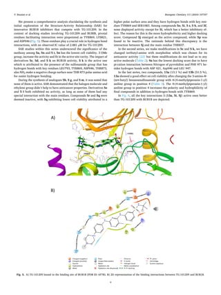
context of docking studies involving TG-101209 and BUBIB, pivotal
residues facilitating interaction were pinpointed as TYR869, LYS821,
and ASP946 (Fig. 5). These residues play a crucial role in hydrogen bond
interactions, with an observed IC value of 2.881 µM for TG-101209.
SAR studies within this series underscored the significance of the
methoxy among 5a, 5n and 5 t, 5n has the lowest cell viability. 3 OMe
group, increase the activity and fit in the active site cavity. The impact of
derivatives 5c, 5d, and 5 k on BUB1B activity, 5 k is the active one
which is attributed to the presence of the sulfonamide group that has
hydrogen bonds with key residues LEU793, TYR869, ASP946, THR873,
also NH2 make a negative charge surface near THR 873 polar amino acid
for easier hydrogen bonding.
During the synthesis of analogues 5b, 5 g, and 5 m, it was noted that
none of them is active. SAR demonstrated that the halogen molecule and
ethylene group didn’t help to have anticancer properties. Derivatives 5e
and 5 l both exhibited no activity, as long as none of them had any
special interaction with the main residues. Compounds 5r and 5q were
deemed inactive, with 5q exhibiting lower cell viability attributed to a
higher polar surface area and they have hydrogen bonds with key resi­
dues TYR869 and SER1083. Among compounds 5o, 5i, 5 s, 5 h, and 5f,
none displayed activity except for 5i, which has a better inhibitory ef­
fect. The reason for this is the more hydrophobicity and higher docking
score. Compound 5j emerged as the active compound, while 5p was
found to be inactive. The rationale behind this discrepancy is the
interaction between 5j and the main residue THR837.
In the second series, we make modifications in 5c and 5 k, we have
changed tertbutyl-amine with morpholine which was chosen for its
anticancer activity [22] but these modifications do not lead us to any
active molecule (Table 2). 9a has the lowest docking score due to have
pi-cation interaction between Nitrogen of pyrrolidine and PHE 971 be­
sides hydrogen bonds with ASP 921, Asp946 and LEU 947.
In the last series, two compounds, 13a (13.1 %) and 13b (51.5 %),
13a showed a good effect on cell viability after changing the 3-amino-N-
(tert-butyl) benzenesulfonamide group with 4-(4-methylpiperazin-1-yl)
aniline group in position 4 (Table 3). The 4-(4-methylpiperazin-1-yl)
aniline group in position 4 increases the polarity and hydrophilicity of
final compounds in addition to hydrogen bonds with TYR869.
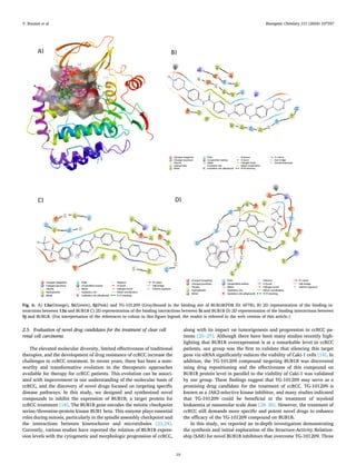
In Fig. 6, all the key interactions 3 (13a, 5i, 5j) active ones better
than TG-101209 with BUB1B are depicted.
Fig. 5. A) TG-101209 bound in the binding site of BUB1B (PDB ID: 6F7B). B) 2D representation of the binding interactions between TG-101209 and BUB1B.
Y. Bouzian et al.
Bioorganic Chemistry 151 (2024) 107597
10
2.5. Evaluation of novel drug candidates for the treatment of clear cell
renal cell carcinoma
The elevated molecular diversity, limited effectiveness of traditional
therapies, and the development of drug resistance of ccRCC increase the
challenges in ccRCC treatment. In recent years, there has been a note­
worthy and transformative evolution in the therapeutic approaches
available for therapy for ccRCC patients. This evolution can be associ­
ated with improvement in our understanding of the molecular basis of
ccRCC, and the discovery of novel drugs focused on targeting specific
disease pathways. In this study, we designed and synthesized novel
compounds to inhibit the expression of BUB1B, a target protein for
ccRCC treatment [18]. The BUB1B gene encodes the mitotic checkpoint
serine/threonine-protein kinase BUB1 beta. This enzyme plays essential
roles during mitosis, particularly in the spindle assembly checkpoint and
the interactions between kinetochores and microtubules [23,24].
Currently, various studies have reported the relation of BUB1B expres­
sion levels with the cytogenetic and morphologic progression of ccRCC,
along with its impact on tumorigenesis and progression in ccRCC pa­
tients [25–27]. Although there have been many studies recently high­
lighting that BUB1B overexpression is at a remarkable level in ccRCC
patients, our group was the first to validate that silencing this target
gene via siRNA significantly reduces the viability of Caki-1 cells [18]. In
addition, the TG-101209 compound targeting BUB1B was discovered
using drug repositioning and the effectiveness of this compound on
BUB1B protein level in parallel to the viability of Caki-1 was validated
by our group. These findings suggest that TG-101209 may serve as a
promising drug candidate for the treatment of ccRCC. TG-101209 is
known as a JAK2-selective kinase inhibitor, and many studies indicated
that TG-101209 could be beneficial in the treatment of myeloid
leukaemia at nanomolar scale dose [28–30]. However, the treatment of
ccRCC still demands more specific and potent novel drugs to enhance
the efficacy of the TG-101209 compound on BUB1B.
In this study, we reported an in-depth investigation demonstrating
the synthesis and initial exploration of the Structure-Activity Relation­
ship (SAR) for novel BUB1B inhibitors that overcome TG-101209. Three
Fig. 6. A) 13a(Orange), 5i(Green), 5j(Pink) and TG-101209 (Gray)bound in the binding site of BUB1B(PDB ID: 6F7B), B) 2D representation of the binding in­
teractions between 13a and BUB1B C) 2D representation of the binding interactions between 5i and BUB1B D) 2D representation of the binding interactions between
5j and BUB1B. (For interpretation of the references to colour in this figure legend, the reader is referred to the web version of this article.)
Y. Bouzian et al.
Bioorganic Chemistry 151 (2024) 107597
11
hit compounds (13a, 5i and 5j) among 27 compounds have exhibited
promising results in terms of deceased BUB1B expression and inducing
necrosis and apoptosis on the Caki-1 cell line. IC50 values of 13a, 5i and
5j were determined as 2.047 µM, 10.046 µM and 6.985 µM, respectively.
In literature, a variety of treatment approaches, including tyrosine ki­
nase inhibitors (Axitinib, Sorafenib, Pazopanib, and Sunitinib)[1–3],
immune checkpoint inhibitors (Ipilimumab, Nivolumab, Pem­
brolizumab, and Atezolizumab etc.)(4, 5), and mammalian target of
rapamycin inhibitors (Everolimus and Tesirolimus)[6,7] have been re­
ported for the treatment of ccRCC patients. Atsushi M. et al reported that
the IC50 value of Sunitinib, a receptor tyrosine kinase inhibitor widely
used in the treatment of ccRCC, was 2.22 uM for the Caki-1 cell line [31].
Sorafenib, another tyrosine kinase inhibitor, has been proven to improve
survival in advanced ccRCC patients [32]. Chu-Che L. et al indicated that
the IC50 value of Sorafenib was 6.23 µM for Caki-1 cells [33]. Com­
pound 13a among our hit compounds seems more effective with a low
IC50 value compared to commonly used tyrosine kinase inhibitors for
ccRCC treatment.
3. Conclusion
The effectiveness of clinical chemotherapies for ccRCC is often
inadequate due to its high molecular diversity, the generally low
response rates to traditional therapies, and the prevalent drug resistance
associated with ccRCC.
As predicted by the computational drug repositioning study, TG-
101209 decreased cellular BUB1B protein level, bound to BUB1B, and
decreased its enzymatic activity. Our most effective compounds 13a 5j
and 5i showed better cytotoxicity against ccRCC cell lines by inducing
additional necrotic cell death. Compounds 13a and 5j showed similar
cellular activity to TG-101209, decreasing BUB1B cellular protein levels
and inhibiting BUB1B enzymatic activity more effectively. However, 5i,
one of the top 3 anti-ccRCC compounds, showed a distinct effect. It binds
to BUB1B but does not decrease the protein level of BUB1B nor inhibit its
enzymatic activity. We hypothesize that 5i has a different mode of ac­
tion compared to other TG-101209 derivatives. Further studies are
warranted for compounds 13a and 5j the unique compound 5i.
Based on docking and SAR studies, compound 5i has greater hy­
drophobicity and a high docking score with the receptor among similar
molecules. Compound 13a increases the polarity and hydrophilicity of
the final compound due to the presence of 4-(4-methylpiperazin-1-yl)
aniline, in addition to forming a hydrogen bond with TYR869. Com­
pound 5j interacts with the main residue THR837.
We advocate for ongoing research and development to fully realize
the therapeutic potential of these compounds and to address the demand
for more targeted and potent drugs in the treatment of ccRCC.
3.1. Materials and methods
3.1.1. Cell culture experiments
Caki-1, a human clear cell renal cell carcinoma (ccRCC) line, was
purchased from CLS Cell Lines Service (Germany). The cells were
maintained in McCoy’s 5A (Modified) Medium (16600082, Thermo
Fisher Scientific, USA) and supplemented with 10 % fetal bovine serum
(FBS, F7524, Sigma-Aldrich), 1 % Penicillin/Streptomycin (P4333,
Sigma-Aldrich). After 24 h of seeding cells, the drugs dissolved in 0.1 %
Dimethyl sulfoxide (DMSO, 41639, Sigma-Aldrich) were treated in the
cells.
3.2. Western blot
To test the effect of the drugs on the protein expression level of
BUB1B, 3x10⁵
Caki-1 cells were seeded onto per well in a 6-well plate.
After two days of 5 µM drug treatments, the cells were washed with
phosphate-buffered saline (PBS) solution and then lysed with CelLytic M
(C2978, Sigma-Aldrich) lysis buffer containing protease inhibitors
(11836170001, Roche). The cell lysates were centrifuged at 12.000 rpm
for 10 min and supernatants were collected. Protein Assay Dye Reagent
(5000006, Bio-Rad) was used to determine protein concentration. The
absorbance of the proteins was measured spectrophotometrically at 595
nm by a microplate reader (Hidex Sense Beta Plus). Protein electro­
phoresis was performed using Mini-PROTEAN® TGXTM
Precast Gels
(4561086, Bio-Rad) and then the separated proteins were transferred to
a Trans-Blot Turbo Mini 0.2 um PVDF Transfer Packs membrane
(1704158, Bio-Rad) by using Trans-Blot® TurboTM
Transfer System
(Bio-Rad). The membrane was blocked with 5 % skim milk for 30 min at
4 ◦
C with gentle rocking. After blocking, the membrane was treated with
primary antibodies: Anti-BUB1B (HPA008419, Human Protein Atlas,
Sweden), GAPDH (ab8245, Abcam), Bcl2 (ab182858, Abcam), PARP
(95425, Cell signalling), and active canspase3 (ab32042, abcam) over­
night at 4 ◦
C on a rocking platform. After the treatment of primary an­
tibodies, the membrane was washed three times with TBS-T buffer (A09-
7500–100, Medicago). Then, Goat anti-Rabbit IgG-HRP (ab205718,
Abcam) was treated as the secondary antibody for 30 min at 4 ◦
C with
gentle rocking. The protein bands on the membrane were revealed using
enhanced chemiluminescence substrate (WBLUF0500, Merck) and
detected with ImageQuant™ LAS 500 (GE Healthcare).
3.3. MTT and LDH assay
10,000 cells per well of Caki-1 cells were seeded into a 96-well plate
as triplicated for MTT assay and LDH assay. Cells were treated with the
compounds at 10 µM the next day for 1 day. Cell viability was measured
by MTT assay (M6494, ThermoFisher) and cytotoxicity was measured by
LDH assay kit (ab65393, Abcam) by following the manufacturer’s in­
structions. Optical density (O.D) had been enumerated with a micro­
plate reader (Hidex Sense Meta Plus).
3.4. Chemistry general methods
3.4.1. General information
Reactions were conducted under nitrogen in oven-dried glassware
using Sigma-Aldrich reagents and solvents without further purification.
Organic solutions were concentrated under reduced pressure with a
Heidolph rotary evaporator. Liquid chromatography-mass spectrometry
(LC-MS) on a Thermo Fisher TSQ Series instrument with an Athena C18-
WP column was employed for reaction monitoring. Alternatively, Thin-
Layer Chromatography (TLC) on silica gel plates was utilized. Chro­
matograms were visualized under 254 and 366 nm UV light, and stained
with vanillin dips or aqueous potassium permanganate. 1
H NMR spectra,
recorded on a Bruker 500 MHz spectrometer in DMSO‑d6, reported
δ/ppm from tetramethyl silane, detailing multiplicity, protons, and
coupling constant (J/Hz). 13
C NMR spectra, recorded at 126 MHz on a
Bruker spectrometer, used the deuterated solvent peak as the internal
standard. Chemical shifts were reported in ppm relative to the solvent
peak, employing signal pattern abbreviations (s, d, t, q, m, br). Flash
column chromatography utilized a PuriFlash XS 520 Plus system
(Interchim) with UV and ELSD detectors, and a fraction collector,
employing interchim silica gel columns.
3.4.2. 3-bromo-N-(tert-butyl)benzenesulfonamide (2)
To a solution of 3-bromobenzenesulfonyl chloride (4 g, 15.65 mmol)
in anhydrous tetrahydrofuran (20 mL), N, N-diisopropylethylamine
(2.88 mL, 16.43 mmol) and tert-butylamine (1.73 mL, 16.43 mmol) were
added. The resulting mixture was stirred at room temperature, after
completion of the reaction, the solvent was removed under reduced
pressure, water (15 mL) was added, and the resulting aqueous phase was
extracted with CH2Cl2 (3 x 20 mL). The organic layer was dried over
anhydrous sodium sulphate and concentrated to give 3-bromo-N-(tert-
butyl) benzenesulfonamide (2) (4.1 g, yield: 90 %) as a white solid. MS
(ESI): m/z 293.0 [M + H]+
.
Y. Bouzian et al.
Bioorganic Chemistry 151 (2024) 107597
12
3.4.3. N-(tert-butyl)-3-((2-chloro-5-methylpyrimidin-4-yl) amino)
benzenesulfonamide (3)
To a solution of 3- bromo-N- (tert butyl) benzenesulfonamide 1 (1.96
g, 6.7 mmol) and 2-chloro-5-methylpyrimidin-4-amine (0.8 g, 5.6
mmol) in anhydrous dioxane (15 mL), cesium carbonate (3.64 g, 11.2
mmol), tri(dibenzylacetone)diphenyl palladium (0.26 g, 0.28 mmol)
and 4,5- diphenylphosphine-9,9-dimethoxyanthracene (0.48 g, 0.84
mmol) were added. The mixture was stirred at 105 ◦
C for 6 h under N2
atmosphere. Then, the reaction mixture was cooled to room temperature
and diluted with chloroform (20 mL). The mixture was filtered and the
filtrate was concentrated under vacuum. The residue was dissolved in
EtOAc and hexanes were added until a solid precipitated. After filtra­
tion, the title compound 3 (1.6 g, 82 %) was obtained as a yellow solid.
MS (ESI): m/z 355. 04 and 357.03 [M + H]+
. It was used in the next step
without purification.
3.4.4. 4-((4-((3-(N-(tert-butyl) sulfamoyl) phenyl) amino)-5-
methylpyrimidin-2-yl) amino) benzoic acid (4)
A mixture of N-(tert-butyl)-3-((2-chloro-5-methylpyrimidin-4-yl)
amino) benzenesulfonamide 3 (1.0 g, 2.8 mmol), 4-aminobenzoic acid
(0.4 g, 2.9 mmol) and trifluoroacetic acid (0.5 mL) in isopropanol (15
mL) was stirred at 83 ◦
C overnight. After completion of the reaction, the
solvent was removed, then saturated NaHCO3 solution (20 mL) was
added and the aqueous layer was extracted with EtOAc (30 mL x 3); the
combined organic layers were dried over MgSO4; filtered and concen­
trated under reduced pressure to give the desired product 4 (0.8 g, yield:
63 %) as a yellow solid, MS (ESI): m/z 456.13 [M + H]+
. It was used in
the next step without purification.
3.4.5. 4-((4-((3-(N-(tert-butyl) sulfamoyl) phenyl) amino)-5-
methylpyrimidin-2-yl) amino)-N-(3,5-dimethoxyphenyl) benzamide (5a)
A solution of intermediate 4 (0.15 g, 0.33 mmol), EDCl.HCl (0.11 g,
0.6 mmol), HOBt (0.067 g, 0.5 mmol), and triethylamine (TEA) (0.07
mL, 0.52 mmol) in DMF (5 mL) was stirred for 30 min at 0 0
C, then, 3,5-
dimethoxyaniline (0.61 g, 0.4 mmol) was added. The mixture was stir­
red at room temperature for 20 h, after completion of the reaction, the
mixture was poured into ice water and extracted with DCM. The com­
bined organic layers were concentrated and the resulting residue was
purified by chromatography on a silica gel column (Ethyl acetate: hex­
ane = 6:4) to give the desired compound 5a (20 mg, yield: 10 %) as a
white solid M.p = 121 ◦
C; 1
H NMR (500 MHz, DMSO‑d6) δ 9.91 (s, 1H),
9.42 (s, 1H), 8.72 (s, 1H), 8.18 – 8.16 (m, 1H), 8.11 (s, 1H), 8.03 (s, 1H),
7.96 (s, 1H), 7.82 (s, 3H), 7.60 (s, 1H), 7.57 –7.55 (m, 2H), 7.09 (d, J =
2.2 Hz, 2H), 6.24 (t, J = 2.2 Hz, 1H), 3.74 (s, 6H), 2.17 (s, 3H), 1.14 (s,
9H).13
C NMR (126 MHz, DMSO‑d6) δ 172.49, 165.64, 162.76, 160.79,
159.56, 157.99, 156.31, 144.99, 144.73, 141.63, 140.73, 129.40,
128.85, 126.62, 125.69, 120.90, 119.73, 117.53, 107.67, 98.81, 95.84,
55.54, 53.72, 30.25, 14.05. MS (ESI): m/z 591.12 [M + H]+
.
3.4.6. N-(3-bromophenyl)-4-((4-((3-(N-(tert-butyl) sulfamoyl) phenyl)
amino)-5-methylpyrimidin-2-yl) amino) benzamide (5b)
Compound 5b was synthesized as a white solid (55 mg, yield: 27.5
%) following the same procedure as that described for compound 5a,
after column chromatography (Ethyl acetate: hexane = 7:3). M.p =
148 ◦
C; 1
H NMR (500 MHz, DMSO‑d6) δ 10.06 (s, 1H), 9.37 (s, 1H), 8.64
(s, 1H), 8.11 – 8.10 (m, 1H), 8. 04 – 8.03 (m, 2H), 7.96 (s, 1H), 7.76 (s,
3H), 7.67 (d, J = 8.4 Hz, 1H), 7.53 (s, 1H), 7.49– 7.48 (m, 2H), 7.26 –
7.19 (m, 3H), 2.10 (s, 3H), 1.06 (s, 9H).13
C NMR (126 MHz, DMSO‑d6) δ
165.80, 159.54, 158.02, 156.41, 144.99, 141.60, 140.74, 131.02,
129.40, 128.98, 126.26, 126.13, 125.68, 122.84, 121.84, 120.88,
119.72, 119.31, 117.53, 107.72, 53.72, 30.26, 14.05. MS (ESI): m/z
610.97 [M + H]+
.
3.4.7. N-(3-(N-(tert-butyl)sulfamoyl)phenyl)-4-((4-((3-(N-(tert-butyl)
sulfamoyl)phenyl)amino)-5-methylpyrimidin-2-yl)amino)benzamide (5c)
Compound 5c was synthesized as a white solid (15 mg, yield: 7 %)
following the same procedure as that described for compound 5a, after
column chromatography (Ethyl acetate: hexane = 8:2). M.p = 190 ◦
C.1
H
NMR (500 MHz, DMSO‑d6) δ 10.29 (s, 1H), 9.45 (s, 1H), 8.73 (s, 1H),
8.37 (s, 1H), 8.18 – 8.16 (m, 1H), 8.10 (s, 1H), 8.03 (s, 1H), 7.95 – 7.94
(m, 1H), 7.85 – 7.82 (m, 4H), 7.60 (s, 1H), 7.56 – 7.53 (m, 5H), 2.18 (s,
3H), 1.13 (s, 9H). 1.13 (s, 9H).13
C NMR (126 MHz, DMSO‑d6) δ 165.82,
159.56, 157.97, 156.31, 145.07, 145.00, 144.95, 140.72, 140.30,
129.71, 129.40, 129.02, 126.13, 125.71, 123.52, 121.43, 120.91,
119.73, 118.36, 117.55, 107.73, 53.72, 30.26, 30.24, 14.05. MS (ESI):
m/z 624.20 [M + H]+
.
3.4.8. 4-((4-((3-(N-(tert-butyl)sulfamoyl)phenyl)amino)-5-
methylpyrimidin-2-yl)amino)-N-(4-(3-morpholinopropoxy)phenyl)
benzamide (5d)
Compound 5d was synthesized as a white solid (80 mg, yield: 36 %)
following the same procedure as that described for compound 5a, after
column (chloroform: methanol = 9:1). M.p = 148 ◦
C; 1
H NMR (500
MHz, DMSO‑d6) δ 9.87 (s, 1H), 9.39 (s, 1H), 8.70 (s, 1H), 9.19 –8.17 (m,
1H), 8.11 (s, 1H), 8.02 (s, 1H), 7.81 (s, 4H), 7.64 (d, J = 9.0 Hz, 2H),
7.60 (s, 1H), 7.56 – 7.55 (m, 2H), 6.90 (d, J = 9.0 Hz, 2H), 3.99 (s, 2H),
3.59 (s, 4H), 2.46 – 2.36 (m, 6H), 2.17 (s, 3H), 1.90 – 1.87 (m, 2H), 1.13
(s, 9H).13
C NMR (126 MHz, DMSO‑d6) δ 165.20, 159.53, 158.07,
156.41, 155.09, 144.98, 144.53, 140.76, 132.96, 129.39, 128.71,
126.79, 125.64, 122.34, 120.84, 119.67, 117.53, 114.74, 107.60, 66.58,
66.29, 55.32, 53.72, 30.26, 14.05. MS (ESI): m/z 674.45 [M + H]+
.
3.4.9. 4-((4-((3-(N-(tert-butyl) sulfamoyl) phenyl) amino)-5-
methylpyrimidin-2-yl) amino)-N-(quinolin-6-yl) benzamide (5e)
Compound 5e was synthesized as a white solid (30 mg, yield: 15 %)
following the same procedure as that described for compound 5a, after
column (Ethyl acetate: hexane = 6:4). M.p = 175 ◦
C; 1
H NMR (500 MHz,
DMSO‑d6) δ 10.38 (s, 1H), 9.30 (s, 1H), 8.86 (dd, J = 1.7, 4.7 Hz, 1H),
8.78 (s, 1H), 8.58 (d, J = 2.3 Hz, 1H), 8.37 (d, J = 10.0 Hz, 1H), 8.25 –
8.22 (m, 1H), 8.16 (s, 1H), 8.12 – 8.04 (m, 3H), 7.96 – 7.90 (m, 4H), 766
(s, 1H), 7.64 – 7.61 (m, 2H), 7.56 (dd, J = 4.2, 8.3 Hz, 1H), 2.11 (s, 3H),
1.06 (s, 9H); 13
C NMR (126 MHz, DMSO‑d6) δ 165.97, 159.57, 158.01,
156.35, 149.47, 145.20, 145.00, 144.91, 140.74, 137.92, 136.00,
129.64, 129.41, 129.01, 128.72, 126.39, 125.73, 124.83, 122.19,
120.91, 119.75, 117.60, 116.41, 107.71, 53.73, 30.26, 14.05. MS (ESI):
m/z 582.22 [M + H]+
.
3.4.10. 4-((4-((3-(N-(tert-butyl) sulfamoyl) phenyl) amino)-5-
methylpyrimidin-2-yl) amino)-N-(3-ethynylphenyl) benzamide (5f)
Compound 5f was synthesized as a white solid (45 mg, yield: 25 %)
following the same procedure as that described for compound 5a, after
column chromatography (Ethyl acetate). M.p = 201 ◦
C; 1
H NMR (500
MHz, DMSO‑d6) δ 9.99 (s, 1H), 9.34 (s, 1H), 8.62 (s, 1H), 8.10 – 8.08 (m,
1H), 8.02 (s, 1H), 7.94 (s, 1H), 7.87 (s, 1H), 7.75 (s, 3H), 7.71 (d, J = 5.3
Hz, 1H), 7.52 (s, 1H), 7.48 – 7.47 (m, 2H), 7.27 (t, J = 7.9 Hz, 2H), 7.10
(d, J = 7.6 Hz, 1H), 4.10 (s, 1H), 2.09 (s, 3H), 1.05 (s, 9H). 13
C NMR
(126 MHz, DMSO‑d6) δ 172.49, 165.78, 159.54, 158.03, 156.40, 144.99,
140.75, 140.17, 129.48, 129.39, 128.94, 126.93, 126.32, 125.67,
123.47, 122.31, 121.21, 120.87, 119.71, 117.54, 107.69, 83.96, 80.93,
53.72, 30.22, 14.05.MS (ESI): m/z 555.13 [M + H]+
.
3.4.11. 4-((4-((3-(N-(tert-butyl)sulfamoyl)phenyl)amino)-5-
methylpyrimidin-2-yl)amino)-N-(4-methyl-3-((4-(pyridin-3-yl)thiazol-2-yl)
amino)phenyl)benzamide (5 g)
Compound 5 g was synthesized as a yellow solid (40 mg, yield: 17 %)
following the same procedure as that described for compound 5a, after
column chromatography (Ethyl acetate: hexane = 9:1). M.p = 233 ◦
C;
1
H NMR (500 MHz, DMSO‑d6) δ 9.99 (s, 1H), 9.46 (s, 1H), 9.41 (s, 1H),
9.17 (d, J = 2.2 Hz, 1H), 8.72 (s, 1H), 8.61 (d, J = 2.2 Hz, 1H), 8.50 (d, J
= 3.2 Hz, 1H), 8.35 – 8.33 (m, 1H), 8.19 – 8.17 (m, 1H), 8.11 (s, 1H),
8.03 (s, 1H), 7.84 – 7.82 (m, 3H), 7.60 (s, 1H), 7.58 – 7.55 (m, 3H), 7.47
(s, 1H), 7.45 –7,42 (m, 1H), 7.37 (d, J = 2.1, 8.2 Hz, 1H), 7.18 (s, 1H),
Y. Bouzian et al.
Bioorganic Chemistry 151 (2024) 107597
13
2.17 (s, 3H), 1.92 (s, 3H), 1.13 (s, 9H).13
C NMR (126 MHz, DMSO‑d6) δ
172.49, 166.32, 165.43, 159.55, 158.02, 156.33, 148.70, 147.65,
147.55, 144.99, 144.62, 140.75, 139.66, 138.50, 133.50, 130.89,
130.77, 129.39, 128.85, 126.83, 125.70, 124.31, 124.13, 120.87,
119.71, 117.57, 116.11, 113.77, 107.62, 104.91, 53.72, 30.25, 21.53,
14.05. MS (ESI): m/z 720.16 [M + H]+
.
3.4.12. 4-((4-((3-(N-(tert-butyl) sulfamoyl) phenyl) amino)-5-
methylpyrimidin-2-yl) amino)-N-(pyridin-4-ylmethyl) benzamide (5 h)
Compound 5 h was synthesized as a white solid (40 mg, yield: 18 %)
following the same procedure as that described for compound 5a, after
column chromatography (Ethyl acetate: hexane = 8:2). M.p = 133 ◦
C;
1
H NMR (500 MHz, DMSO‑d6) δ 9.37 (s, 1H), 8.95 (d, J = 5.9 Hz, 1H),
8.74 (s, 1H), 8.50 – 8.59 (m, 2H), 8.18 – 8.16 (m, 1H), 8.10 (s, 1H), 8.01
(s, 1H), 7.77 (s, 3H), 7.62 (s, 1H), 7.54 – 5.53 (m, 2H), 7.29 (d, J = 4.0
Hz, 2H), 4.47 (d, J = 5.9 Hz, 2H), 1.62 (s, 3H), 1.12 (s, 9H).13
C NMR
(126 MHz, DMSO‑d6) δ 172.49, 166.67, 159.56, 157.99, 156.24, 151.09,
149.78, 149.69, 144.98, 144.47, 140.73, 129.36, 128.41, 125.96,
125.75, 123.21, 122.62, 120.87, 119.73, 117.62, 107.56, 53.72, 42.12,
30.25, 14.04.MS (ESI): m/z 546.10 [M + H]+
.
3.4.13. 4-((4-((3-(N-(tert-butyl) sulfamoyl) phenyl) amino)-5-
methylpyrimidin-2-yl) amino)-N-(4-(4-methylpiperazin-1-yl) phenyl)
benzamide (5i)
Compound 5i was synthesized as a white solid (85 mg, yield: 41 %)
following the same procedure as that described for compound 5a, after
column (chloroform: methanol = 9:1). M.p = 148 ◦
C; 1
H NMR (500
MHz, DMSO‑d6) δ 9.79 (s, 1H), 9.38 (s, 1H), 8.70 (s, 1H), 8.32 (s, 1H),
8.19 – 8.17 (m, 1H), 8.11 (s, 1H), 8.02 (s, 1H), 7.81 (s, 3H), 7.60 – 7.59
(m, 3H), 7.57 – 7.55 (m, 2H), 6.91 (d, J = 9.1 Hz, 2H), 3.10 (t, J = 5.0
Hz, 4H), 2.47 (t, J = 5.0 Hz, 4H), 2.24 (s, 3H), 2.17 (s, 3H), 1.13 (s, 9H).
13
C NMR (126 MHz, DMSO‑d6) δ 165.06, 159.52, 158.08, 156.41,
147.67, 144.98, 144.45, 140.77, 131.83, 129.39, 128.67, 126.91,
125.64, 121.88, 120.83, 119.66, 117.53, 115.97, 107.58, 55.03, 53.72,
48.92, 46.11, 30.26, 14.05.MS (ESI): m/z 629.21 [M + H]+
.
3.4.14. 4-((4-((3-(N-(tert-butyl) sulfamoyl) phenyl) amino)-5-
methylpyrimidin-2-yl) amino)-N-(4-(2-(pyrrolidin-1-yl) ethoxy) phenyl)
benzamide (5j)
Compound 5j was synthesized as a white solid (80 mg, yield: 38 %)
following the same procedure as that described for compound 5a, after
column (chloroform: methanol = 9:1). M.p = 185 ◦
C; 1
H NMR (500
MHz, DMSO‑d6) δ 9.90 (s, 1H), 9.39 (s, 1H), 8.71 (s, 1H), 8.18 – 8.17 (m,
1H), 8.11 (s, 1H), 8.02 (s, 1H), 7.82 (s, 4H), 7.67 (d, J = 6.9 Hz, 2H),
7.61 (s, 1H), 7.56 – 7.55(m, 2H), 6.94 (d, J = 6.9 Hz, 2H), 4.13 – 4.12 (t,
J = 5.6 Hz, 2H), 3.03 – 3.02 (t, J = 5.6 Hz, 2H), 2.79 – 2.78 (m, 4H), 2.17
(s, 3H), 1.79– 1.75 (m, 4H), 1.13 (s, 9H); 13
C NMR (126 MHz, DMSO‑d6)
δ 165.24, 159.53, 158.06, 156.41, 154.68, 144.97, 144.56, 140.77,
133.23, 129.39, 128.73, 126.74, 125.65, 122.34, 120.84, 119.68,
117.53, 114.85, 107.61, 66.23, 54.39, 54.35, 53.72, 30.25, 23.42,
14.06. MS (ESI): m/z 644.29 [M + H]+
.
3.4.15. 4-((4-((3-(N-(tert-butyl) sulfamoyl) phenyl) amino)-5-
methylpyrimidin-2-yl) amino)-N-(4-methyl-3-sulfamoylphenyl) benzamide
(5 k)
Compound 5 k was synthesized as a white solid (70 mg, yield: 34 %)
following the same procedure as that described for compound 5a, after
column (chloroform: methanol = 8:2). M.p = 185 ◦
C; 1
H NMR (500
MHz, DMSO‑d6) δ 10.22 (s, 1H), 9.43 (s, 1H), 8.72 (s, 1H), 8.37 (d, J =
2.2 Hz, 1H), 8.19 – 8.17 (m, 1H), 8.11 (s, 1H), 8.03 (s, 1H), 7.92 – 7.90
(m, 1H), 7.87 – 7.82 (m, 4H), 7.60 (s, 1H), 7.57 – 7.55 (m, 2H), 7.37 (s,
2H), 7.32 (d, J = 8.5 Hz, 1H), 2.56 (s, 3H), 2.17 (s, 3H), 1.13 (s, 9H). 13
C
NMR (126 MHz, DMSO‑d6) δ 165.62, 159.55, 158.01, 156.36, 144.99,
144.85, 142.49, 140.74, 137.92, 132.76, 130.62, 129.40, 128.94,
126.23, 125.69, 123.57, 120.89, 119.72, 119.58, 117.52, 107.69, 53.72,
30.26, 19.73, 14.05. MS (ESI): m/z 624.20 [M + H]+
.
3.4.16. 4-((4-((3-(N-(tert-butyl) sulfamoyl) phenyl) amino)-5-
methylpyrimidin-2-yl) amino)-N-(3-chloro-4-fluorophenyl) benzamide (5 l)
Compound 5 l was synthesized as a white solid (35 mg, yield: 18 %)
following the same procedure as that described for compound 5a, after
column chromatography (Ethyl acetate: hexane = 6:4). M.p = 250 ◦
C;
1
H NMR (500 MHz, DMSO‑d6) δ 10.18 (s, 1H), 9.44 (s, 1H), 8.71 (s, 1H),
8.18 – 8.16 (m, 1H), 8.10 (s, 1H), 8.08 (dd, J = 2.6, 9.9 Hz, 1H), 8.03 (s,
1H), 7.84 – 7.81 (m, 4H), 7.72 – 7.70 (m, 1H), 7.60 (s, 1H), 7.56 – 7.54
(m, 2H), 7.41 (t, J = 10 Hz, 1H), 2.17 (s, 3H), 1.13 (s, 9H). 13
C NMR
(126 MHz, DMSO‑d6) δ 165.73, 159.54, 158.01, 156.40, 154.52, 152.60,
145.01, 144.99, 140.74, 137.23, 137.21, 129.39, 128.94, 126.00,
125.68, 121.96, 120.89, 120.83, 119.72, 119.44, 119.29, 117.55,
117.28, 117.11, 107.73, 53.72, 30.25, 14.05. MS (ESI): m/z 584.11 [M
+ H]+
.
3.4.17. 4-((4-((3-(N-(tert-butyl) sulfamoyl) phenyl) amino)-5-
methylpyrimidin-2-yl) amino)-N-(tetrahydro-2H-pyran-4-yl) benzamide (5
m)
Compound 5 m was synthesized as a white solid (35 mg, yield: 20 %)
following the same procedure as that described for compound 5a, after
column chromatography (Ethyl acetate: hexane = 8:2). M.p = 240 ◦
C;
1
H NMR (500 MHz, DMSO‑d6) δ 9.30 (s, 1H), 8.68 (s, 1H), 8.18 – 8.16
(m, 1H), 8.11 (s, 1H), 8.03– 8.01 (m, 2H), 7.76 – 7.69 (m, 4H), 7.60 (s,
1H), 7.53 (d, J = 1.4 Hz, 2H), 3.98 (m, 1H), 3.89 – 3.86 (m, 2H), 3.40 –
3.38 (m, 2H), 2.16 (s, 3H), 1.76 – 1.76 (m, 2H), 1.59 – 1.56 (m, 2H), 1.13
(s, 9H). 13
C NMR (126 MHz, DMSO‑d6) δ 165.76, 159.49, 158.09,
156.38, 144.96, 144.19, 140.79, 129.37, 128.37, 126.65, 125.52,
120.76, 119.58, 117.43, 107.51, 66.71, 53.71, 46.02, 33.04, 30.25,
14.03.MS (ESI): m/z 539.24 [M + H]+
.
3.4.18. 4-((4-((3-(N-(tert-butyl) sulfamoyl) phenyl) amino)-5-
methylpyrimidin-2-yl) amino)-N-(3,4,5-trimethoxyphenyl) benzamide (5n)
Compound 5n was synthesized as a white solid (60 mg, yield: 29 %)
following the same procedure as that described for compound 5a, after
column chromatography (Ethyl acetate: hexane = 7:3). M.p = 144 ◦
C;
1
H NMR (500 MHz, DMSO‑d6) δ 9.89 (s, 1H), 9.41 (s, 1H), 8.71 (s, 1H),
8.19 – 8.17 (m, 1H), 8.11 (s, 1H), 8.03 (s, 1H), 7.96 (s, 1H), 7.83 (s, 3H),
7.60 (s, 1H), 7.57 – 7.53 (m, 2H), 7.24 (s, 2H), 3.78 (s, 6H), 3.64 (s, 3H),
2.73 (s, 3H), 2.17 (s, 3H), 1.13 (s, 9H);13
C NMR (126 MHz, DMSO‑d6) δ
170.80, 165.39, 162.76, 159.54, 158.05, 156.41, 153.02, 144.99,
144.71, 140.76, 136.09, 133.84, 129.39, 128.74, 126.60, 125.67,
120.87, 119.71, 117.53, 107.65, 98.31, 60.58, 56.16, 53.72, 30.25,
14.05.MS (ESI): m/z 621.24 [M + H]+
.
3.4.19. 4-((4-((3-(N-(tert-butyl) sulfamoyl) phenyl) amino)-5-
methylpyrimidin-2-yl) amino)-N-(4-morpholinophenyl) benzamide (5o)
Compound 5o was synthesized as a white solid (55 mg, yield: 27 %)
following the same procedure as that described for compound 5a, after
column chromatography (Ethyl acetate: hexane = 8:2). M.p = 250 ◦
C;
1
H NMR (500 MHz, DMSO‑d6) δ 9.82 (s, 1H), 9.39 (s, 1H), 8.70 (s, 1H),
8.20 – 8.18 (m, 1H), 8.12 (s, 1H), 8.02 (s, 1H), 7.82 (s, 4H), 7.64 – 7.60
(m, 3H), 7.57 – 7.54 (m, 2H), 6.93 (d, J = 9.1 Hz, 2H), 3.74 (t, J = 4 Hz,
4H), 3.07 (t, J = 4 Hz, 4H), 2.17 (s, 3H), 1.14 (s, 9H).13
C NMR (126 MHz,
DMSO‑d6) δ 165.09, 162.76, 159.53, 158.08, 156.40, 147.74, 144.98,
144.47, 140.77, 132.13, 129.39, 128.68, 126.90, 125.64, 121.88,
120.84, 119.67, 117.53, 115.71, 107.58, 66.60, 53.72, 49.40, 30.26,
14.05.MS (ESI):m/z 616.26 [M + H]+
.
3.4.20. 4-((4-((3-(N-(tert-butyl) sulfamoyl) phenyl) amino)-5-
methylpyrimidin-2-yl) amino)-N-(6-(4-chlorophenoxy)pyridin-3-yl)
benzamide (5p)
Compound 5p was synthesized as a white solid (54 mg, yield: 25 %)
following the same procedure as that described for compound 5a, after
column chromatography (Ethyl acetate: hexane = 6:4). M.p = 112 ◦
C;
1
H NMR (500 MHz, DMSO‑d6) δ 10.18 (s, 1H), 9.43 (s, 1H), 8.71 (s, 1H),
8.51 (d, J = 2.7 Hz, 1H), 8.23 (dd, J = 2.7, 8.8 Hz, 1H), 8.19 – 8.17 (m,
Y. Bouzian et al.
Bioorganic Chemistry 151 (2024) 107597
14
1H), 8.10 (s, 1H), 8.03 (s, 1H), 7.84 (s, 4H), 7.60 (s, 1H), 7.57 – 7.54 (m,
1H), 7.47 (d, J = 2.2 Hz, 2H), 7.46 (d, J = 2.2 Hz, 2H), 7.15 (d, J = 2.2
Hz, 2H), 7.10 (d, J = 8.8 Hz, 1H), 2.17 (s, 3H), 1.13 (s, 9H).13
C NMR
(126 MHz, DMSO‑d6) δ 165.68, 159.54, 158.68, 158.00, 156.36, 153.78,
144.99, 144.96, 140.74, 139.58, 133.11, 132.97, 130.00, 129.40,
128.93, 128.52, 125.92, 125.67, 122.94, 120.88, 119.72, 117.55,
112.02, 107.73, 53.72, 30.26, 29.50, 14.06.MS (ESI): m/z 659.18 [M +
H]+
.
3.4.21. 4-((4-((3-(N-(tert-butyl) sulfamoyl) phenyl) amino)-5-
methylpyrimidin-2-yl) amino)-N-(3-chloro-4-((3-fluorobenzyl)oxy)phenyl)
benzamide (5q)
Compound 5q was synthesized as a white solid (58 mg, yield: 25.5
%) following the same procedure as that described for compound 5a,
after column chromatography (Ethyl acetate: hexane = 7:3). M.p =
245 ◦
C; 1
H NMR (500 MHz, DMSO‑d6) δ 10.01 (s, 1H), 9.43 (s, 1H), 8.73
(s, 1H), 8.16 – 8.18 (m, 1H), 8.10 (s, 1H), 8.02 (s, 1H), 7.95 (d, J = 4 Hz,
1H), 7.82 (s, 3H), 7.65 (dd, J = 2.4, 9.0 Hz, 1H), 7.60 (s, 1H), 7.56 – 7.55
(m, 2H), 7.46 – 7.47 (m, 1H), 7.31 – 7.29 (m, 2H), 7.23 (d, J = 9.1 Hz,
1H), 7.20 – 7.16 (m, 2H), 5.23 (s, 2H), 2.17 (s, 3H), 1.13 (s, 9H). 13
C
NMR (126 MHz, DMSO‑d6) δ 165.42, 163.61, 161.68, 159.58, 157.92,
149.70, 144.99, 144.73, 140.70, 140.17, 140.11, 134.11, 131.04,
130.97, 129.39, 128.81, 126.37, 125.72, 123.80, 123.78, 122.28,
121.59, 120.92, 120.40, 119.75, 117.60, 115.22, 115.06, 114.94,
114.58, 114.41, 107.69, 69.82, 60.22, 53.72, 30.25; MS (ESI): m/z
690.17 [M + H]+
.
3.4.22. 4-((4-((3-(N-(tert-butyl) sulfamoyl) phenyl) amino)-5-
methylpyrimidin-2-yl) amino)-N-(5-nitropyridin-2-yl) benzamide (5r)
Compound 5r was synthesized as a white solid (43 mg, yield: 23 %)
following the same procedure as that described for compound 5a, after
column chromatography (Ethyl acetate: hexane = 7:3). M.p = 243 ◦
C;
1
H NMR (500 MHz, DMSO‑d6) δ 9.48 (s, 1H), 8.73 (s, 1H), 8.12 – 8.10
(m, 1H), 8.08 (s, 1H), 8.03 – 7.98 (m, 2H), 7.77 (s, 3H), 7.73 (d, J = 8.4
Hz, 1H), 7.59 (s, 1H), 7.57 – 7.53 (m, 4H), 7.44 – 7.40 (m, 1H), 2.17 (s,
3H), 1.13 (s, 9H).13
C NMR (126 MHz, DMSO‑d6) δ 167.67, 159.65,
157.92, 156.26, 145.75, 145.00, 140.69, 130.65, 129.37, 128.27,
127.81, 126.12, 124.98, 122.43, 121.06, 120.03, 119.61, 117.54,
110.06, 107.74, 53.71, 30.23, 14.03. MS (ESI): m/z 577.19 [M + H]+
.
3.4.23. 4-((4-((3-(N-(tert-butyl) sulfamoyl) phenyl) amino)-5-
methylpyrimidin-2-yl) amino)-N-(4-methoxybenzyl) benzamide (5 s)
Compound 5 s was synthesized as a white solid (47 mg, yield: 25 %)
following the same procedure as that described for compound 5a, after
column chromatography (Ethyl acetate: hexane = 7:3). M.p = 170 ◦
C;
1
H NMR (500 MHz, DMSO‑d6) δ 9.35 (s, 1H), 8.79 – 8.77 m, 1H), 8.71 (s,
1H), 8.19 – 8.17 (m, 1H), 8.13 (s, 1H), 8.02 (s, 1H), 7.78 (s, 4H), 7.61 (s,
1H), 7.54 – 7.57 (m, 2H), 7.26 (d, J = 8.7 Hz, 2H), 6.90 (d, J = 8.7 Hz,
2H), 4.42 (d, J = 5.9 Hz, 2H), 3.73 (s, 3H), 2.17 (s, 3H), 1.14 (s, 9H). 13
C
NMR (126 MHz, DMSO‑d6) δ 166.36, 159.54, 158.56, 158.07, 156.36,
144.95, 144.29, 140.77, 132.41, 129.35, 128.99, 128.35, 126.40,
125.72, 120.84, 119.66, 117.58, 114.08, 107.52, 55.48, 53.73, 30.23,
14.02; MS (ESI): m/z 575.28 [M + H]+
.
3.4.24. 4-((4-((3-(N-(tert-butyl) sulfamoyl) phenyl) amino)-5-
methylpyrimidin-2-yl) amino)-N-(3,4-dimethoxyphenyl) benzamide (5 t)
Compound 5 t was synthesized as a white solid (42 mg, yield: 21.5
%) following the same procedure as that described for compound 5a,
after column chromatography (Ethyl acetate: hexane = 7:3). M.p =
165 ◦
C; 1
H NMR (500 MHz, DMSO‑d6) δ 9.87 (s, 1H), 9.41 (s, 1H), 8.73
(s, 1H), 8.21 – 8.15 (m, 1H), 8.12 (s, 1H), 8.03 (s, 1H), 7.83 (s, 4H), 7.61
(s, 1H), 7.57 – 7.56 (m, 2H), 7.49 (d, J = 2.3 Hz, 1H), 7.32 (dd, J = 2.3,
8.7 Hz, 1H), 6.93 (d, J = 8.8 Hz, 1H), 3.77 (s, 3H), 3.75 (s, 3H), 2.18 (s,
3H), 1.14 (s, 9H). 13
C NMR (126 MHz, DMSO‑d6) δ 165.21, 162.78,
159.56, 158.00, 156.27, 148.84, 145.34, 144.98, 144.52, 140.73,
133.47, 129.40, 128.70, 126.81, 125.71, 120.90, 119.72, 117.57,
112.66, 112.31, 107.62, 105.92, 56.17, 55.82, 53.73, 30.25, 14.04; MS
(ESI): m/z 591.16 [M + H]+
.
3.4.25. 4-((3-bromophenyl) sulfonyl) morpholine (6)
To a solution of 3-bromobenzenesulfonyl chloride (3.1 g, 12.1
mmol), in anhydrous tetrahydrofuran (20 mL) N, N-diisopropylethyl­
amine (2.25 mL, 12.7 mmol) and Morpholine (1.1 mL, 12.7 mmol) were
added. The resulting mixture was stirred at room temperature, after
completion of the reaction, the solvent was removed under reduced
pressure, water (15 mL) was added, and the resulting aqueous phase was
extracted with CH2Cl2 (3 x 20 mL). The organic layer was dried over
anhydrous sodium sulphate and concentrated to give 4-((3-bromo­
phenyl) sulfonyl) morpholine 6 (3.4 g, yield: 90 %) as a white solid. MS
(ESI): m/z 307.89[M + H]+
.
3.4.26. 2-chloro-5-methyl-N-(3-(morpholinosulfonyl) phenyl) pyrimidin-4-
amine (7)
To a solution of 4-((3-bromophenyl) sulfonyl) morpholine 6 (0.77 g,
2.5 mmol) and 2-chloro-5-methylpyrimidin-4-amine (0.3 g, 2.1 mmol)
in anhydrousdioxane (10 mL), cesium carbonate (1.36 g, 4.2 mmol), tri
(dibenzylacetone)diphenyl palladium (0.1 g, 0.1 mmol) and 4,5-
diphenylphosphine-9,9-dimethoxyanthracene (0.18 g, 0.3 mmol) were
added. The mixture was stirred at 105 ◦
C for 5 h under N2 atmosphere.
Then, the reaction mixture was cooled to room temperature and diluted
with CHCl3 (15 mL). The mixture was filtered, and the filtrate was
concentrated in vacuo. The residue was dissolved in EtOAc, and hexanes
were added until a solid precipitated. After filtration, the title compound
(0.7 g, 90 %) was obtained as a yellow solid; MS (ESI): m/z 369.05 and
370.06 [M + H]+
. It was used in the next step without purification.
3.4.27. 4-((5-methyl-4-((3-(morpholinosulfonyl) phenyl) amino)
pyrimidin-2-yl) amino) benzoic acid (8)
Compound 8 was synthesized as a white solid (0.8 g, yield: 63 %)
following the same procedure as that described for compound 4. M.p =
277 ◦
C; 1
H NMR (500 MHz, DMSO‑d6) δ 10.69 (s, 1H), 9.90 (s, 1H), 8.01
– 7.99 (m, 2H), 7.75 – 7.74 (m, 1H), 7.69 (d, J = 2.0 Hz, 2H), 7.67 – 7.63
(m, 1H), 7.58 – 7.54 (m, 2H), 7.45 (d, J = 8.8 Hz, 2H), 6.51 (d, J = 8.6
Hz, 1H), 3.54 – 3.52 (m, 4H), 2.80 – 2.78 (m, 4H), 2.13 (s, 3H).13
C NMR
(126 MHz, DMSO‑d6) δ 167.89, 167.28, 161.79, 152.31, 142.37, 139.14,
135.48, 131.65, 130.68, 130.20, 125.60, 125.04, 123.92, 119.70,
113.56, 108.63, 72.74, 65.69, 60.70, 46.39, 13.87.MS (ESI): m/z 470.09
[M + H]+
.
3.4.28. 4-((5-methyl-4-((3-(morpholinosulfonyl) phenyl) amino)
pyrimidin-2-yl) amino)-N-(4-(2-(pyrrolidin-1-yl) ethoxy) phenyl)
benzamide (9a)
A solution of intermediate 8 (0.15 g, 0.32 mmol), EDCI.HCl (0.074 g,
0.48 mmol), HOBt (0.064 g, 0.48 mmol), and triethylamine (TEA) (65
µL, 0.48 mmol) in DMF (5 mL) was stirred for 30 min at 0 0
C, then, 4-(2-
(pyrrolidin-1-yl) ethoxy) aniline (0.078 g, 0.38 mmol) was added. The
mixture was stirred at room temperature overnight, after completion of
the reaction; the mixture was poured into ice water and extracted with
DCM. The combined organic layers were concentrated, and the resulting
residue was purified by chromatography on a silica gel column (chlo­
roform: methanol = 9:1) to give the desired compound 9a (45 mg, yield:
21 %) as a white solid. M.p = 169 ◦
C; 1
H NMR (500 MHz, DMSO‑d6) δ
9.94 (s, 1H), 9.49 (s, 1H), 8.84 (s, 1H), 8.54 (d, J = 10.3 Hz, 1H), 8.10 (s,
1H), 8.00 – 7.82 (m, 5H), 7.73 – 7.70 (m, 3H), 7.49 (d, J = 10.0, 1H),
6.98 (d, J = 9.1 Hz, 2H), 4.13 (t, J = 5.8 Hz, 2H), 3.70 (t, J = 4.7 Hz, 4H),
3.05 – 2.83 (m, 6H), 2.75 – 2.60 (m, 4H), 2.23 (s, 3H), 1.82 – 1.73 (m,
4H). 13
C NMR (126 MHz, DMSO‑d6) δ 165.20, 159.40, 158.08, 156.65,
154.88, 144.53, 141.33, 135.00, 133.05, 129.86, 128.70, 126.96,
126.82, 122.36, 121.91, 120.74, 117.62, 114.77, 107.63, 66.88, 65.75,
54.67, 54.46, 46.44, 23.53, 14.01. MS (ESI): m/z 658.13 [M + H]+
.
Y. Bouzian et al.
Bioorganic Chemistry 151 (2024) 107597
15
3.4.29. 4-((5-methyl-4-((3-(morpholinosulfonyl) phenyl) amino)
pyrimidin-2-yl) amino)-N-(4-(4-methylpiperazin-1-yl) phenyl) benzamide
(9b)
Compound 9b was synthesized as a white solid (48 mg, yield: 23 %)
following the same procedure as that described for compound 9a, after
column (chloroform: methanol = 9:1). M.p = 250 ◦
C; 1
H NMR (500
MHz, DMSO‑d6) δ 9.76 (s, 1H), 9.36 (s, 1H), 8.73 (s, 1H), 8.40 (d, J =
8.3 Hz, 1H), 7.97 (s, 1H), 7.79 – 7.71 (m, 5H), 7.61 – 7.58 (m, 1H), 7.53
(d, J = 9.0 Hz, 2H), 7.35 (d, J = 8.3 Hz, 1H), 6.83 (d, J = 9.1 Hz, 2H),
3.58 – 3.56 (m, 4H), 3.03 – 3.01 (m, 4H), 2.84 – 2.83 (m, 4H), 2.19 –
2.17 (m 4H), 2.15 (s, 3H), 2.11 (s, 3H). 13
C NMR (126 MHz, DMSO‑d6) δ
165.07, 159.41, 158.07, 156.64, 147.76, 144.43, 141.31, 134.98,
131.72, 129.88, 128.65, 127.00, 126.92, 121.95, 120.75, 117.62,
115.94, 107.62, 65.75, 55.10, 49.04, 48.99, 46.43, 46.23, 14.00.MS
(ESI): m/z 643.22 [M + H]+
.
3.4.30. N-(3-(N-(tert-butyl) sulfamoyl) phenyl)-4-((5-methyl-4-((3-
(morpholinosulfonyl) phenyl) amino) pyrimidin-2-yl) amino) benzamide
(9c)
Compound 9c was synthesized as a white solid (55 mg, yield: 25 %)
following the same procedure as that described for compound 9a, after
column (Ethyl acetate: hexane = 7:3). M.p = 178 ◦
C; 1
H NMR (500 MHz,
DMSO‑d6) δ 10.29 (s, 1H), 9.49 (s, 1H), 8.79 (s, 1H), 8.48 (d, J = 9.2 Hz,
1H), 8.38 (s, 1H), 8.05 (s, 1H), 7.96 – 7.94 (m, 1H), 7.88 – 7.82 (m, 5H),
7.69 – 7.65 (m, 1H), 7.55 – 7.53 (m, 3H), 7.43 (d, J = 7.8 Hz, 1H, 1H),
3.65 – 3.64 (m, 4H), 2.92 – 2.89 (m, 4H), 2.18 (s, 3H), 1.13 (s, 9H). 13
C
NMR (126 MHz, DMSO‑d6) δ 165.80, 159.42, 158.03, 156.64, 145.06,
144.97, 141.30, 140.28, 135.01, 129.88, 129.71, 128.99, 127.00,
126.14, 123.55, 121.96, 121.44, 120.80, 118.38, 117.61, 107.75, 65.75,
53.74, 46.43, 30.23, 22.67, 20.82, 14.01; MS (ESI): m/z 680.18 [M +
H]+
.
3.4.31. N-(4-methyl-3-sulfamoylphenyl)-4-((5-methyl-4-((3-
(morpholinosulfonyl) phenyl) amino) pyrimidin-2-yl) amino) benzamide
(9d)
Compound 9d was synthesized as a white solid (61 mg, yield: 30 %)
following the same procedure as that described for compound 9a, after
column (chloroform: methanol = 9:1). M.p = 191 ◦
C; 1
H NMR (500
MHz, DMSO‑d6) δ 10.22 (s, 1H), 9.48 (s, 1H), 8.79 (s, 1H), 8.48 (d, J =
8.2 Hz, 1H), 8.38 (d, J = 1.9 Hz, 1H), 8.05 (s, 1H), 7.92 (dd, J = 2.1, 8.2
Hz, 1H), 7.89 – 7.80 (m, 5H), 7.70 – 7.65 (m, 1H), 7.43 (d, J = 8.3 Hz,
1H), 7.37 (s, 2H), 7.33 (d, J = 8.5 Hz, 1H), 3.69 – 3.62 (m, 4H), 2.96 –
2.84 (m, 4H), 2.56 (s, 3H), 2.18 (s, 3H). 13
C NMR (126 MHz, DMSO‑d6) δ
165.60, 159.41, 158.05, 156.64, 144.86, 142.48, 141.31, 137.91,
135.01, 132.77, 130.63, 129.87, 128.92, 128.79, 128.25, 127.49,
126.99, 126.26, 124.84, 123.60, 121.95, 120.80, 119.59, 119.52,
117.60, 110.18, 107.72, 79.64, 65.75, 46.43, 19.73, 14.01.MS (ESI): m/
z 638.14 [M + H]+
.
3.4.32. 2-chloro-5-methyl-N-(4-(4-methylpiperazin-1-yl) phenyl)
pyrimidin-4-amine (11)
A solution of 2,4-dichloro-5-methylpyrimidine 10 (1 g, 6.1 mmol), 4-
(4-methylpiperazin-1-yl) aniline) (1.17 g, 6.1 mmol), Na2CO3 (0.71 g,
6.7 mmol) in EtOH (20 mL) was heated at 78◦
C for 16 h, to afford the
title compound 11 as a brown powder, after 10 trituration (Hex/EtOAc)
(1.4 g, 72 %). 1
H NMR (500 MHz, DMSO‑d6) δ 8.71 (s, 1H), 7.95 (s, 1H),
7.39 (d, J = 9.0 Hz, 2H), 6.94 (d, J = 9.0 Hz, 2H), 3.13 –3.11 (m, 4H),
2.46 –2.44 (m, 4H), 2.22 (s, 3H), 2.12 (s, 3H).13
C NMR (126 MHz,
DMSO‑d6) δ 160.98, 157.46, 156.01, 148.40, 130.31, 124.44, 115.70,
114.31, 55.10, 48.77, 46.25, 13.94. m/z 318. 11 and 319.11 [M + H]+
.
3.4.33. 4-((5-methyl-4-((4-(4-methylpiperazin-1-yl)-phenyl) amino)
pyrimidin-2-yl) amino) benzoic acid (12)
A mixture of intermediate 11 (1 g, 3.15 mmol), 4-aminobenzoic acid
(0.43 g, 3.2 mmol) and trifluoroacetic acid (0.9 mL) in isopropanol (20
mL) was stirred at 84 ◦
C for 20 h. After completion of the reaction, the
solvent was removed, then saturated NaHCO3 solution (15 mL) was
added, and the aqueous layer was extracted with EtOAc (25 mL x 3); the
combined organic layers were dried over MgSO4; filtered and concen­
trated under reduced pressure. The residue was purified by column
chromatography (Ethyl acetate: hexane = 7:3) to give the desired
product 12 as a yellow solid. 1
H NMR (500 MHz, DMSO‑d6) δ 12.70 (s,
1H), 10.94 (s, 1H), 9.81 (s, 1H), 7.97 (s, 1H), 7.76 (d, J = 4.7 Hz, 2H),
7.56 (d, J = 4.7 Hz, 2H), 7.40 (d, J = 4.6 Hz, 2H), 7.11 (d, J = 4.6 Hz,
2H), 3.91 – 3.84 (m, 4H), 3.55 – 3.49 (m, 4H), 2.85 (s, 3H), 2.16 (s,
3H).13
C NMR (126 MHz, DMSO‑d6) δ 167.29, 161.77, 160.05, 159.79,
159.53, 159.27, 148.12, 142.50, 130.59, 126.74, 119.69, 116.37,
108.11, 52.52, 46.05, 42.45, 13.85, MS (ESI): m/z 419.15 [M + H]+
.
3.4.34. 4-((5-methyl-4-((4-(4-methylpiperazin-1-yl)phenyl)amino)
pyrimidin-2-yl)amino)-N-(4-(4-methylpiperazin-1-yl)phenyl)benzamide
(13a)
Compound 13a was synthesized as a white solid (45 mg, yield: 21 %)
following the same procedure as that described for compound 5a, after
column chromatography (chloroform: methanol = 9:1). M.p = 250 0
C;
1
H NMR (DMSO‑d6, 500 MHz) δ 9.79 (s, 1H), 9.26 (s. 1H), 8.25 (s, 1H),
7.87 (s, 1H), 7.78 (d, J = 8.6 Hz, 2H), 7.74 (), 7.59 (d, J = 8.6 Hz, 2H),
7.46 (d, J = 8.6 Hz, 2H), 6.97 (d, J = 8.7 Hz, 2H), 6.91 (d, J = 8.7 Hz,
2H), 3.16 – 3.14 (m, 4H), 3.10 – 3.08 (m, 4H), 2.47 – 2.45 (m, 8H), 2.22
(s, 3H), 2.21 (s, 3H), 2.10 (s, 3H).13
C NMR (126 MHz, DMSO) δ 165.15,
160.14, 158.31, 155.44, 147.99, 147.69, 144.77, 131.91, 131.64,
128.55, 126.56, 124.88, 121.75, 117.44, 115.96, 115.86, 106.46, 55.12,
49.08, 49.03, 46.23, 14.05; MS (ESI): m/z 592.27 [M + H]+
.
3.4.35. N-(4-methyl-3-sulfamoylphenyl)-4-((5-methyl-4-((4-(4-
methylpiperazin-1-yl)phenyl)amino)pyrimidin-2-yl)amino)benzamide
(13b)
Compound 13b was synthesized as a white solid (55 mg, yield: 26 %)
following the same procedure as that described for compound 5a, after
column chromatography (chloroform: methanol = 9:1). M.p = 157 0
C.
1
H NMR (500 MHz, DMSO‑d6) δ 10.24 (s, 1H), 9.33 (s, 1H), 8.38 (d, J =
2.2 Hz, 1H), 8.31 (s, 1H), 7.92 (dd, J = 1.9, 8.3 Hz, 2H), 7.89 (s, 1H),
7.81 (s, 3H), 7.52 (d, J = 8.9 Hz, 2H), 7.37 (s, 2H), 7.33 (d, J = 8.5 Hz,
1H), 7.01 (d, J = 9.0 Hz, 2H), 3.34 – 3.32 (m, 4H), 3.00 – 2.98 (m, 4H),
2.58 (s, 3H), 2.55 (s, 3H), 2.11 (s, 3H). 13
C NMR (126 MHz, DMSO‑d6) δ
165.69, 160.06, 158.22, 155.51, 147.00, 145.16, 143.29, 142.47,
137.98, 132.77, 132.36, 130.57, 128.84, 128.22, 125.84, 124.76,
123.51, 119.51, 119.36, 117.44, 116.31, 110.42, 106.67, 53.71, 47.57,
45.92, 19.73, 14.07; MS (ESI): m/z 587. 18 [M + H]+
.
CRediT authorship contribution statement
Younos Bouzian: Writing – original draft. Mohamed El Hafi:
Writing – original draft. Negar Parvizi: Writing – original draft.
Woonghee Kim: Writing – original draft. Mine Subaşioğlu: Writing –
original draft. Mehmet Ozcan: Writing – original draft. Hasan Turkez:
Project administration, Funding acquisition. Adil Mardinoglu: Project
administration, Funding acquisition.
Declaration of competing interest
The authors declare that they have no known competing financial
interests or personal relationships that could have appeared to influence
the work reported in this paper.
Acknowledgments
The authors gratefully acknowledge the financial support from
Trustlife Therapeutics and The Knut and Alice Wallenberg Foundation
(No.72254).
Y. Bouzian et al.
Bioorganic Chemistry 151 (2024) 107597
16
References
[1] R.J. Motzer, E. Jonasch, S. Boyle, M.I. Carlo, B. Manley, N. Agarwal, A. Alva,
K. Beckermann, R.K. Choueiri, B.A. Costello, I.H. Derweesh, A. Desai, S. George, J.
L. Gore, N. Haas, S.L. Hancock, C. Kyriakopoulos, E.T. Lam, C. Lau, B. Lewis, D.
C. Madoff, B. McCreery, M.D. Michaelson, A. Mortazavi, L. Nandagopal, P.
M. Pierorazio, E.R. Plimack, L. Ponsky, S. Ramalingam, B. Shuch, Z.L. Smith,
B. Somer, J. Sosman, M.A. Dwyer, A.D. Motter, NCCN guidelines insights: kidney
cancer, version 1.2021: featured updates to the NCCN guidelines, J. Natl. Compr.
Canc. Netw. 18 (9) (2020) 1160–1170.
[2] F.Z. Bielecka, M.A. Czarnecka, W. Solarek, A. Kornakiewicz, C. Szczylik,
Mechanisms of acquired resistance to tyrosine kinase inhibitors in clear-cell renal
cell carcinoma (ccRCC), Curr. Signal Transduct. Ther. 8 (3) (2013) 219–228.
[3] M.J. Méndez-Vidal, Á. Molina, U. Anido, I. Chirivella, O. Etxaniz, E. Fernández-
Parra, M. Guix, C. Hernández, J. Lambea, A. Montesa, A. Pinto, S. Ros, E. Gallardo,
Pazopanib: Evidence review and clinical practice in the management of advanced
renal cell carcinoma, BMC Pharmacol. Toxicol. 19 (2018) 1–12.
[4] Y.F. Liu, Z.C. Zhang, S.Y. Wang, S.Q. Fu, X.F. Cheng, R. Chen, T. Sun, Immune
checkpoint inhibitor-based therapy for advanced clear cell renal cell carcinoma: a
narrative review, Int. Immunopharmacol. 110 (2022) 108900.
[5] R.J. Motzer, B. Escudier, D.F. McDermott, S. George, H.J. Hammers, S. Srinivas, S.
S. Tykodi, J.A. Sosman, G. Procopio, E.R. Plimack, D. Castellano, T.K. Choueiri,
H. Gurney, F. Donskov, P. Bono, J. Wagstaff, T.C. Gauler, T. Ueda, Y. Tomita, F.
A. Schutz, K. Cr, J. Larkin, A. Ravaud, J.S. Simon, L.A. Xu, I.S. Waxman, M. p.
Nivolumab versus everolimus in advanced renal-cell carcinoma, N. Engl. J. Med.
373 (19) (2015) 1803–1813.
[6] Ahrens M, Scheich S, Hartmann A, Bergmann L, Society I-NIWGKCotGC. Non-clear
cell renal cell carcinoma-pathology and treatment options. Oncology research and
treatment. 2019;42(3):128-35.
[7] R.J. Motzer, B. Escudier, S. Oudard, T.E. Hutson, C. Porta, S. Bracarda,
V. Grünwald, J.A. Thompson, R.A. Figlin, N. Hollaender, G. Urbanowitz, W.J. Berg,
A. Kay, D. Lebwohl, A. Ravaud, Efficacy of everolimus in advanced renal cell
carcinoma: a double-blind, randomised, placebo-controlled phase III trial, Lancet
372 (9637) (2008) 449–456.
[8] 8. de Joode K, van de Geer W, van Leenders G, Hamberg P, Westgeest H, Beeker.
Oosting S. F, van Rooijen J. M, Beerepoot L. V, Labots M, Mathijssen R. H. J,
Lolkema M. P, Cuppen E, Sleijfer S,. van de Werken H. J. G, der Veldt A. A. M. The
genomic and transcriptomic landscape of advanced renal cell cancer for
individualized treatment strategies. Scientific reports. 2023;13(1):10720.
[9] R. Flippot, B. Escudier, L. Albiges, Immune checkpoint inhibitors: toward new
paradigms in renal cell carcinoma, Drugs 78 (2018) 1443–1457.
[10] B. Turanli, O. Altay, J. Borén, H. Turkez, J. Nielsen, M. Uhlen, K. Yalcin Arga,
A. Mardinoglu, Systems biology based drug repositioning for development of
cancer therapy, in: Seminars in Cancer Biology, Academic Press, 2021, pp. 47–58.
[11] M. Yuan, K. Shong, X. Li, S. Ashraf, M. Shi, W. Kim, J. Nielsen, H. Turkez, S. Shoaie,
M. Uhlen, et al., A Gene Co-Expression Network-Based Drug Repositioning
Approach Identifies Candidates for Treatment of Hepatocellular Carcinoma,
Cancers 14 (6) (2022) 1573, https://doi.org/10.3390/cancers14061573.
[12] X. Li, K. Shong, W. Kim, M. Yuan, H. Yang, Y. Sato, H. Kume, S. Ogawa, H. Turkez,
S. Shoaie, J. Boren, J. Nielsen, M. Uhlen, C. Zhang, A. Mardinoglu, Prediction of
drug candidates for clear cell renal cell carcinoma using a systems biology-based
drug repositioning approach, EBioMedicine (2022) 78.
[13] A. Mardinoglu, J. Nielsen, Systems medicine and metabolic modelling, J. Intern.
Med. 271 (2) (2012) 142–154.
[14] A. Mardinoglu, J. Boren, U. Smith, M. Uhlen, J. Nielsen, Systems biology in
hepatology: approaches and applications, Nat. Rev. Gastroenterol. Hepatol. 15 (6)
(2018) 365–377.
[15] C. Zhang, Y. Wei, A. Mardinoglu, P. Zhang, Application of Systems Biology in
Molecular Characterization and Diagnosis of Cancer, Front. Mol. Biosci. 8 (2021)
668146.
[16] A. Mardinoglu, J. Nielsen, The impact of systems medicine on human health and
disease, Front. Physiol. 7 (2016) 235967.
[17] S. Pushpakom, F. Iorio, P.A. Eyers, K.J. Escott, S. Hopper, A. Wells, A. Doig,
T. Guilliams, J. Latimer, C. McNamee, A. Norris, P. Sanseau, D. Cavalla,
M. Pirmohamed, Drug repurposing: progress, challenges and recommendations,
Nat. Rev. Drug Discov. 18 (1) (2019) 41–58.
[18] X. Li, K. Shong, W. Kim, M. Yuan, H. Yang, Y. Sato, Prediction of drug candidates
for clear cell renal cell carcinoma using a systems biology-based drug repositioning
approach, EBioMedicine (2022) 78.
[19] Schrödinger Release 2023-3: Schrödinger, LLC, New York, NY, 2023.
[20] [Schrödinger Release 2023-3: Protein Preparation Wizard; Epik, Schrödinger, LLC,
New York, NY, 2023; Impact, Schrödinger, LLC, New York, NY; Prime,
Schrödinger, LLC, New York, NY, 2023.
[21] [Schrödinger Release 2023-3: LigPrep, Schrödinger, LLC, New York, NY, 2023.
[22] A. Kumari, R.K. Singh, Morpholine as ubiquitous pharmacophore in medicinal
chemistry: Deep insight into the structure-activity relationship (SAR), Bioorg.
Chem. (2020) 103578–103596.
[23] R.E. Karess, K. Wassmann, Z. Rahmani, New insights into the role of BubR1 in
mitosis and beyond, Int. Rev. Cell Mol. Biol. 306 (2013) 223–273.
[24] G. Manic, F. Corradi, A. Sistigu, S. Siteni, I. Vitale, Molecular regulation of the
spindle assembly checkpoint by kinases and phosphatases, Int. Rev. Cell Mol. Biol.
328 (2017) 105–161.
[25] Tian X, Xu W-H, Wang H-K, Wan F-N, Zhu Y, Cao D-L, et al. Genome-wide profiling
identification of prognostic novel signatures in papillary renal cell carcinoma based
on large-scale sequencing data. 2020.
[26] B. Wan, Y. Yang, Z. Zhang, Identification of Differentially Methylated Genes
Associated with Clear Cell Renal Cell Carcinoma and Their Prognostic Values,
J. Environ. Public Health 2023 (2023).
[27] D. Xu, Y. Xu, Y. Lv, F. Wu, Y. Liu, M. Zhu, et al., Identification of four pathological
stage-relevant genes in association with progression and prognosis in clear cell
renal cell carcinoma by integrated bioinformatics analysis, Biomed Res. Int. (2020;
2020.).
[28] S. Demyanets, E. Jaeger, E. Pablik, G. Greiner, S. Herndlhofer, P. Valent,
I. Schwarzinger, The JAK 2 blocker TG 101209 is a potent inhibitor of clonogenic
progenitor cell growth in patients with chronic myeloid leukaemia, Br. J.
Haematol. 181 (1) (2018) 137–139.
[29] W. Fiskus, R.R. Manepalli, R. Balusu, K.N. Bhalla, Synergistic activity of
combinations of JAK2 kinase inhibitor with PI3K/mTOR, MEK or PIM kinase
inhibitor against human myeloproliferative neoplasm cells expressing JAK2V617F,
Blood. 116 (21) (2010) 798.
[30] K. Geissler, E. Jäger, A. Barna, T. Sliwa, P. Knöbl, I. Schwarzinger, H. Gisslinger,
P. Valent, In vitro and in vivo effects of JAK 2 inhibition in chronic
myelomonocytic leukemia, Eur. J. Haematol. 97 (6) (2016) 562–567.
[31] A. Mizumoto, K. Yamamoto, Y. Nakayama, K. Takara, T. Nakagawa, T. Hirano,
M. Hirai, Induction of epithelial-mesenchymal transition via activation of
epidermal growth factor receptor contributes to sunitinib resistance in human
renal cell carcinoma cell lines, J. Pharmacol. Exp. Ther. 355 (2) (2015) 152–158.
[32] R.C. Kane, A.T. Farrell, H. Saber, S. Tang, G. Williams, J.M. Jee, C. Liang, B. Booth,
N. Chidambaram, D. Morse, R. Sridhara, P. Garvey, R. Justice, R. Pazdur, Sorafenib
for the treatment of advanced renal cell carcinoma, Clin. Cancer Res. 12 (24)
(2006) 7271–7278.
[33] C.C. Lee, P.Y. Huang, Y.H. Hsieh, Y.S. Chen, J.P. Tsai, Melatonin combined with
sorafenib synergistically M the invasive ability through targeting metastasis-
associated protein 2 expression in human renal cancer cells, Tzu-Chi Medical
Journal. 34 (2) (2022) 192.
Y. Bouzian et al.