JAI NARAIN VYAS
UNIVERSITY
DEPARTMENT OF ZOOLOGY
Molecular Physiology of vision
SUBMITTED BY :
Mitali Singh Panwar
Overview
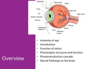
• Anatomy of eye
• Introduction
• Function of retina
• Photoceptor structure and function
• Phototransduction cascade
• Neural Pathways to the brain
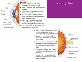
Anatomy of eye
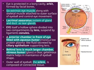
• Eye is protected in a bony cavity, orbit,
formed by facial bones.
• Six extrinsic eye muscle along with
skeletal muscle attached to outer surface
of eyeball and control eye movement.
• Lacrimal apparatus,system of gland
and duct as tear glands.
• EYE itself a hollow sphere divided into
two compartments by lens, suspend by
ligaments zonules.
• A anterior chamber in front of eye
filled with aqueous humor (moist),a low
protein plasma -like fluid secreted by
ciliary epithelium supporting lens.
• Behind lens is much larger chamber,
vitreous humor (glass) a clear, gelatinous
matrix helps in maintaince of eyeball
shape.
• Outer wall of eyeball, the sclera , is
composed of connective tissue.
INTRODUCTION
• The eye is a sensory receptor that focuses light
on a light-sensitive surface (the retina) and using
a lens and an aperture or opening (pupil) .
• VISION is the process through which light
reflected from objects in our environment is
translated into a mental image.
THIS PROCESS CAN BE DIVIDED
INTO 3 PARTS:
1. Light enters the eye and the lens focus it on the
retina.
2. Photoreceptors of the retina transduce light energy
into an electrical signal.
3. Neural pathways from the retina to brain process
electrical signals into visual image.
LIGHT ENTERS THE EYE THROUGH
PUPIL:
• Light enters the eye through cornea , which refracts it , then
passes the pupil (controlled by iris) is further focused by the
lens onto the retina.
• Light enter the eye is refracted twice : first it passes
cornea ;again when it passes lens. Two-third total refraction
occurs at cornea and remaining one third at the lens.
• Refraction occur through lens because it is capable of changing
its shape, and keep in focus is known as accommodation.
• Change in lens shape are controlled by ciliary muscle.
FUNCTION OF RETINA
• The retina is responsible for transducing light energy into
electrical impulses.It has following key functions:
1. Capture photons: the retina contains millions of light sensitive
cells called photorecptors. Two types of photoreceptors are
found in the retina of a human eye : rod and cones. Photon
hits rods and cones and start the process of transduction .
2. Produce electrical signals: The photoreceptors together with
other layers of cells in the retina convert into electrical energy.
3. Process visual signals: There are layers of cells that perform
various processing functions such as motion detection,image
sharpening and balancing the colour.
PHOTORECEPTOR
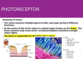
•Anatomy of retina:
• The retina contains multiple layers of cells, each layer perform different
functions.
• At the centre of the of the retina is a pitted region known as the fovea. The
fovea contains only cones which are photoreceptors sensitive to bright
colour lights.
• The fovea is responsible for detailed colour vision.
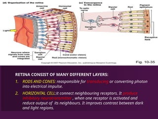
RETINA CONSIST OF MANY DIFFERENT LAYERS:
1. RODS AND CONES: reasponsible for transducing or converting photon
into electrical impulse.
2. HORIZONTAL CELL:it connect neighbouring receptors. It produce
inhibitory neurotransmitter , when one receptor is activated and
reduce output of its neighbours. It improves contrast between dark
and light regions.
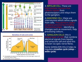
3. BIPOLAR CELL: These are
intermediate neurons that
collects signals from
photoreceptors and relays them
to the ganglion.
4.AMACRINE CELL: these are
interneurons which refine signals
from cells. They perform aspect
of spatial processing including
sharpening edges , identifying
changes such as movement, help
processing colours .
5. GANGLION CELLS: this is
output layer of cells that combine
electrical signals from bipolar
and amacrine cell and send this
to brain via optic nerve fibres.
Some GANGLION CELLS helps to
regulate circadian cycle (sleep –
wake cycle)
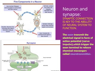
Neuron and
synapse:
SYNAPTIC CONNECTION
IS KEY TO THE ABILLITY
OF NEURAL SYSTEM TO
FUNCTION.
The axon transmit the
electrical signal in form of
action potential (nerve
impulse),which trigger the
axon terminal to release
chemical messengers
called neurotransmitter.
TRANSDUCTION CASCADE
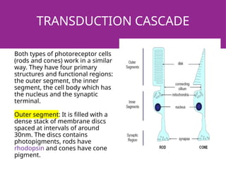
Both types of photoreceptor cells
(rods and cones) work in a similar
way. They have four primary
structures and functional regions:
the outer segment, the inner
segment, the cell body which has
the nucleus and the synaptic
terminal.
Outer segment: It is filled with a
dense stack of membrane discs
spaced at intervals of around
30nm. The discs contains
photopigments, rods have
rhodopsin and cones have cone
pigment.
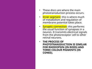
• These discs are where the main
phototransduction process occurs.
• Inner segment: this is where much
of metabolism and regulation of
membrane potential takes place.
• Synaptic connection: this performs
the usual function of synapses in a
neuron. It transmits electrical signals
from the photoreceptor cell to other
retinal neurons.
• THE PROCESS OF
PHOTOTRANSDUCTION IS SIMILAR
FOR RHODOPSIN (IN RODS) AND
THREE COLOUR PIGMENTS (IN
CONES).
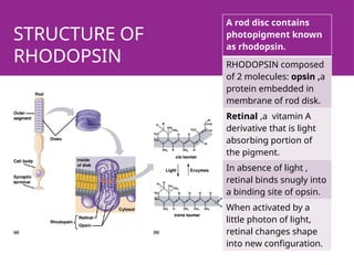
STRUCTURE OF
RHODOPSIN
A rod disc contains
photopigment known
as rhodopsin.
RHODOPSIN composed
of 2 molecules: opsin ,a
protein embedded in
membrane of rod disk.
Retinal ,a vitamin A
derivative that is light
absorbing portion of
the pigment.
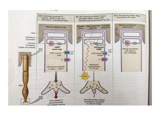
In absence of light ,
retinal binds snugly into
a binding site of opsin.
When activated by a
little photon of light,
retinal changes shape
into new configuration.
Rods contain 3
types of
channels:
1. cyclic
nucleotide
channel (cng
channels)
2. potassium
channel.
3. calcium
channel in
synaptic
terminal.
1. THE ACTIVATED RETINAL NO
LONGER BIND TO OPSIN
AND IS RELEASED FROM THE
PIGMENT IN THE PROCESS
KNOWN AS BLEACHING.
1. When there is no light
received, the sodium channel
kept opened by a chemical
called cyclic GMP (cGMP).
2. Na+ ions flows freely across
the membrane through the
channel and constantly
depolarizing.
3. When even a photon hits the
rhodopsin molecule, it
changes its shape and releases
a messenger called
transducin.
VISUAL PATHWAY
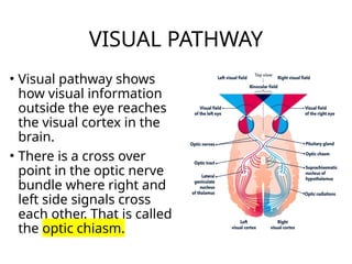
• Visual pathway shows
how visual information
outside the eye reaches
the visual cortex in the
brain.
• There is a cross over
point in the optic nerve
bundle where right and
left side signals cross
each other. That is called
the optic chiasm.
Thank
you