Comprehensive Principles and Applications of Various Microscopy Techniques
Explore different microscopy types including simple, compound, phase contrast, fluorescence, and electron microscopes, their principles, components, and scientific uses in biological and medical research.
Comprehensive Principles and Applications of Various Microscopy Techniques
1.
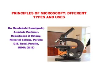
PRINCIPLES OF MICROSCOPY:DFFERENT
TYPES AND USES
Dr. Nandadulal Sannigrahi,
Associate Professor,
Department of Botany,
Nistarini College, Purulia
D.B. Road, Purulia,
INDIA (W.B)
2.
INTRODUCTION
Observation delicatelyin any scientific enquiries is very important for
study and research,
The observation of biological objects in general and small one in particular
needs much magnifications for the same,
Microscope is a device is used for getting the magnification of the object
supposed to be observed before study and research,
Light microscopy is a general term used for any type of microscopy where
light is being transmitted from a source which is on the opposite side of the
sample, to the objective lens.
Generally, the light is passed through a condenser to focus it on the sample
to have maximum brightness to form an image of the object,
After the light has passed through the sample, it goes through the objective
lens to magnify the image of the sample & then to the oculars, where the
enlarged image is viewed for the different purposes in scientific research,
The delicate observation through images can give deep insight about the
objects under study.
3.
SIMPLE MICROSCOPE
• Themost common type of microscope is simple microscope,
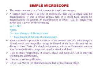
A simple microscope is a type of microscope that uses a single lens for
magnification. It uses a single convex lens of a small focal length for
magnification. In general, its magnification is about 10X. Its magnifying
power (m) is given by the formulae;
m=1+ D/F
where,
D = least distance of distinct vision
F = focal length of the lens of a microscope.
when a sample is placed in the focus of the convex lens of a microscope, a
virtual, erect, and magnified image is formed at the least distance of the
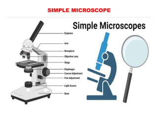
distinct vision. Parts of a simple microscope; mirror as illuminator, convex
lens for magnification, stage and metallic stand with base.
Used to study morphology of insects, algae, and fungi & Used in studying
soil type and components,
Have very low magnification,
Up to 10X Mirror for illumination and lack of mechanical stage.
COMPOUND MICROSCOPE
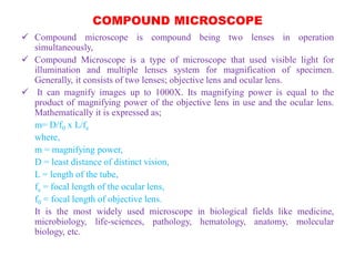
Compoundmicroscope is compound being two lenses in operation
simultaneously,
Compound Microscope is a type of microscope that used visible light for
illumination and multiple lenses system for magnification of specimen.
Generally, it consists of two lenses; objective lens and ocular lens.
It can magnify images up to 1000X. Its magnifying power is equal to the
product of magnifying power of the objective lens in use and the ocular lens.
Mathematically it is expressed as;
m= D/f0 x L/fe
where,
m = magnifying power,
D = least distance of distinct vision,
L = length of the tube,
fe = focal length of the ocular lens,
f0 = focal length of objective lens.
It is the most widely used microscope in biological fields like medicine,
microbiology, life-sciences, pathology, hematology, anatomy, molecular
biology, etc.
COMPOUND MICROSCOPE
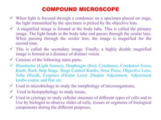
Whenlight is focused through a condenser on a specimen placed on stage,
the light transmitted by the specimen is picked by the objective lens.
A magnified image is formed at the body tube. This is called the primary
image. The light bends in the body tube and passes through the ocular lens.
When passing through the ocular lens, the image is magnified for the
second time.
This is called the secondary image. Finally, a highly double magnified
image is formed at a distance of distinct vision.
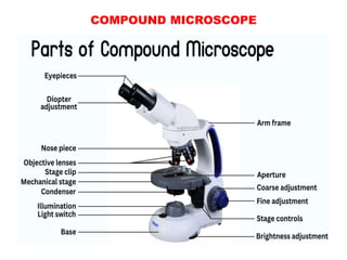
Consists of the following main parts-
Illuminator (Light Source), Diaphragm (Iris), Condenser, Condenser Focus
Knob, Rack Stop Stage, Stage Control Knobs, Nose Piece, Objective Lens,
Tube (Head), Eyepiece (Ocular Lens) ,Diopter Adjustment, Adjustment
knobs-coarse and fine etc.
Used in microbiology to study the morphology of microorganisms,
Used in histopathology to study tissue,
Used in cytology to study cellular structure of different types of cells and to
Use by biologist to observe slides of cells, tissues or segments of biological
components during the different purposes.
8.
PHASE CONTRAST MICROSCOPE
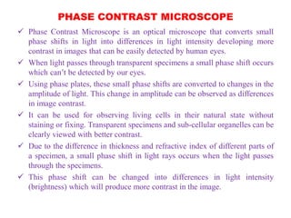
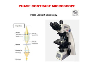
Phase Contrast Microscope is an optical microscope that converts small
phase shifts in light into differences in light intensity developing more
contrast in images that can be easily detected by human eyes.
When light passes through transparent specimens a small phase shift occurs
which can’t be detected by our eyes.
Using phase plates, these small phase shifts are converted to changes in the
amplitude of light. This change in amplitude can be observed as differences
in image contrast.
It can be used for observing living cells in their natural state without
staining or fixing. Transparent specimens and sub-cellular organelles can be
clearly viewed with better contrast.
Due to the difference in thickness and refractive index of different parts of
a specimen, a small phase shift in light rays occurs when the light passes
through the specimens.
This phase shift can be changed into differences in light intensity
(brightness) which will produce more contrast in the image.
PHASE CONTRAST MICROSCOPE
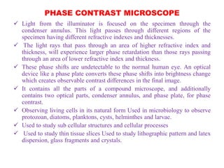
Light from the illuminator is focused on the specimen through the
condenser annulus. This light passes through different regions of the
specimen having different refractive indexes and thicknesses.
The light rays that pass through an area of higher refractive index and
thickness, will experience larger phase retardation than those rays passing
through an area of lower refractive index and thickness.
These phase shifts are undetectable to the normal human eye. An optical
device like a phase plate converts these phase shifts into brightness change
which creates observable contrast differences in the final image.
It contains all the parts of a compound microscope, and additionally
contains two optical parts, condenser annulus, and phase plate, for phase
contrast.
Observing living cells in its natural form Used in microbiology to observe
protozoan, diatoms, planktons, cysts, helminthes and larvae.
Used to study sub cellular structures and cellular processes
Used to study thin tissue slices Used to study lithographic pattern and latex
dispersion, glass fragments and crystals.
11.
FLUORESCENCE MICROSCOPE
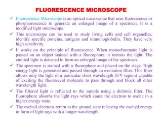
FluorescenceMicroscope is an optical microscope that uses fluorescence or
phosphorescence to generate an enlarged image of a specimen. It is a
modified light microscope.
This microscope can be used to study living cells and cell organelles,
identify specific proteins, antigens and immunoglobulin. They have very
high sensitivity.
It works on the principle of fluorescence. When monochromatic light is
passed on an object stained with a fluorophore, it reemits the light. The
emitted light is detected to form an enlarged image of the specimen.
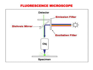
The specimen is stained with a fluorophore and placed on the stage. High
energy light is generated and passed through an excitation filter. This filter
allows only the light of a particular short wavelength (UV region) capable
of exciting the fluorescent molecule to pass through and block all other
wavelength light.
The filtered light is reflected to the sample using a dichroic filter. The
fluorophore absorbs the light rays which cause the electron to excite in a
higher energy state.
The excited electrons return to the ground state releasing the excited energy
in form of light rays with a longer wavelength.
FLUORESCENCE MICROSCOPE
Theemitted light passes through the dichroic mirror and hits the emission filter.
This filter blocks the short-wavelength light and allows longer wavelength light
to pass through ocular lenses to a detector system.
In the detector, an enlarged image is formed. The background is observed as
dark and the image appears as bright.
• Fluorophore (Fluorescent Dye)
These are the chemical compounds that possess the property of fluorescence
i.e. re-emit the light upon excitation by light. These are combinations of several
aromatic or planar compounds with several pi (π) bonds. Most of them are
organic compounds. They stain a wide range of bio-molecules and cellular
structures.
Some common fluorophores used are fluorescein, rhodamine, cyanine,
antraquinone, acridine orange, acridine yellow, auramine, malachite green, etc.
• Light Source
Commonly mercury vapor lamp is used for generating UV light. Besides,
xenon arc lamps, high-power LEDs, and lasers are also used. They emit the
light of high energy.
14.
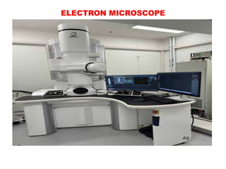
ELECTRON MICROSCOPE
Itis a microscope that uses accelerated electron, beams instead of light
rays, to illuminate the specimen and get the highly magnified image. In this
microscope, glass lenses are replaced by electromagnets.
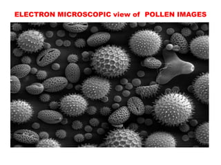
Due to the very short wavelength of electrons, this microscope produces a
very high-resolution image with magnification up to 10,000,000X. Very
high-quality images with very high contrast, revealing detailed structures
are produced.
Specimen up to 0.2 nm can be clearly viewed using an electron
microscope.
An electron microscope uses accelerated electrons with a wavelength of
about 100,000 times shorter than visible light to illuminate specimens and
produce images.
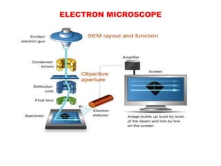
The electron gun, usually a heated tungsten or field emission filament, is
used to generate a stream of high voltage (100 – 1000 kV) electrons.
These electrons are accelerated using an anode plate in a vacuum system
and focused on the specimen using aperture and electromagnetic lenses.
15.
ELECTRON MICROSCOPE
Theelectron beam passes through the specimen and interacts with sample
components. Upon striking the specimen, the electrons are scattered. The
degree of scattering depends on the refractive index or thickness of the
specimen.
The scattered electrons from the sample are collected and passed through
objective and ocular electromagnetic lenses. These scattered beams are
detected and transformed into highly magnified images by the magnetic
lenses.
• The Electron Microscope consists of the following parts for its high
resolution and degree of accuracy:
Electron Gun- Source of electron,
Electromagnetic lens- for high magnifications by allowing electrons of
specific energy to pass through and block other electrons with three type of
lens-condenser lens, projector lens & objective lens,
Aperture system- pinholes to filter unwanted electrons.
ELECTRON MICROSCOPEs
Sampleholder- mechanical arm for holding and positioning specimens in
fixed position,
Vacuum system- Highly sensitive vacuum system contains to allow
electrons to generate and travel without colliding and scattering from their
path,
Imaging system- electromagnetic lenses used for refocusing, enlarging and
projecting images of the specimen to form an electron micrograph on a
phosphorescent plate of monitor.
• There are different types of electron microscopes-
Transmission Electron Microscope ( TEM),
Scanning electron Microscope ( SEM),
Reflection Electron Microscope ( REM),
Scanning Transmission Electron Microscope ( STEM),
Scanning Tunneling Microscope ( STM).
ELECTRON MICROSCOPE-USES ANDLIMITATIONS
Used in microbiology to study structure of viruses, flagella, pili, and
bacterial cells.
Used in crystallography, and nanotechnology,
To study morphology of cellular organelles ,
Used in forensics for ballistic study of gunshots,
Used in geology for studying rocks, minerals and gems,
Used in quality control, detection of fracture and cracks, drug development
and analysis of atomic structure.
• LIMITATIONS
Highly expensive and complex system,
Images are in black and white,
TEM requires very thin specimen,
Need of vacuum system.
21.
OTHER TYPES OFMICROSCOPES
• Besides these microscopes, different other type of microscopes are used as
follows:
Dark field microscope- uses light scattered by the specimen,
Dissecting microscope- to view 3D images during dissection by using reflected
light
Digital Microscope-lack ocular lens, uses digital camera and screen for image
observation,
Atomic Force Microscope ( ATM)- scanning probe electron microscope
Inverted Microscope- objective lens and turret below the stage to see upward
Acoustic Microscope-High frequency ultrasound wave instead of light,
X-ray Microscope-uses cathode ray tubes to produce electron beams,
Polarizing Microscope-used polarized light for illumination,
Metallurgical Microscope-uses reflected light instead of transmitted light for
imaging
Pocket Microscope-Portable , LED light source, camera having 100X
magnification,
USB Microscope-Digital camera with high power macro lens connected to
USB port.
22.
THANKS FOR YOURVISIT
• INFORMATION SOURCES:
https://microbenotes.com/types-of-microscopes/#compound-microscope-
working –principle
Microscope- University of Edinburgh,
Microscope and types- braincart.com ,
Microbe online,
Microscope Clarity,
Microscopy- Khan Academy,
DISCLAIMER:
• This presentation has been designed to address the academic fraternity
without any financial interest. This is absolutely free to use . The author
does not claim any kind of financial benefits from this content.